

Search Count: 42
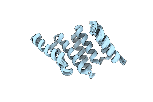 |
Apo Solution Structure Of Hop Tpr2A
|
Organism: Homo sapiens
Method: SOLUTION NMR
Release Date: 2017-03-29
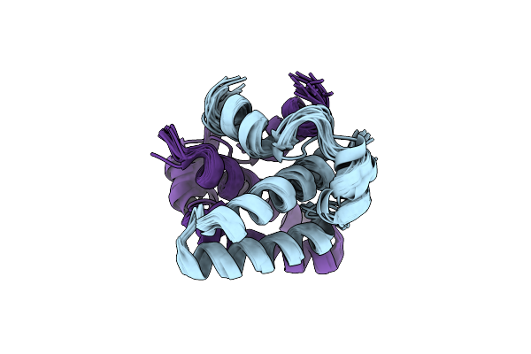 |
Solution Structure Of The Sgta N-Terminal Domain
|
Organism: Homo sapiens
Method: SOLUTION NMR
Release Date: 2014-12-03
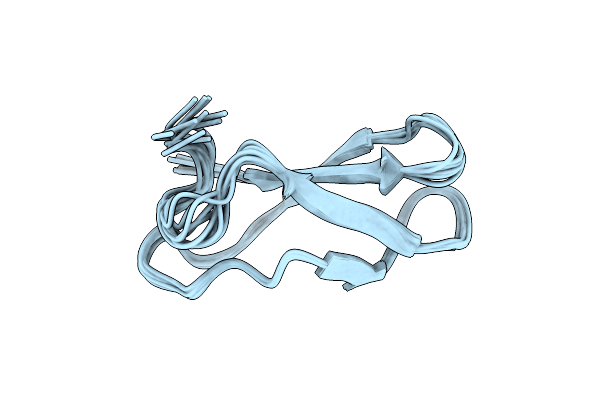 |
The Actinobacterial Transcription Factor Rbpa Binds To The Principal Sigma Subunit Of Rna Polymerase
Organism: Streptomyces coelicolor
Method: SOLUTION NMR Release Date: 2013-05-08 Classification: TRANSCRIPTION |
Organism: Streptomyces coelicolor
Method: SOLUTION NMR
Release Date: 2013-05-08
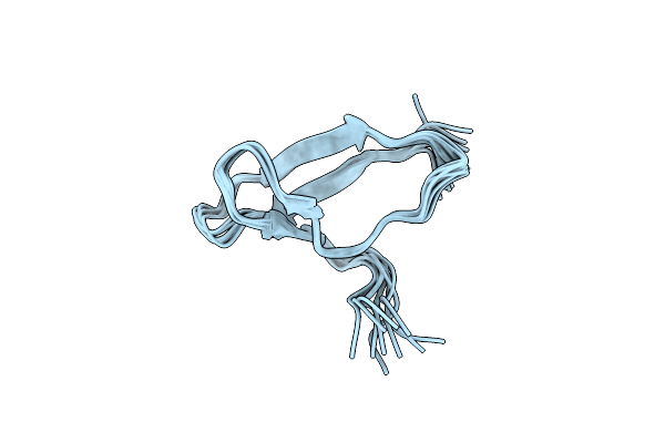 |
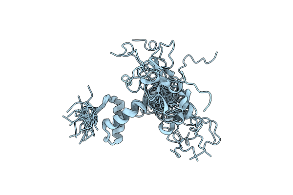
The Actinobacterial Transcription Factor Rbpa Binds To The Principal Sigma Subunit Of Rna Polymerase
Organism: Mycobacterium bovis
Method: SOLUTION NMR Release Date: 2013-05-08 Classification: TRANSCRIPTION |
Organism: Mycobacterium bovis
Method: SOLUTION NMR
Release Date: 2013-05-08
 |
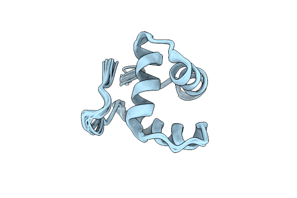
Solution Structure Of The Core Domain (11-85) Of The Murine Norovirus Vpg Protein
Organism: Murine norovirus 1
Method: SOLUTION NMR Release Date: 2013-03-27 Classification: VIRAL PROTEIN |
Organism: Murine norovirus 1
Method: SOLUTION NMR
Release Date: 2013-03-27
 |
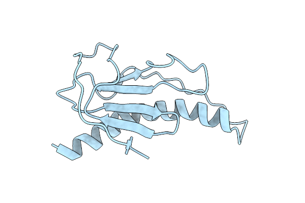
Solution Structure Of The Core Domain (10-76) Of The Feline Calicivirus Vpg Protein
Organism: Feline calicivirus
Method: SOLUTION NMR Release Date: 2013-03-27 Classification: VIRAL PROTEIN |
Organism: Feline calicivirus
Method: SOLUTION NMR
Release Date: 2013-03-27
 |
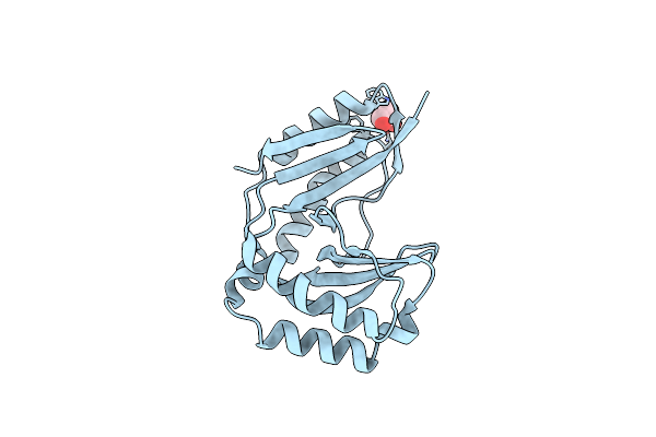
Global Fold Of The Type Iv Pilin Comp From Neisseria Meningitidis
Organism: Neisseria meningitidis
Method: SOLUTION NMR Release Date: 2013-02-13 Classification: MOTOR PROTEIN |
Organism: Neisseria meningitidis
Method: SOLUTION NMR
Release Date: 2013-02-13
 |
Crystal Structure Of Human C16Orf57
Organism: Homo sapiens
Method: X-RAY DIFFRACTION Resolution:1.10 Å Release Date: 2013-02-06 Classification: UNKNOWN FUNCTION Ligands: GOL, CL |
Organism: Homo sapiens
Method: X-RAY DIFFRACTION
Release Date: 2013-02-06
Ligands: GOL, CL
 |
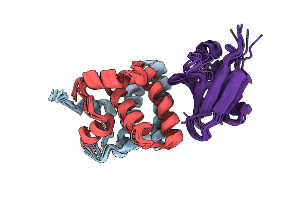
Solution Structure Of The N-Terminal Dimerisation Domain Of Sgt2
Organism: Saccharomyces cerevisiae
Method: SOLUTION NMR Release Date: 2013-01-16 Classification: CHAPERONE |
Organism: Saccharomyces cerevisiae
Method: SOLUTION NMR
Release Date: 2013-01-16
 |
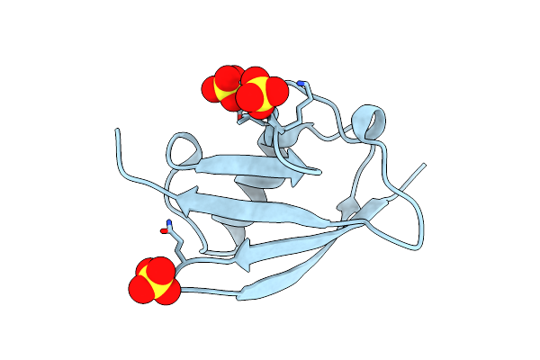
Structure Of The Complex Between The N-Terminal Dimerisation Domain Of Sgt2 And The Ubl Domain Of Get5
Organism: Saccharomyces cerevisiae
Method: SOLUTION NMR Release Date: 2013-01-16 Classification: CHAPERONE |
Organism: Saccharomyces cerevisiae
Method: SOLUTION NMR
Release Date: 2013-01-16
 |
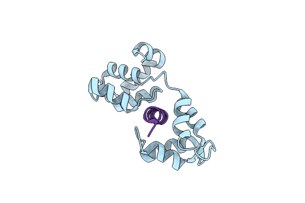
Crystal Structure Of The Ubl Domain Of Mdy2 (Get5) At 1.78A
Organism: Saccharomyces cerevisiae
Method: X-RAY DIFFRACTION Resolution:1.78 Å Release Date: 2012-11-14 Classification: PROTEIN BINDING Ligands: SO4 |
Organism: Saccharomyces cerevisiae
Method: X-RAY DIFFRACTION
Release Date: 2012-11-14
Ligands: SO4
 |
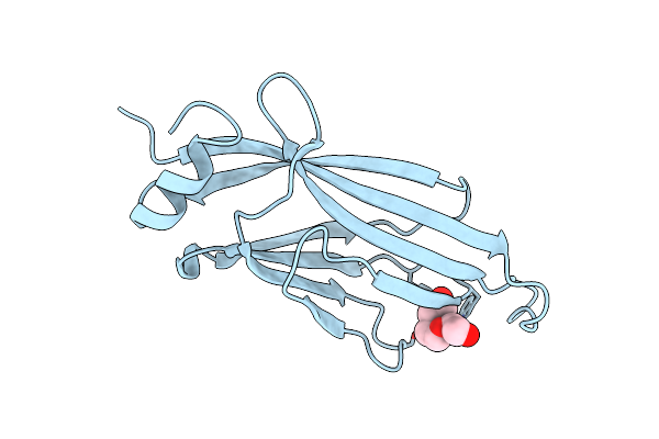
Mtip And Myoa Complex
Organism: Plasmodium falciparum
Method: X-RAY DIFFRACTION Resolution:1.94 Å Release Date: 2012-09-05 Classification: MEMBRANE PROTEIN/MOTOR PROTEIN |
Organism: Plasmodium falciparum
Method: X-RAY DIFFRACTION
Release Date: 2012-09-05
 |
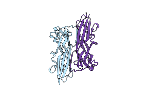
Crystal Structure Of The Taf14 Yeats Domain
Organism: Saccharomyces cerevisiae
Method: X-RAY DIFFRACTION Resolution:1.70 Å Release Date: 2012-02-29 Classification: NUCLEAR PROTEIN Ligands: PGE |
Organism: Saccharomyces cerevisiae
Method: X-RAY DIFFRACTION
Release Date: 2012-02-29
Ligands: PGE
 |
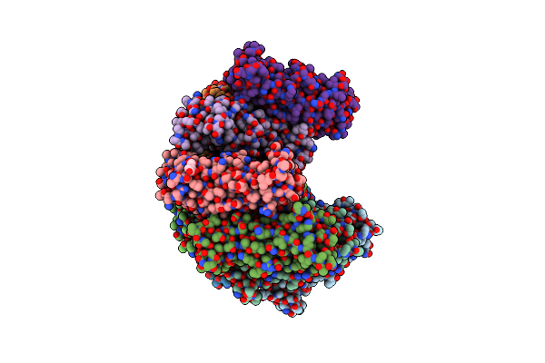
Crystal Structure Of The Biofilm Forming Subunit Of The E. Coli Common Pilus: Full Length Domain Swapped Dimer Of Ecpa
Organism: Escherichia coli
Method: X-RAY DIFFRACTION Resolution:1.78 Å Release Date: 2012-02-22 Classification: CELL ADHESION Ligands: IOD |
Organism: Escherichia coli
Method: X-RAY DIFFRACTION
Release Date: 2012-02-22
Ligands: IOD
 |
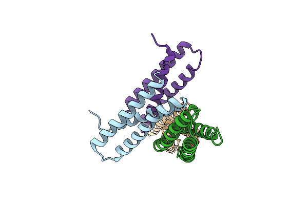
Crystal Structure Of The Biofilm Forming Subunit Of The E. Coli Common Pilus: Donor Strand Complemented (Dsc) Ecpa
Organism: Escherichia coli
Method: X-RAY DIFFRACTION Resolution:2.10 Å Release Date: 2012-02-22 Classification: CELL ADHESION Ligands: FLC |
Organism: Escherichia coli
Method: X-RAY DIFFRACTION
Release Date: 2012-02-22
Ligands: FLC
 |
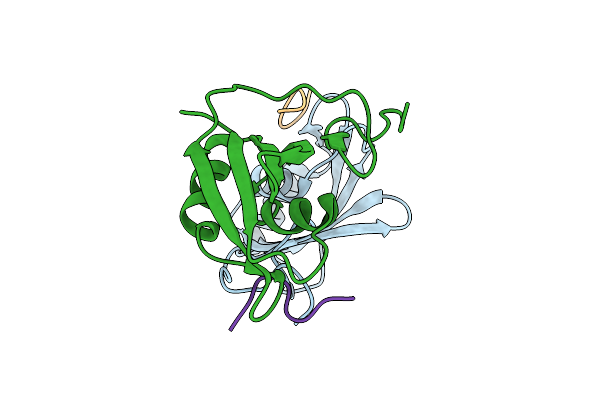
Structure Of Fap-Nra At Ph 5.0
Organism: Streptococcus parasanguinis
Method: X-RAY DIFFRACTION Resolution:3.00 Å Release Date: 2011-12-28 Classification: STRUCTURAL PROTEIN Ligands: GLC |
Organism: Streptococcus parasanguinis
Method: X-RAY DIFFRACTION
Release Date: 2011-12-28
Ligands: GLC
 |
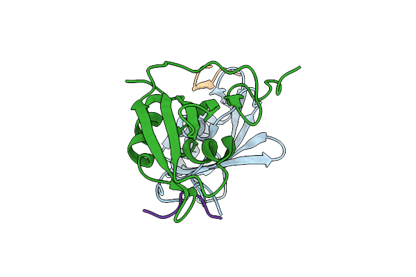
Crystal Structure Of A Raver1 Pri3 Peptide In Complex With Polypyrimidine Tract Binding Protein Rrm2
Organism: Homo sapiens, Mus musculus
Method: X-RAY DIFFRACTION Resolution:1.40 Å Release Date: 2011-12-28 Classification: PROTEIN BINDING |
Organism: Homo sapiens, Mus musculus
Method: X-RAY DIFFRACTION
Release Date: 2011-12-28
 |
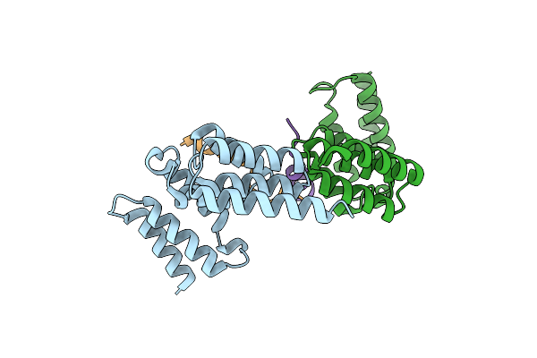
Crystal Structure Of A Raver1 Pri4 Peptide In Complex With Polypyrimidine Tract Binding Protein Rrm2
Organism: Homo sapiens, Mus musculus
Method: X-RAY DIFFRACTION Resolution:1.55 Å Release Date: 2011-12-28 Classification: PROTEIN BINDING Ligands: IOD |
Organism: Homo sapiens, Mus musculus
Method: X-RAY DIFFRACTION
Release Date: 2011-12-28
Ligands: IOD
 |
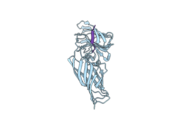
Kinetochore-Bubr1 Kinase Complex
Organism: Homo sapiens
Method: X-RAY DIFFRACTION Resolution:2.20 Å Release Date: 2011-10-26 Classification: CELL CYCLE |
Organism: Homo sapiens
Method: X-RAY DIFFRACTION
Release Date: 2011-10-26
 |
Structure Of N-Terminal Domain Of Candida Albicans Als9-2 In Complex With Human Fibrinogen Gamma Peptide
Organism: Candida albicans, Homo sapiens
Method: X-RAY DIFFRACTION Resolution:1.49 Å Release Date: 2011-10-05 Classification: CELL ADHESION |
Organism: Candida albicans, Homo sapiens
Method: X-RAY DIFFRACTION
Release Date: 2011-10-05