

Search Count: 54
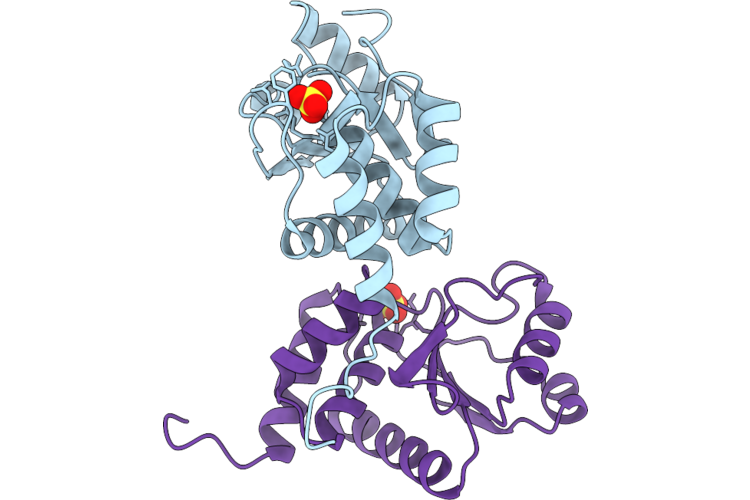 |
Crystal Structure Of Bacterial Dusp From Candidatus Chlorohelix Allophototropha
Organism: Candidatus chlorohelix allophototropha
Method: X-RAY DIFFRACTION Resolution:2.40 Å Release Date: 2026-08-12 Classification: HYDROLASE Ligands: SO4, CL |
Organism: Candidatus chlorohelix allophototropha
Method: X-RAY DIFFRACTION
Release Date: 2026-08-12
Ligands: SO4, CL
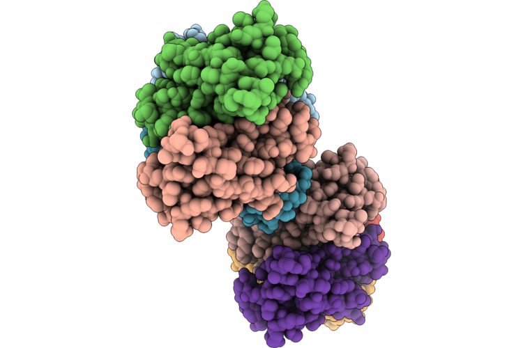 |
Crystal Structure Of Human Transthyretin (Ttr) With Pryazole-Based Stabilizer
Organism: Homo sapiens
Method: X-RAY DIFFRACTION Resolution:1.58 Å Release Date: 2026-05-06 Classification: TRANSPORT PROTEIN Ligands: A1MB7 |
Organism: Homo sapiens
Method: X-RAY DIFFRACTION
Release Date: 2026-05-06
Ligands: A1MB7
 |
Crystal Structure Of The Mu2 Subunit Of The Clathrin-Adaptor Protein 2 (Ap2) Bound To Hpv16 E7(Residues 22-39)
Organism: Rattus norvegicus, Human papillomavirus 16
Method: X-RAY DIFFRACTION Resolution:3.70 Å Release Date: 2025-09-10 Classification: ENDOCYTOSIS |
Organism: Rattus norvegicus, Human papillomavirus 16
Method: X-RAY DIFFRACTION
Release Date: 2025-09-10
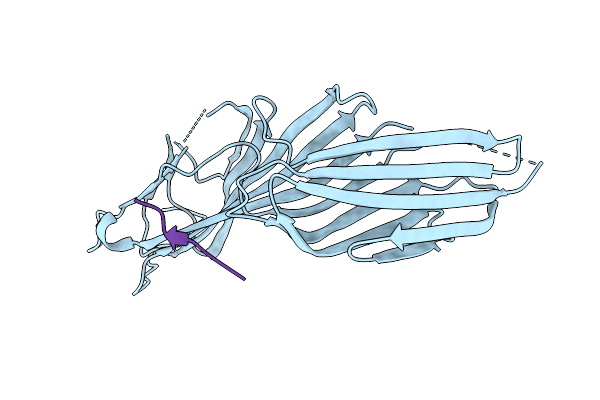 |
Crystal Structure Of The Mu2 Subunit Of The Clathrin-Adaptor Protein 2 (Ap2) Bound To Hpv16 E7(Residues 22-32; S31E And S32E)
Organism: Rattus norvegicus, Human papillomavirus 16
Method: X-RAY DIFFRACTION Resolution:3.20 Å Release Date: 2025-09-10 Classification: ENDOCYTOSIS |
Organism: Rattus norvegicus, Human papillomavirus 16
Method: X-RAY DIFFRACTION
Release Date: 2025-09-10
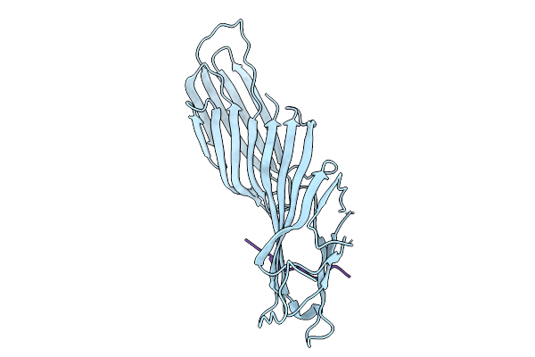 |
Crystal Structure Of The Mu2 Subunit Of The Clathrin-Adaptor Protein 2 (Ap2) Bound To Hpv16 E7(Residues 22-39; S31E And S32E)
Organism: Rattus norvegicus, Human papillomavirus 16
Method: X-RAY DIFFRACTION Resolution:3.30 Å Release Date: 2025-09-10 Classification: ENDOCYTOSIS |
Organism: Rattus norvegicus, Human papillomavirus 16
Method: X-RAY DIFFRACTION
Release Date: 2025-09-10
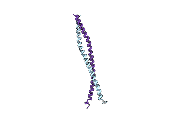 |
Crystal Structure Of Hook3(553-624)
Organism: Homo sapiens
Method: X-RAY DIFFRACTION Resolution:2.70 Å Release Date: 2025-02-26 Classification: TRANSPORT PROTEIN |
Organism: Homo sapiens
Method: X-RAY DIFFRACTION
Release Date: 2025-02-26
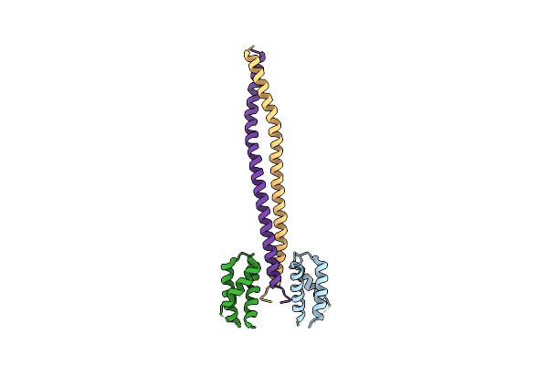 |
Crystal Structure Of Hook3(553-624) Bound To Kif1C(714-809)
Organism: Homo sapiens
Method: X-RAY DIFFRACTION Resolution:3.00 Å Release Date: 2025-02-26 Classification: TRANSPORT PROTEIN |
Organism: Homo sapiens
Method: X-RAY DIFFRACTION
Release Date: 2025-02-26
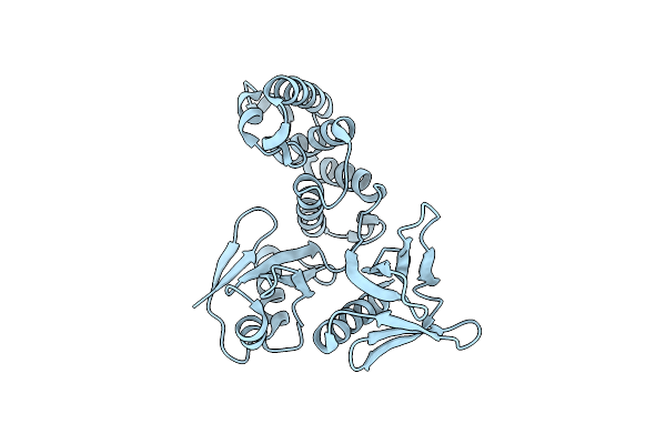 |
Crystal Structure Of The Ptpn21 Ferm Domain
Organism: Homo sapiens
Method: X-RAY DIFFRACTION Resolution:2.10 Å Release Date: 2024-07-10 Classification: PROTEIN BINDING Ligands: CL |
Organism: Homo sapiens
Method: X-RAY DIFFRACTION
Release Date: 2024-07-10
Ligands: CL
 |
Crystal Structure Of Ubr Box Of Ubr4 Apo
Organism: Homo sapiens
Method: X-RAY DIFFRACTION Resolution:2.18 Å Release Date: 2023-12-06 Classification: LIGASE Ligands: ZN |
Organism: Homo sapiens
Method: X-RAY DIFFRACTION
Release Date: 2023-12-06
Ligands: ZN
 |
Crystal Structure Of Ubr Box Of Yifs-Ubr4
Organism: Homo sapiens
Method: X-RAY DIFFRACTION Resolution:1.65 Å Release Date: 2023-12-06 Classification: LIGASE Ligands: ZN |
Organism: Homo sapiens
Method: X-RAY DIFFRACTION
Release Date: 2023-12-06
Ligands: ZN
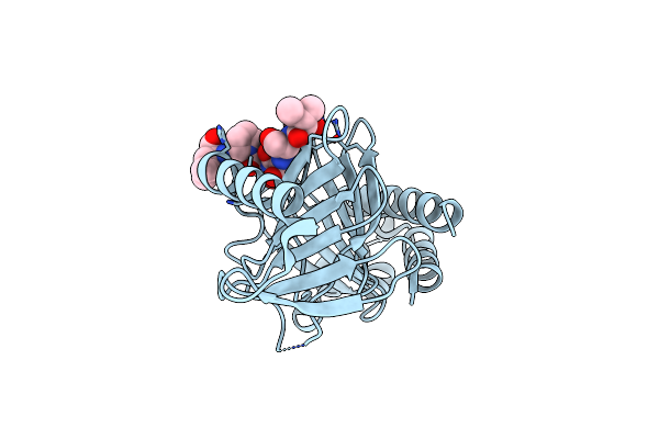 |
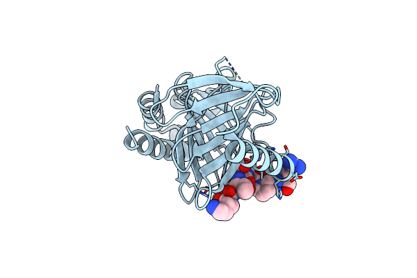
Plk1 Polo-Box Domain Bound To Hpv18 L2 Residues 209-215 With Pthr213
Organism: Homo sapiens, Human papillomavirus 18
Method: X-RAY DIFFRACTION Resolution:1.80 Å Release Date: 2023-10-11 Classification: CELL CYCLE |
Organism: Homo sapiens, Human papillomavirus 18
Method: X-RAY DIFFRACTION
Release Date: 2023-10-11
 |
Plk1 Polo-Box Domain Bound To Hpv4 L2 Residues 251-257 With Pthr255
Organism: Homo sapiens, Human papillomavirus 4
Method: X-RAY DIFFRACTION Resolution:2.61 Å Release Date: 2023-10-11 Classification: CELL CYCLE |
Organism: Homo sapiens, Human papillomavirus 4
Method: X-RAY DIFFRACTION
Release Date: 2023-10-11
 |
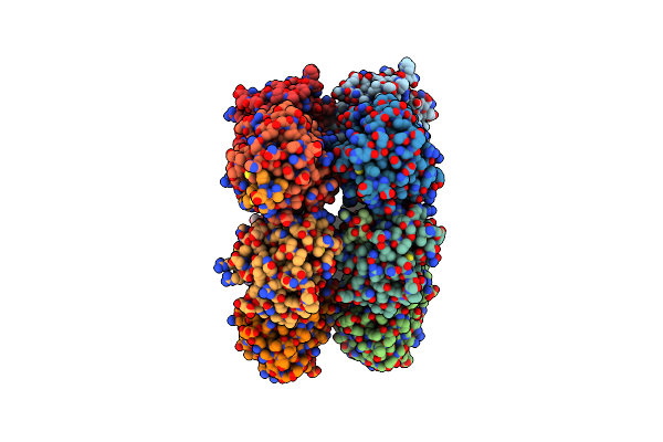
Crystal Structure Of Bak Bound To Bnip5 Bh3
Organism: Homo sapiens
Method: X-RAY DIFFRACTION Resolution:1.70 Å Release Date: 2023-09-20 Classification: APOPTOSIS |
Organism: Homo sapiens
Method: X-RAY DIFFRACTION
Release Date: 2023-09-20
 |
Crystal Structure Of Human Bak In Complex With The Pxt1 Bh3 Domain
Organism: Homo sapiens
Method: X-RAY DIFFRACTION Resolution:2.20 Å Release Date: 2023-05-10 Classification: APOPTOSIS |
Organism: Homo sapiens
Method: X-RAY DIFFRACTION
Release Date: 2023-05-10
 |
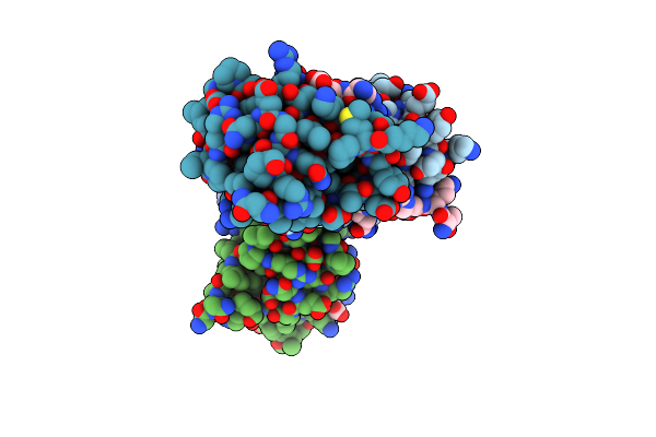
Crystal Structure Of Bcl-Xl Bound To The Bh3 Domain Of Human Pxt1
Organism: Mus musculus, Homo sapiens
Method: X-RAY DIFFRACTION Resolution:1.70 Å Release Date: 2022-09-14 Classification: APOPTOSIS Ligands: SO4, MG |
Organism: Mus musculus, Homo sapiens
Method: X-RAY DIFFRACTION
Release Date: 2022-09-14
Ligands: SO4, MG
 |
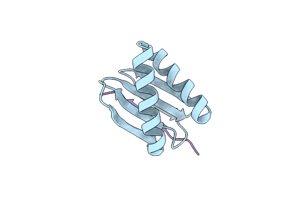
Crystal Structure Of Ptpn4 Pdz Bound To The Pbm Of Hpv16 E6
Organism: Homo sapiens, Human papillomavirus type 16
Method: X-RAY DIFFRACTION Resolution:2.88 Å Release Date: 2022-03-02 Classification: PROTEIN BINDING |
Organism: Homo sapiens, Human papillomavirus type 16
Method: X-RAY DIFFRACTION
Release Date: 2022-03-02
 |
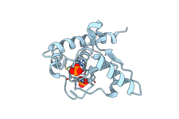
Human Lc8 Bound To Ebola Virus Vp35(67-76)
Organism: Homo sapiens, Ebola virus
Method: X-RAY DIFFRACTION Resolution:2.40 Å Release Date: 2021-04-14 Classification: UNKNOWN FUNCTION |
Organism: Homo sapiens, Ebola virus
Method: X-RAY DIFFRACTION
Release Date: 2021-04-14
 |
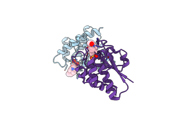
Crystal Structure Of Darp, Drosophila Arginine Phosphatase
Organism: Drosophila melanogaster
Method: X-RAY DIFFRACTION Resolution:2.19 Å Release Date: 2021-01-13 Classification: SIGNALING PROTEIN Ligands: PO4, CL |
Organism: Drosophila melanogaster
Method: X-RAY DIFFRACTION
Release Date: 2021-01-13
Ligands: PO4, CL
 |
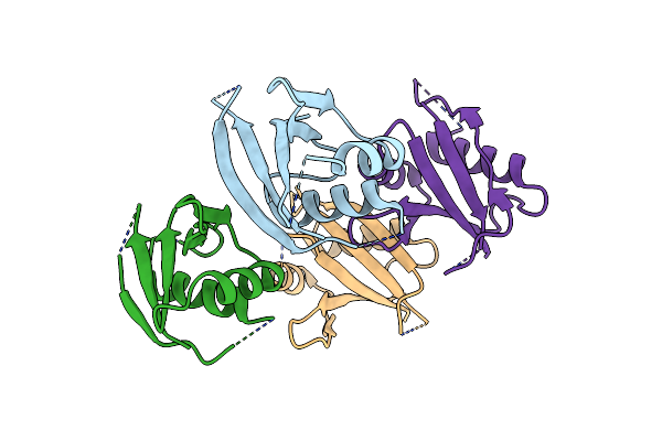
Crystal Structure Of Primo-1
Organism: Drosophila melanogaster
Method: X-RAY DIFFRACTION Resolution:2.08 Å Release Date: 2021-01-13 Classification: HYDROLASE Ligands: EPE |
Organism: Drosophila melanogaster
Method: X-RAY DIFFRACTION
Release Date: 2021-01-13
Ligands: EPE
 |
The Crystal Structure Of Pb1 Homo-Dimer Of Human P62/Sqstm1
Organism: Homo sapiens
Method: X-RAY DIFFRACTION Resolution:3.20 Å Release Date: 2020-01-15 Classification: SIGNALING PROTEIN |
Organism: Homo sapiens
Method: X-RAY DIFFRACTION
Release Date: 2020-01-15