

Search Count: 39
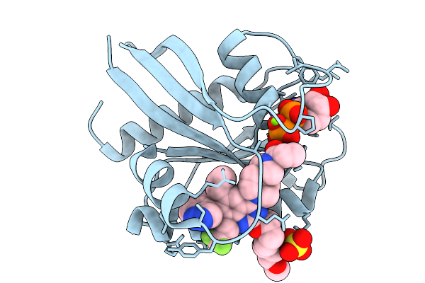 |
Krasg12C In Complex With Asp2453
Organism: Homo sapiens
Method: X-RAY DIFFRACTION Resolution:1.75 Å Release Date: 2026-03-04 Classification: HYDROLASE Ligands: GDP, MG, A1L9E, SO4 |
Organism: Homo sapiens
Method: X-RAY DIFFRACTION
Release Date: 2026-03-04
Ligands: GDP, MG, A1L9E, SO4
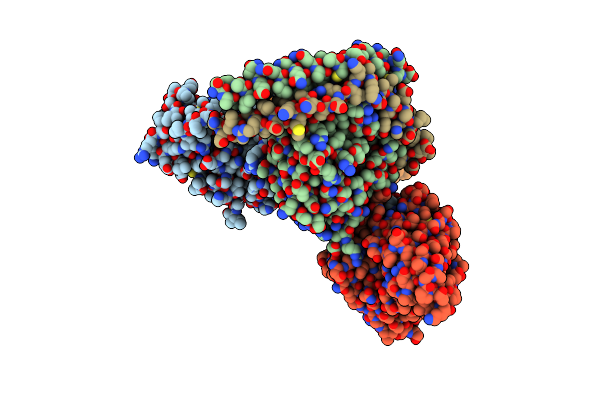 |
Full Agonist-Bound Mu-Type Opioid Receptor-G Protein Complex
Organism: Escherichia coli, Homo sapiens, Macaca mulatta, Synthetic construct
Method: ELECTRON MICROSCOPY Release Date: 2024-05-29 Classification: SIGNALING PROTEIN |
Organism: Escherichia coli, Homo sapiens, Macaca mulatta, Synthetic construct
Method: ELECTRON MICROSCOPY
Release Date: 2024-05-29
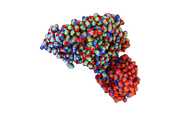 |
Full Agonist- And Positive Allosteric Modulator-Bound Mu-Type Opioid Receptor-G Protein Complex
Organism: Escherichia coli, Homo sapiens, Macaca mulatta, Synthetic construct
Method: ELECTRON MICROSCOPY Release Date: 2024-05-29 Classification: SIGNALING PROTEIN Ligands: VV9 |
Organism: Escherichia coli, Homo sapiens, Macaca mulatta, Synthetic construct
Method: ELECTRON MICROSCOPY
Release Date: 2024-05-29
Ligands: VV9
 |
Krasg12C In Complex With Inhibitor
Organism: Homo sapiens
Method: X-RAY DIFFRACTION Resolution:1.85 Å Release Date: 2024-01-17 Classification: HYDROLASE/INHIBITOR Ligands: GDP, MG, Y9D |
Organism: Homo sapiens
Method: X-RAY DIFFRACTION
Release Date: 2024-01-17
Ligands: GDP, MG, Y9D
 |
Cryo-Em Structure Of The J-K-St Region Of Emcv Ires In Complex With Eif4G-Heat1 And Eif4A
Organism: Homo sapiens, Encephalomyocarditis virus
Method: ELECTRON MICROSCOPY Release Date: 2023-08-02 Classification: TRANSLATION Ligands: MG |
Organism: Homo sapiens, Encephalomyocarditis virus
Method: ELECTRON MICROSCOPY
Release Date: 2023-08-02
Ligands: MG
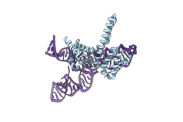 |
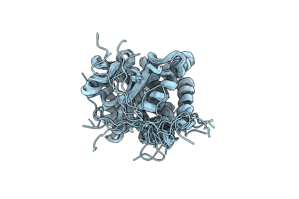
Cryo-Em Structure Of The J-K-St Region Of Emcv Ires In Complex With Eif4G-Heat1 And Eif4A (J-K-St/Eif4G Focused)
Organism: Homo sapiens, Encephalomyocarditis virus
Method: ELECTRON MICROSCOPY Resolution:3.70 Å Release Date: 2023-08-02 Classification: TRANSLATION Ligands: MG |
Organism: Homo sapiens, Encephalomyocarditis virus
Method: ELECTRON MICROSCOPY
Release Date: 2023-08-02
Ligands: MG
 |
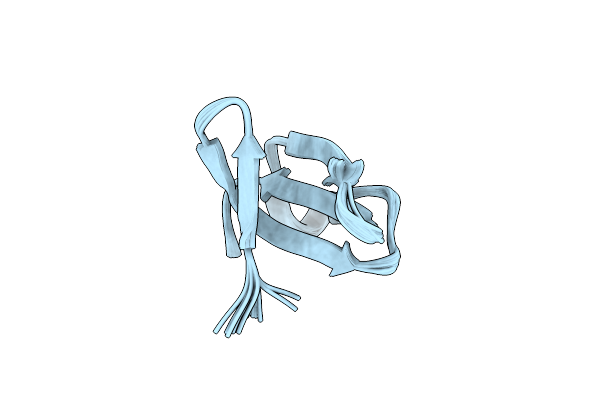
Multi-State Structure Determination And Dynamics Analysis Elucidate A New Ubiquitin-Recognition Mechanism Of Yeast Ubiquitin C-Terminal Hydrolase.
Organism: Saccharomyces cerevisiae
Method: SOLUTION NMR Release Date: 2022-04-20 Classification: HYDROLASE |
Organism: Saccharomyces cerevisiae
Method: SOLUTION NMR
Release Date: 2022-04-20
 |
Solution Structure Of Recombinant Apetx1
Organism: Anthopleura elegantissima
Method: SOLUTION NMR Release Date: 2020-12-23 Classification: TOXIN |
Organism: Anthopleura elegantissima
Method: SOLUTION NMR
Release Date: 2020-12-23
 |
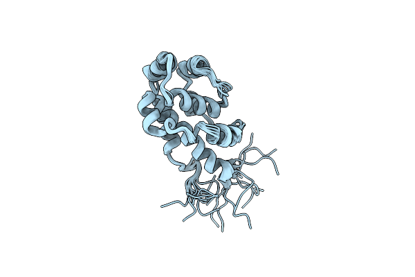
The Microtubule-Binding Domains Of Yeast Cytoplasmic Dynein In The High Affinity State
Organism: Saccharomyces cerevisiae (strain atcc 204508 / s288c)
Method: SOLUTION NMR Release Date: 2020-03-18 Classification: MOTOR PROTEIN |
Organism: Saccharomyces cerevisiae (strain atcc 204508 / s288c)
Method: SOLUTION NMR
Release Date: 2020-03-18
 |
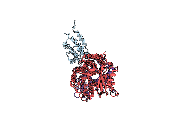
The Microtubule-Binding Domains Of Yeast Cytoplasmic Dynein In The Low Affinity State
Organism: Saccharomyces cerevisiae (strain atcc 204508 / s288c)
Method: SOLUTION NMR Release Date: 2020-03-18 Classification: MOTOR PROTEIN |
Organism: Saccharomyces cerevisiae (strain atcc 204508 / s288c)
Method: SOLUTION NMR
Release Date: 2020-03-18
 |
Complex Of Yeast Cytoplasmic Dynein Mtbd-High And Mt Without Dtt
Organism: Saccharomyces cerevisiae s288c, Sus scrofa
Method: ELECTRON MICROSCOPY Release Date: 2020-03-04 Classification: MOTOR PROTEIN/STRUCTURAL PROTEIN |
Organism: Saccharomyces cerevisiae s288c, Sus scrofa
Method: ELECTRON MICROSCOPY
Release Date: 2020-03-04
 |
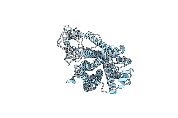
Complex Of Yeast Cytoplasmic Dynein Mtbd-High And Mt With Dtt
Organism: Saccharomyces cerevisiae s288c, Sus scrofa
Method: ELECTRON MICROSCOPY Release Date: 2020-03-04 Classification: MOTOR PROTEIN/STRUCTURAL PROTEIN |
Organism: Saccharomyces cerevisiae s288c, Sus scrofa
Method: ELECTRON MICROSCOPY
Release Date: 2020-03-04
 |
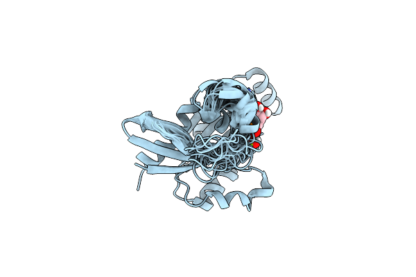
Structure Of The Beta2 Adrenergic Receptor In The Full Agonist Bound State
Organism: Homo sapiens
Method: SOLUTION NMR Release Date: 2020-01-29 Classification: MEMBRANE PROTEIN |
Organism: Homo sapiens
Method: SOLUTION NMR
Release Date: 2020-01-29
 |
Structure Of Rac1 In The Low-Affinity State For Mg2+
Organism: Homo sapiens
Method: SOLUTION NMR Release Date: 2019-03-20 Classification: SIGNALING PROTEIN Ligands: GDP, MG |
Organism: Homo sapiens
Method: SOLUTION NMR
Release Date: 2019-03-20
Ligands: GDP, MG
 |
Crystal Structure Of The Cytosolic Domain Of The Mg2+ Channel Mgte In Complex With Atp
Organism: Thermus thermophilus (strain hb8 / atcc 27634 / dsm 579)
Method: X-RAY DIFFRACTION Resolution:3.00 Å Release Date: 2017-08-16 Classification: METAL TRANSPORT Ligands: ATP, MG |
Organism: Thermus thermophilus (strain hb8 / atcc 27634 / dsm 579)
Method: X-RAY DIFFRACTION
Release Date: 2017-08-16
Ligands: ATP, MG
 |
Crystal Structure Of The Mg2+ Channel Mgte In Complex With Atp
Organism: Thermus thermophilus (strain hb8 / atcc 27634 / dsm 579)
Method: X-RAY DIFFRACTION Resolution:3.60 Å Release Date: 2017-08-16 Classification: METAL TRANSPORT Ligands: ATP, MG |
Organism: Thermus thermophilus (strain hb8 / atcc 27634 / dsm 579)
Method: X-RAY DIFFRACTION
Release Date: 2017-08-16
Ligands: ATP, MG
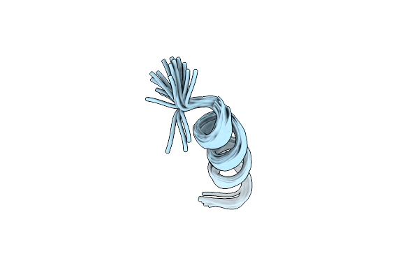 |
Human Ccr2 Membrane-Proximal C-Terminal Region (Pro-C) In A Membrane Bound Form
Organism: Homo sapiens
Method: SOLUTION NMR Release Date: 2014-10-08 Classification: SIGNALING PROTEIN |
Organism: Homo sapiens
Method: SOLUTION NMR
Release Date: 2014-10-08
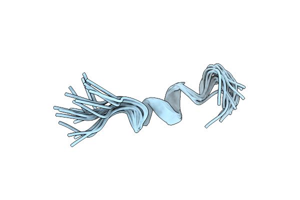 |
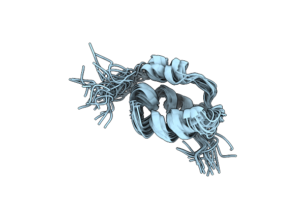
Human Ccr2 Membrane-Proximal C-Terminal Region (Pro-C) In A Frount Bound Form
Organism: Homo sapiens
Method: SOLUTION NMR Release Date: 2014-10-08 Classification: SIGNALING PROTEIN |
Organism: Homo sapiens
Method: SOLUTION NMR
Release Date: 2014-10-08
 |
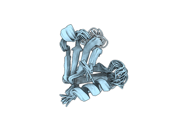
Solution Structure Of Mouse Pheromone Esp1
Organism: Mus musculus
Method: SOLUTION NMR Release Date: 2013-04-17 Classification: SIGNALING PROTEIN |
Organism: Mus musculus
Method: SOLUTION NMR
Release Date: 2013-04-17
 |
Solution Structure Of The Cert Ph Domain
Organism: Homo sapiens
Method: SOLUTION NMR Release Date: 2012-08-15 Classification: LIPID TRANSPORT |
Organism: Homo sapiens
Method: SOLUTION NMR
Release Date: 2012-08-15