

Search Count: 18
 |
The Receptor Binding Domain Of Sars-Cov-2 Omicron Variant Spike Glycoprotein In Complex With Beta-55 And Ey6A Fabs
Organism: Homo sapiens, Severe acute respiratory syndrome coronavirus 2
Method: X-RAY DIFFRACTION Resolution:2.40 Å Release Date: 2022-01-19 Classification: VIRAL PROTEIN Ligands: GOL, ACT, CL |
Organism: Homo sapiens, Severe acute respiratory syndrome coronavirus 2
Method: X-RAY DIFFRACTION
Release Date: 2022-01-19
Ligands: GOL, ACT, CL
 |
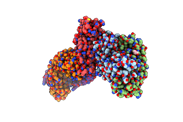
The Receptor Binding Domain Of Sars-Cov-2 Spike Glycoprotein In Complex With Beta-55 And Ey6A Fabs
Organism: Homo sapiens, Severe acute respiratory syndrome coronavirus 2
Method: X-RAY DIFFRACTION Resolution:2.92 Å Release Date: 2022-01-19 Classification: VIRAL PROTEIN |
Organism: Homo sapiens, Severe acute respiratory syndrome coronavirus 2
Method: X-RAY DIFFRACTION
Release Date: 2022-01-19
 |
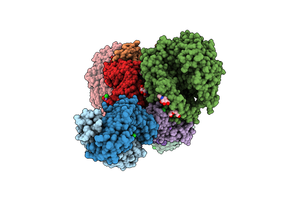
The Receptor Binding Domain Of Sars-Cov-2 Spike Glycoprotein In Complex With Covox-58 And Covox-158 Fabs
Organism: Homo sapiens, Severe acute respiratory syndrome coronavirus 2
Method: X-RAY DIFFRACTION Resolution:2.84 Å Release Date: 2022-01-19 Classification: VIRAL PROTEIN Ligands: GOL, NAG, NA |
Organism: Homo sapiens, Severe acute respiratory syndrome coronavirus 2
Method: X-RAY DIFFRACTION
Release Date: 2022-01-19
Ligands: GOL, NAG, NA
 |
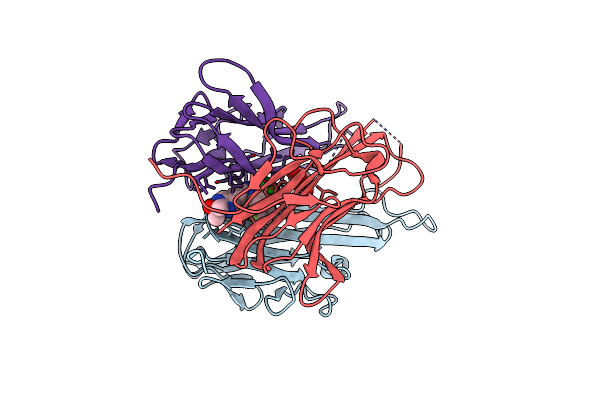
Circulating Sars-Cov-2 Spike N439K Variants Maintain Fitness While Evading Antibody-Mediated Immunity
Organism: Homo sapiens, Severe acute respiratory syndrome coronavirus 2
Method: X-RAY DIFFRACTION Resolution:2.78 Å Release Date: 2021-02-17 Classification: VIRAL PROTEIN/HYDROLASE Ligands: CL, SO4, NA, PG5, PG4, ZN, NAG, PGE |
Organism: Homo sapiens, Severe acute respiratory syndrome coronavirus 2
Method: X-RAY DIFFRACTION
Release Date: 2021-02-17
Ligands: CL, SO4, NA, PG5, PG4, ZN, NAG, PGE
 |
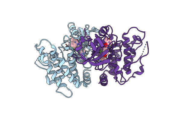
Human Tnf-Alpha In Complex With 2-[5-(3-Chloro-4-{[(1R)-1-(2-Fluorophenyl)Ethyl]Amino}Quinolin-6-Yl)Pyrimidin-2-Yl]Propan-2-Ol
Organism: Homo sapiens
Method: X-RAY DIFFRACTION Resolution:2.10 Å Release Date: 2020-12-09 Classification: CYTOKINE Ligands: VGY, GOL |
Organism: Homo sapiens
Method: X-RAY DIFFRACTION
Release Date: 2020-12-09
Ligands: VGY, GOL
 |
Crystal Structure Of Mripk3 Complexed With N-(3-Fluoro-4-{1H-Pyrrolo[2,3-B]Pyridin-4-Yloxy}Phenyl)-1-(4-Fluorophenyl)-2-Oxo-1,2-Dihydropyridine-3-Carboxamide
Organism: Mus musculus
Method: X-RAY DIFFRACTION Resolution:2.10 Å Release Date: 2019-07-17 Classification: TRANSFERASE Ligands: 1FN |
Organism: Mus musculus
Method: X-RAY DIFFRACTION
Release Date: 2019-07-17
Ligands: 1FN
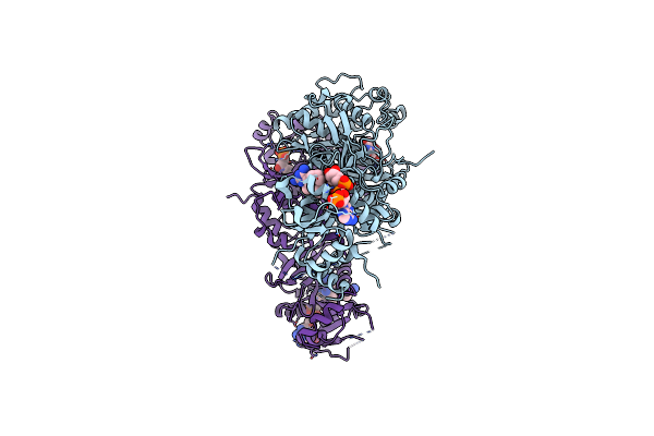 |
Crystal Structure Of Wild Type Plasmodium Falciparum Dhfr-Ts Complexed With Bt1, Nadph, And Dump
Organism: Plasmodium falciparum
Method: X-RAY DIFFRACTION Resolution:2.38 Å Release Date: 2019-04-24 Classification: OXIDOREDUCTASE Ligands: 9QO, NAP, UMP |
Organism: Plasmodium falciparum
Method: X-RAY DIFFRACTION
Release Date: 2019-04-24
Ligands: 9QO, NAP, UMP
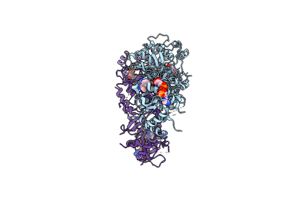 |
Crystal Structure Of Quadruple Mutant (N51I+C59R+S108N+I164L) Plasmodium Falciparum Dhfr-Ts Complexed With Bt1, Nadph, And Dump
Organism: Plasmodium falciparum
Method: X-RAY DIFFRACTION Resolution:2.38 Å Release Date: 2019-04-24 Classification: OXIDOREDUCTASE Ligands: 9QO, NAP, UMP |
Organism: Plasmodium falciparum
Method: X-RAY DIFFRACTION
Release Date: 2019-04-24
Ligands: 9QO, NAP, UMP
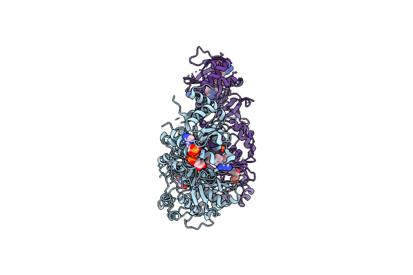 |
Crystal Structure Of Wild Type Plasmodium Falciparum Dhfr-Ts Complexed With Bt2, Nadph, And Dump
Organism: Plasmodium falciparum
Method: X-RAY DIFFRACTION Resolution:2.20 Å Release Date: 2019-04-24 Classification: OXIDOREDUCTASE Ligands: 9QR, NAP, UMP, PO4 |
Organism: Plasmodium falciparum
Method: X-RAY DIFFRACTION
Release Date: 2019-04-24
Ligands: 9QR, NAP, UMP, PO4
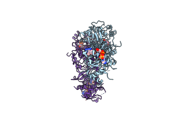 |
Crystal Structure Of Quadruple Mutant (N51I+C59R+S108N+I164L) Plasmodium Falciparum Dhfr-Ts Complexed With Bt2, Nadph, And Dump
Organism: Plasmodium falciparum
Method: X-RAY DIFFRACTION Resolution:2.45 Å Release Date: 2019-04-24 Classification: OXIDOREDUCTASE Ligands: 9QR, NAP, UMP |
Organism: Plasmodium falciparum
Method: X-RAY DIFFRACTION
Release Date: 2019-04-24
Ligands: 9QR, NAP, UMP
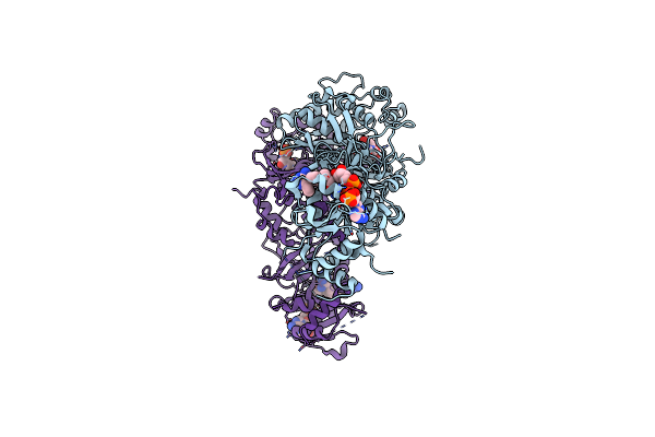 |
Crystal Structure Of Wild Type Plasmodium Falciparum Dhfr-Ts Complexed With Bt3, Nadph, And Dump
Organism: Plasmodium falciparum
Method: X-RAY DIFFRACTION Resolution:2.35 Å Release Date: 2019-04-24 Classification: OXIDOREDUCTASE/OXIDOREDUCTASE INHIBITOR Ligands: 9RR, NAP, UMP |
Organism: Plasmodium falciparum
Method: X-RAY DIFFRACTION
Release Date: 2019-04-24
Ligands: 9RR, NAP, UMP
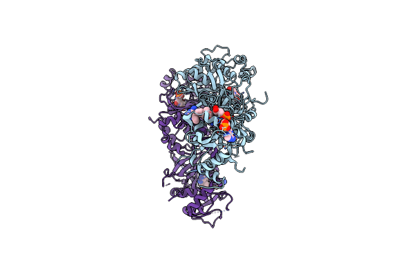 |
Crystal Structure Of Quadruple Mutant (N51I+C59R+S108N+I164L) Plasmodium Falciparum Dhfr-Ts Complexed With Bt3, Nadph, And Dump
Organism: Plasmodium falciparum
Method: X-RAY DIFFRACTION Resolution:2.60 Å Release Date: 2019-04-24 Classification: OXIDOREDUCTASE/OXIDOREDUCTASE INHIBITOR Ligands: 9RR, NAP, UMP |
Organism: Plasmodium falciparum
Method: X-RAY DIFFRACTION
Release Date: 2019-04-24
Ligands: 9RR, NAP, UMP
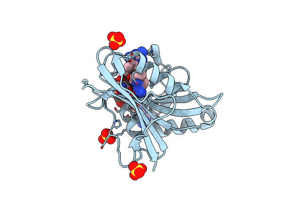 |
Human Dihydrofolate Reductase Complexed With Nadph And Bt1
Organism: Homo sapiens
Method: X-RAY DIFFRACTION Resolution:2.06 Å Release Date: 2019-04-10 Classification: OXIDOREDUCTASE Ligands: NDP, 9QO, SO4 |
Organism: Homo sapiens
Method: X-RAY DIFFRACTION
Release Date: 2019-04-10
Ligands: NDP, 9QO, SO4
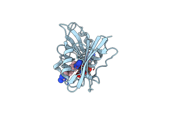 |
Human Dihydrofolate Reductase Complexed With Nadph And Bt2
Organism: Homo sapiens
Method: X-RAY DIFFRACTION Resolution:1.85 Å Release Date: 2019-04-10 Classification: OXIDOREDUCTASE Ligands: 9QR, NDP |
Organism: Homo sapiens
Method: X-RAY DIFFRACTION
Release Date: 2019-04-10
Ligands: 9QR, NDP
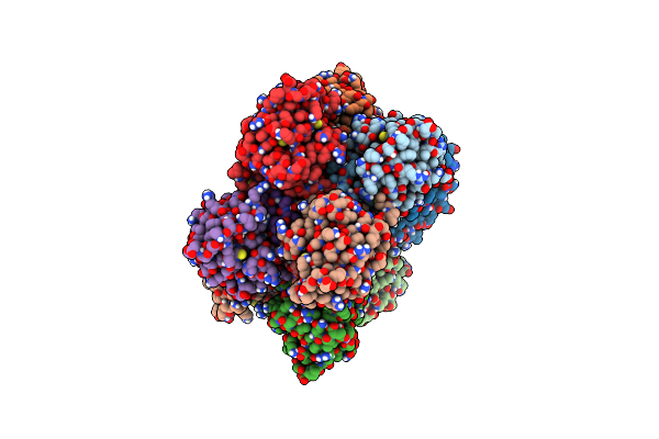 |
Crystal Structures Of Cyclophilin A Complexed With Cyclosporin A And N-Methyl-4-[(E)-2-Butenyl]-4,4-Dimethylthreonine Cyclosporin A
Organism: Homo sapiens, Tolypocladium inflatum
Method: X-RAY DIFFRACTION Resolution:2.10 Å Release Date: 1995-02-07 Classification: ISOMERASE/IMMUNOSUPPRESSANT |
Organism: Homo sapiens, Tolypocladium inflatum
Method: X-RAY DIFFRACTION
Release Date: 1995-02-07
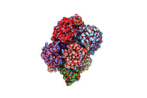 |
Crystal Structures Of Cyclophilin A Complexed With Cyclosporin A And N-Methyl-4-[(E)-2-Butenyl]-4,4-Dimethylthreonine Cyclosporin A
Organism: Homo sapiens, Tolypocladium inflatum
Method: X-RAY DIFFRACTION Resolution:2.10 Å Release Date: 1995-02-07 Classification: ISOMERASE/IMMUNOSUPPRESSANT |
Organism: Homo sapiens, Tolypocladium inflatum
Method: X-RAY DIFFRACTION
Release Date: 1995-02-07
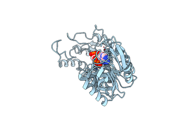 |
The Structure Of Yeast Phosphoglycerate Kinase At 0.25 Nm Resolution
Organism: Saccharomyces cerevisiae
Method: X-RAY DIFFRACTION Resolution:2.50 Å Release Date: 1982-09-24 Classification: TRANSFERASE Ligands: MG, ATP, 3PG |
Organism: Saccharomyces cerevisiae
Method: X-RAY DIFFRACTION
Release Date: 1982-09-24
Ligands: MG, ATP, 3PG
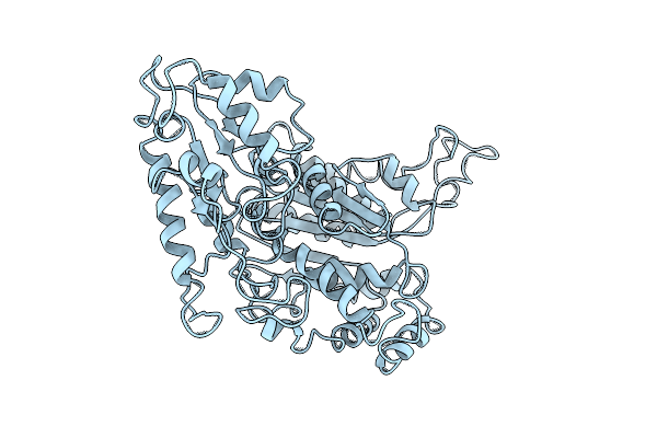 |
Crystallographic Structure Analysis Of Glucose 6-Phosphate Isomerase At 3.5 Angstroms Resolution
Organism: Sus scrofa
Method: X-RAY DIFFRACTION Resolution:3.50 Å Release Date: 1977-09-23 Classification: ISOMERASE(INTRAMOLECULAR ALDOSE/KETOSE) |
Organism: Sus scrofa
Method: X-RAY DIFFRACTION
Release Date: 1977-09-23