

Search Count: 420
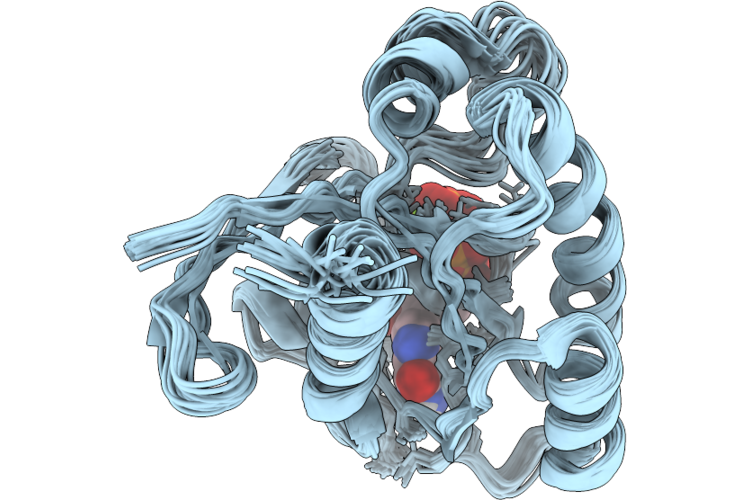 |
Solution Structure Of G12D Mutant Gppnhp-Bound T35S Kras4B
Organism: Homo sapiens
Method: SOLUTION NMR Release Date: 2026-08-19 Classification: SIGNALING PROTEIN/HYDROLASE Ligands: GNP, MG |
Organism: Homo sapiens
Method: SOLUTION NMR
Release Date: 2026-08-19
Ligands: GNP, MG
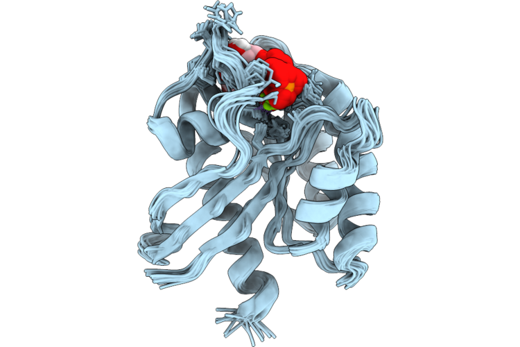 |
Solution Structure Of G12C Mutant Gppnhp-Bound T35S Kras4B
Organism: Homo sapiens
Method: SOLUTION NMR Release Date: 2026-08-19 Classification: SIGNALING PROTEIN/HYDROLASE Ligands: GNP, MG |
Organism: Homo sapiens
Method: SOLUTION NMR
Release Date: 2026-08-19
Ligands: GNP, MG
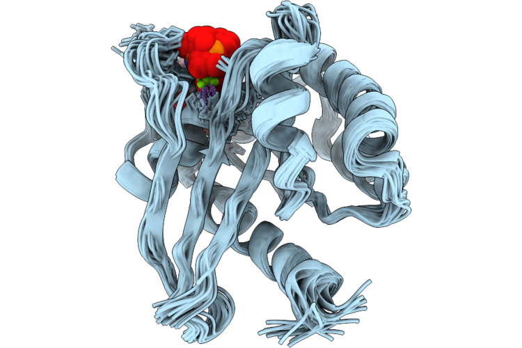 |
Solution Structure Of T35S Mutant Of Kras4B-Gppnhp
Organism: Homo sapiens
Method: SOLUTION NMR Release Date: 2026-08-19 Classification: ONCOPROTEIN/HYDROLASE Ligands: GNP, MG |
Organism: Homo sapiens
Method: SOLUTION NMR
Release Date: 2026-08-19
Ligands: GNP, MG
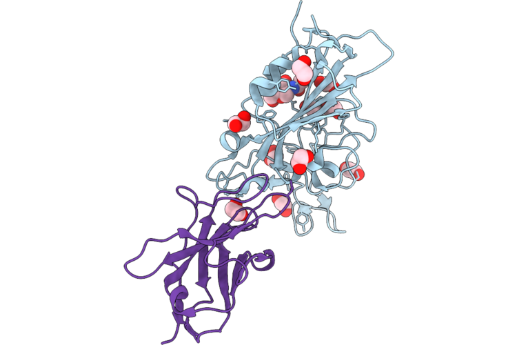 |
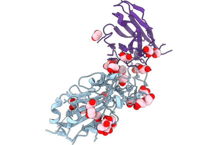
Crystal Structure Of Vhh Mod225 In Complex With Sars-Cov-2 Kp.3 Rbd
Organism: Homo sapiens, Lama glama
Method: X-RAY DIFFRACTION Resolution:2.35 Å Release Date: 2026-08-12 Classification: VIRAL PROTEIN Ligands: NAG, EDO, IOD |
Organism: Homo sapiens, Lama glama
Method: X-RAY DIFFRACTION
Release Date: 2026-08-12
Ligands: NAG, EDO, IOD
 |
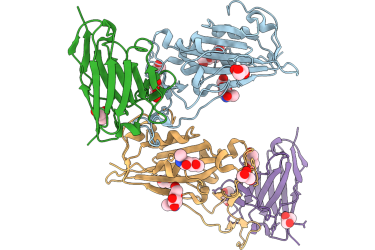
Crystal Structure Of Vhh Mod203 In Complex With Sars-Cov-2 Kp.3 Rbd
Organism: Homo sapiens, Lama glama
Method: X-RAY DIFFRACTION Resolution:1.90 Å Release Date: 2026-08-12 Classification: VIRAL PROTEIN Ligands: NAG, EDO, PEG |
Organism: Homo sapiens, Lama glama
Method: X-RAY DIFFRACTION
Release Date: 2026-08-12
Ligands: NAG, EDO, PEG
 |
Crystal Structure Of Vhh Mod239 In Complex With Sars-Cov-2 Kp.3 Rbd
Organism: Homo sapiens, Lama glama
Method: X-RAY DIFFRACTION Resolution:2.10 Å Release Date: 2026-08-12 Classification: VIRAL PROTEIN Ligands: NAG, GOL |
Organism: Homo sapiens, Lama glama
Method: X-RAY DIFFRACTION
Release Date: 2026-08-12
Ligands: NAG, GOL
 |
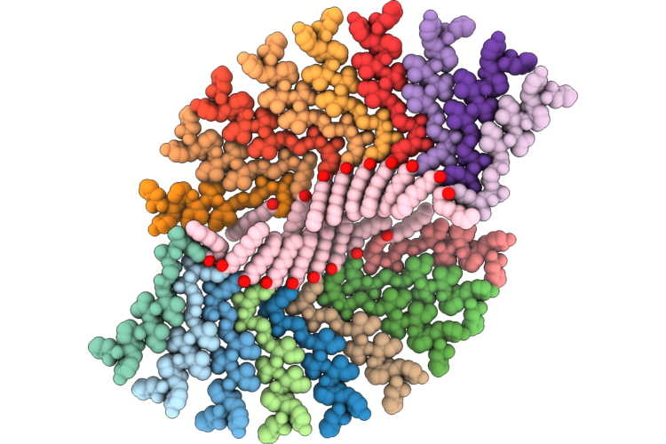
The Cryoem Structure Of T8 Nanofiber
Organism: Synthetic construct
Method: ELECTRON MICROSCOPY Resolution:2.41 Å Release Date: 2026-07-01 Classification: PROTEIN FIBRIL Ligands: OCA |
Organism: Synthetic construct
Method: ELECTRON MICROSCOPY
Release Date: 2026-07-01
Ligands: OCA
 |
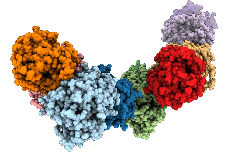
The Cryoem Structure Of T10 Type1 Nanofiber
Organism: Synthetic construct
Method: ELECTRON MICROSCOPY Resolution:3.08 Å Release Date: 2026-07-01 Classification: PROTEIN FIBRIL Ligands: DKA |
Organism: Synthetic construct
Method: ELECTRON MICROSCOPY
Release Date: 2026-07-01
Ligands: DKA
 |
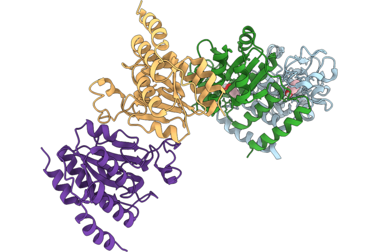
Crystal Structure Of Monoalkyl Phthalate Hydrolase From Rhodococcus Sp. Eg-5 N Complex With Paranitrophenyl Butyrate (Pnpb)
Organism: Rhodococcus sp. eg-5
Method: X-RAY DIFFRACTION Resolution:2.80 Å Release Date: 2026-07-01 Classification: HYDROLASE Ligands: NPO |
Organism: Rhodococcus sp. eg-5
Method: X-RAY DIFFRACTION
Release Date: 2026-07-01
Ligands: NPO
 |
Crystal Structure Of Cysteine-Dependent Hydrolase (Csdh) From Rhodococcus Opacus In Complex With Catechol
Organism: Rhodococcus opacus
Method: X-RAY DIFFRACTION Resolution:2.90 Å Release Date: 2026-07-01 Classification: HYDROLASE Ligands: CAQ |
Organism: Rhodococcus opacus
Method: X-RAY DIFFRACTION
Release Date: 2026-07-01
Ligands: CAQ
 |
Crystal Structure Of Mycobacterium Tuberculosis Rv2514C-Rv2515C Toxin Antitoxin System.
Organism: Mycobacterium tuberculosis h37rv
Method: X-RAY DIFFRACTION Resolution:3.20 Å Release Date: 2026-07-01 Classification: TOXIN Ligands: ZN, MG |
Organism: Mycobacterium tuberculosis h37rv
Method: X-RAY DIFFRACTION
Release Date: 2026-07-01
Ligands: ZN, MG
 |
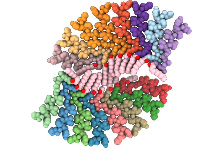
Crystal Structure Of Rv2514C From Mycobacterium Tuberculosis
Organism: Mycobacterium tuberculosis
Method: X-RAY DIFFRACTION Resolution:2.60 Å Release Date: 2026-07-01 Classification: TOXIN Ligands: SO4, GOL |
Organism: Mycobacterium tuberculosis
Method: X-RAY DIFFRACTION
Release Date: 2026-07-01
Ligands: SO4, GOL
 |
The Cryoem Structure Of T10 Type2 Nanofiber
Organism: Synthetic construct
Method: ELECTRON MICROSCOPY Resolution:3.11 Å Release Date: 2026-06-17 Classification: PROTEIN FIBRIL Ligands: DKA |
Organism: Synthetic construct
Method: ELECTRON MICROSCOPY
Release Date: 2026-06-17
Ligands: DKA
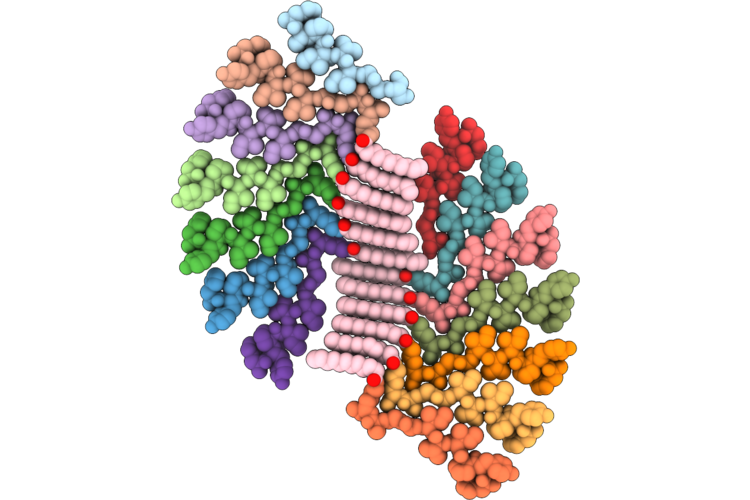 |
The Cryoem Structure Of T12 Type1 Nanofiber
Organism: Synthetic construct
Method: ELECTRON MICROSCOPY Release Date: 2026-06-17 Classification: PROTEIN FIBRIL Ligands: DAO |
Organism: Synthetic construct
Method: ELECTRON MICROSCOPY
Release Date: 2026-06-17
Ligands: DAO
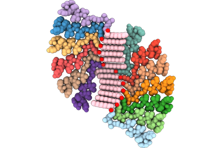 |
The Cryoem Structure Of T12 Type2 Nanofiber
Organism: Synthetic construct
Method: ELECTRON MICROSCOPY Resolution:2.97 Å Release Date: 2026-06-17 Classification: PROTEIN FIBRIL Ligands: DAO |
Organism: Synthetic construct
Method: ELECTRON MICROSCOPY
Release Date: 2026-06-17
Ligands: DAO
 |
Crystal Structure Of Cysteine-Dependent Hydrolase (Csdh) From Rhodococcus Opacus In Complex With Phthalate
Organism: Rhodococcus opacus
Method: X-RAY DIFFRACTION Resolution:2.90 Å Release Date: 2026-06-17 Classification: HYDROLASE Ligands: PHT |
Organism: Rhodococcus opacus
Method: X-RAY DIFFRACTION
Release Date: 2026-06-17
Ligands: PHT
 |
Crystal Structure Of Monoalkyl Phthalate Hydrolase From Rhodococcus Sp. Eg-5
Organism: Rhodococcus sp. eg-5
Method: X-RAY DIFFRACTION Resolution:3.00 Å Release Date: 2026-06-10 Classification: HYDROLASE |
Organism: Rhodococcus sp. eg-5
Method: X-RAY DIFFRACTION
Release Date: 2026-06-10
 |
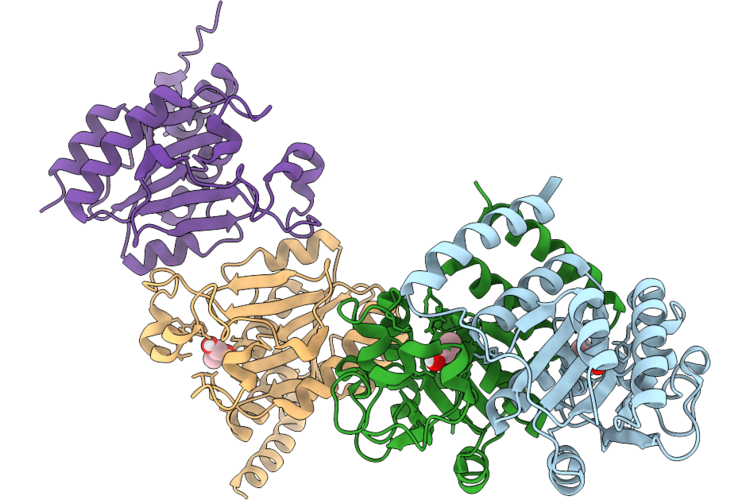
Crystal Structure Of Cysteine-Dependent Hydrolase (Csdh) From Rhodococcus Opacus In Complex With Monobutylphthalate (Mbp)
Organism: Rhodococcus opacus
Method: X-RAY DIFFRACTION Resolution:2.90 Å Release Date: 2026-06-10 Classification: HYDROLASE Ligands: PHT |
Organism: Rhodococcus opacus
Method: X-RAY DIFFRACTION
Release Date: 2026-06-10
Ligands: PHT
 |
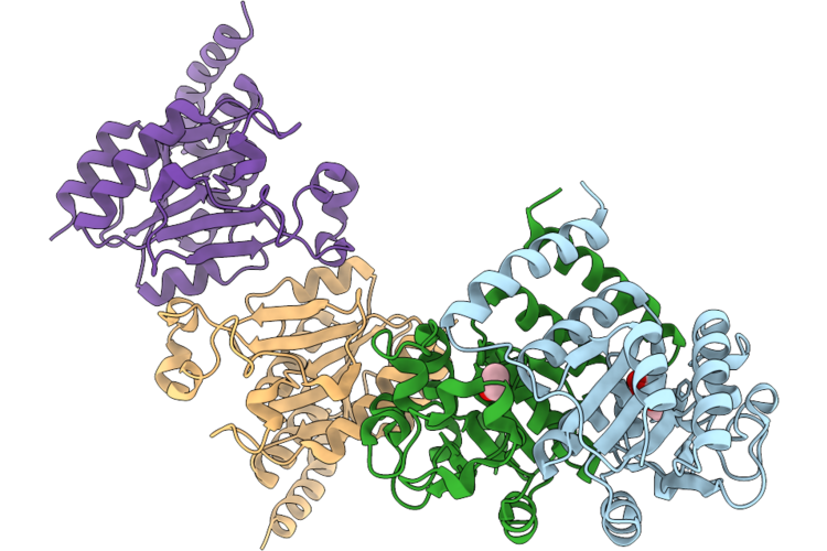
Crystal Structure Of Cysteine-Dependent Hydrolase (Csdh) From Rhodococcus Opacus In Complex With Propylene Glycol
Organism: Rhodococcus opacus
Method: X-RAY DIFFRACTION Resolution:2.80 Å Release Date: 2026-06-10 Classification: HYDROLASE Ligands: PGO |
Organism: Rhodococcus opacus
Method: X-RAY DIFFRACTION
Release Date: 2026-06-10
Ligands: PGO
 |
Crystal Structure Of Cysteine-Dependent Hydrolase (Csdh) From Rhodococcus Opacus In Complex With Dibutylphthalate
Organism: Rhodococcus opacus
Method: X-RAY DIFFRACTION Resolution:2.90 Å Release Date: 2026-06-10 Classification: HYDROLASE Ligands: PHT, EOH |
Organism: Rhodococcus opacus
Method: X-RAY DIFFRACTION
Release Date: 2026-06-10
Ligands: PHT, EOH