

Search Count: 24
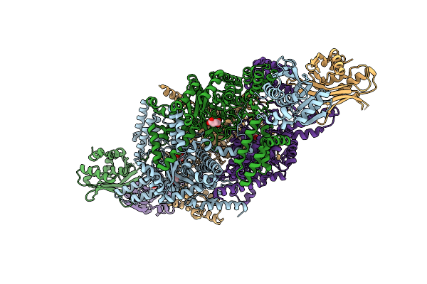 |
Crystal Structure Of Mycobacterium Tuberculosis Isocitrate Lyase 2 Fixed In The Apo Form With Disulfide Bonds
Organism: Mycobacterium tuberculosis cdc1551
Method: X-RAY DIFFRACTION Resolution:2.60 Å Release Date: 2026-03-18 Classification: LYASE Ligands: SIN, EDO |
Organism: Mycobacterium tuberculosis cdc1551
Method: X-RAY DIFFRACTION
Release Date: 2026-03-18
Ligands: SIN, EDO
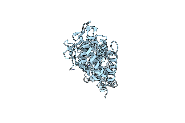 |
Crystal Structure Of Lyssavirus Rabies (Ni-Ce Strain) Nucleoprotein In Complex With Phosphoprotein Chaperone
Organism: Rabies virus (strain nishigahara rceh)
Method: X-RAY DIFFRACTION Resolution:2.00 Å Release Date: 2025-04-23 Classification: VIRAL PROTEIN |
Organism: Rabies virus (strain nishigahara rceh)
Method: X-RAY DIFFRACTION
Release Date: 2025-04-23
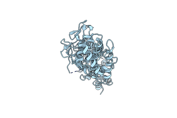 |
Crystal Structure Of Lyssavirus Rabies (Nishigahara Strain) Nucleoprotein In Complex With Phosphomimetic Phosphoprotein S48E
Organism: Lyssavirus rabies
Method: X-RAY DIFFRACTION Resolution:2.10 Å Release Date: 2025-04-23 Classification: VIRAL PROTEIN |
Organism: Lyssavirus rabies
Method: X-RAY DIFFRACTION
Release Date: 2025-04-23
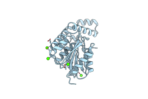 |
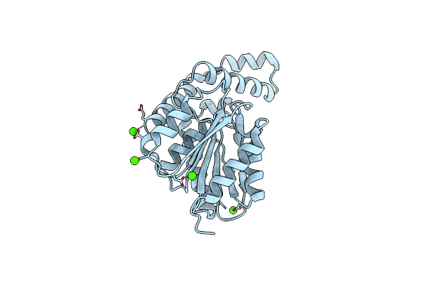
Fphh, Staphylococcus Aureus Fluorophosphonate-Binding Serine Hydrolases H, Apo Crystal Form 2
Organism: Staphylococcus aureus usa300-ca-263
Method: X-RAY DIFFRACTION Resolution:1.79 Å Release Date: 2025-01-08 Classification: HYDROLASE Ligands: CA |
Organism: Staphylococcus aureus usa300-ca-263
Method: X-RAY DIFFRACTION
Release Date: 2025-01-08
Ligands: CA
 |
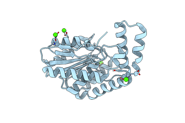
Fphh, Staphylococcus Aureus Fluorophosphonate-Binding Serine Hydrolases H, Apo Form 2 At Room Temperature
Organism: Staphylococcus aureus usa300-ca-263
Method: X-RAY DIFFRACTION Resolution:2.50 Å Release Date: 2025-01-08 Classification: HYDROLASE Ligands: CA |
Organism: Staphylococcus aureus usa300-ca-263
Method: X-RAY DIFFRACTION
Release Date: 2025-01-08
Ligands: CA
 |
Fphi, Staphylococcus Aureus Fluorophosphonate-Binding Serine Hydrolases I, Apo Form At Room Temperature
Organism: Staphylococcus aureus usa300-ca-263
Method: X-RAY DIFFRACTION Resolution:1.70 Å Release Date: 2025-01-08 Classification: HYDROLASE Ligands: CA |
Organism: Staphylococcus aureus usa300-ca-263
Method: X-RAY DIFFRACTION
Release Date: 2025-01-08
Ligands: CA
 |
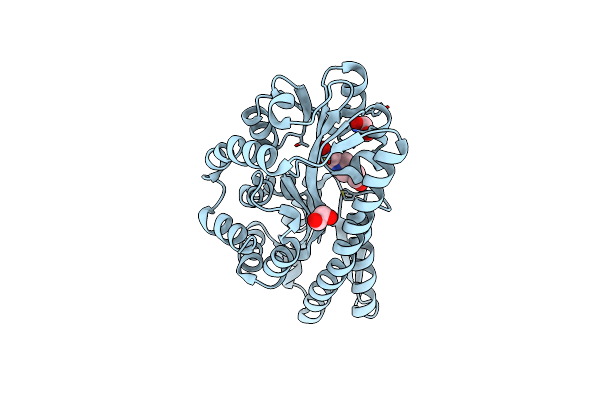
Crystal Structure Of A Mes Bound Substrate Binding Protein (Isep) From An Isethionate Trap Transporter
Organism: Oleidesulfovibrio alaskensis g20
Method: X-RAY DIFFRACTION Resolution:1.65 Å Release Date: 2024-08-28 Classification: TRANSPORT PROTEIN Ligands: MES |
Organism: Oleidesulfovibrio alaskensis g20
Method: X-RAY DIFFRACTION
Release Date: 2024-08-28
Ligands: MES
 |
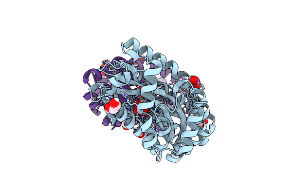
Structure Of A Hepes Bound Trap Transporter Substrate Binding Protein.
Organism: Oleidesulfovibrio alaskensis g20
Method: X-RAY DIFFRACTION Resolution:1.89 Å Release Date: 2024-08-28 Classification: TRANSPORT PROTEIN Ligands: EPE, EDO |
Organism: Oleidesulfovibrio alaskensis g20
Method: X-RAY DIFFRACTION
Release Date: 2024-08-28
Ligands: EPE, EDO
 |
Crystal Structure Of An Isethionate Bound Substrate Binding Protein (Isep) From An Isethionate Trap Transporter
Organism: Oleidesulfovibrio alaskensis g20
Method: X-RAY DIFFRACTION Resolution:1.25 Å Release Date: 2024-07-10 Classification: TRANSPORT PROTEIN Ligands: 8X3, NO3, EDO |
Organism: Oleidesulfovibrio alaskensis g20
Method: X-RAY DIFFRACTION
Release Date: 2024-07-10
Ligands: 8X3, NO3, EDO
 |
Apo Crystal Structure Of A Substrate Binding Protein (Isep) From An Isethionate Trap Transporter
Organism: Oleidesulfovibrio alaskensis g20
Method: X-RAY DIFFRACTION Resolution:1.48 Å Release Date: 2024-06-26 Classification: TRANSPORT PROTEIN Ligands: EDO, CL |
Organism: Oleidesulfovibrio alaskensis g20
Method: X-RAY DIFFRACTION
Release Date: 2024-06-26
Ligands: EDO, CL
 |
Fphi, Staphylococcus Aureus Fluorophosphonate-Binding Serine Hydrolases I, Apo Form
Organism: Staphylococcus aureus usa300-0114
Method: X-RAY DIFFRACTION Resolution:1.14 Å Release Date: 2024-02-14 Classification: HYDROLASE Ligands: MG, CL |
Organism: Staphylococcus aureus usa300-0114
Method: X-RAY DIFFRACTION
Release Date: 2024-02-14
Ligands: MG, CL
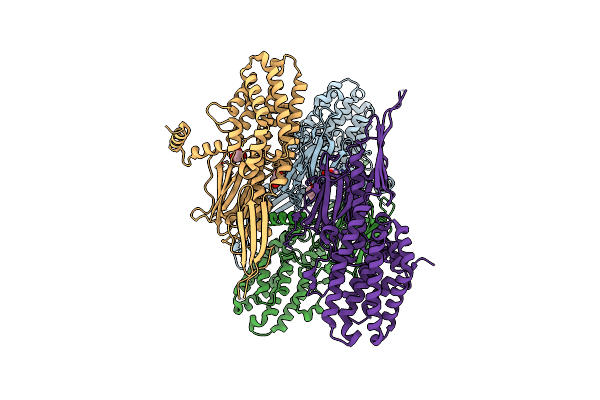 |
A Novel Insecticidal Protein From Ferns Ipd113_Cow.
Organism: Colysis wrightii
Method: X-RAY DIFFRACTION Resolution:2.10 Å Release Date: 2023-11-08 Classification: PLANT PROTEIN Ligands: GOL |
Organism: Colysis wrightii
Method: X-RAY DIFFRACTION
Release Date: 2023-11-08
Ligands: GOL
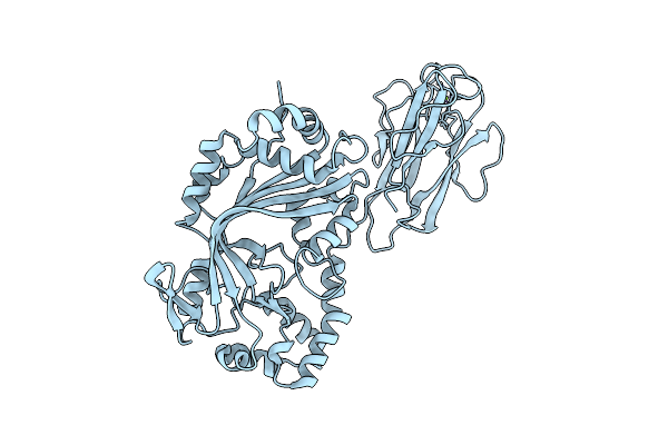 |
Mpf2Ba1 Monomer
Organism: Pseudomonas monteilii
Method: X-RAY DIFFRACTION Resolution:2.13 Å Release Date: 2023-05-31 Classification: TOXIN Ligands: MG |
Organism: Pseudomonas monteilii
Method: X-RAY DIFFRACTION
Release Date: 2023-05-31
Ligands: MG
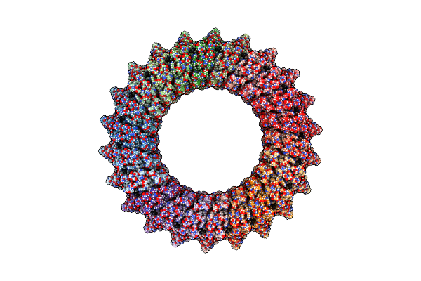 |
Mp2Ba1 Pre-Pore
Organism: Pseudomonas monteilii
Method: ELECTRON MICROSCOPY Release Date: 2023-05-31 Classification: TOXIN Ligands: MG |
Organism: Pseudomonas monteilii
Method: ELECTRON MICROSCOPY
Release Date: 2023-05-31
Ligands: MG
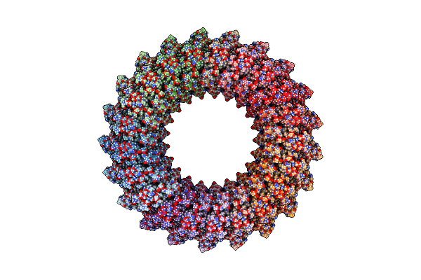 |
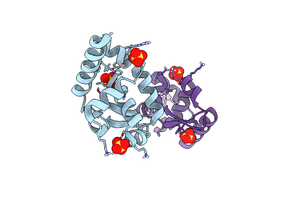
Mpf2Ba1 Pore
Organism: Pseudomonas monteilii
Method: ELECTRON MICROSCOPY Release Date: 2023-05-31 Classification: TOXIN Ligands: MG |
Organism: Pseudomonas monteilii
Method: ELECTRON MICROSCOPY
Release Date: 2023-05-31
Ligands: MG
 |
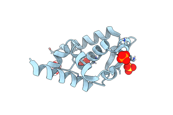
Structure Of Rabies Virus Phosphoprotein C-Terminal Domain, S210E Mutant
Organism: Rabies virus nishigahara rceh
Method: X-RAY DIFFRACTION Resolution:1.70 Å Release Date: 2022-04-20 Classification: VIRAL PROTEIN Ligands: SO4 |
Organism: Rabies virus nishigahara rceh
Method: X-RAY DIFFRACTION
Release Date: 2022-04-20
Ligands: SO4
 |
Structure Of Rabies Virus Phosphoprotein C-Terminal Domain, Wild Type
Organism: Rabies virus nishigahara rceh
Method: X-RAY DIFFRACTION Resolution:1.50 Å Release Date: 2022-04-20 Classification: VIRAL PROTEIN Ligands: EDO, SO4, PO4 |
Organism: Rabies virus nishigahara rceh
Method: X-RAY DIFFRACTION
Release Date: 2022-04-20
Ligands: EDO, SO4, PO4
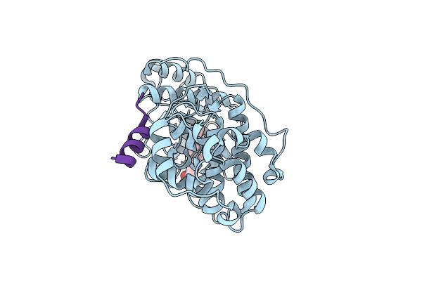 |
Crystal Structure Of Double-Phosphorylated P38Alpha With Atf2(83-102)
Organism: Homo sapiens
Method: X-RAY DIFFRACTION Resolution:1.95 Å Release Date: 2020-11-18 Classification: TRANSCRIPTION Ligands: 3FF |
Organism: Homo sapiens
Method: X-RAY DIFFRACTION
Release Date: 2020-11-18
Ligands: 3FF
 |
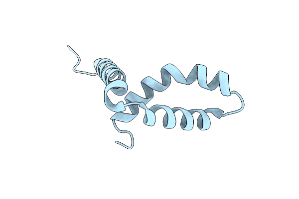
Crystal Structure Of Jnk1 In Complex With Atf2(19-58)
Organism: Homo sapiens
Method: X-RAY DIFFRACTION Resolution:2.70 Å Release Date: 2020-11-18 Classification: TRANSCRIPTION Ligands: MG, ANP, ZN |
Organism: Homo sapiens
Method: X-RAY DIFFRACTION
Release Date: 2020-11-18
Ligands: MG, ANP, ZN
 |
Crystal Structure Of Sox30
Organism: Homo sapiens
Method: X-RAY DIFFRACTION Resolution:1.40 Å Release Date: 2020-08-19 Classification: TRANSCRIPTION |
Organism: Homo sapiens
Method: X-RAY DIFFRACTION
Release Date: 2020-08-19