

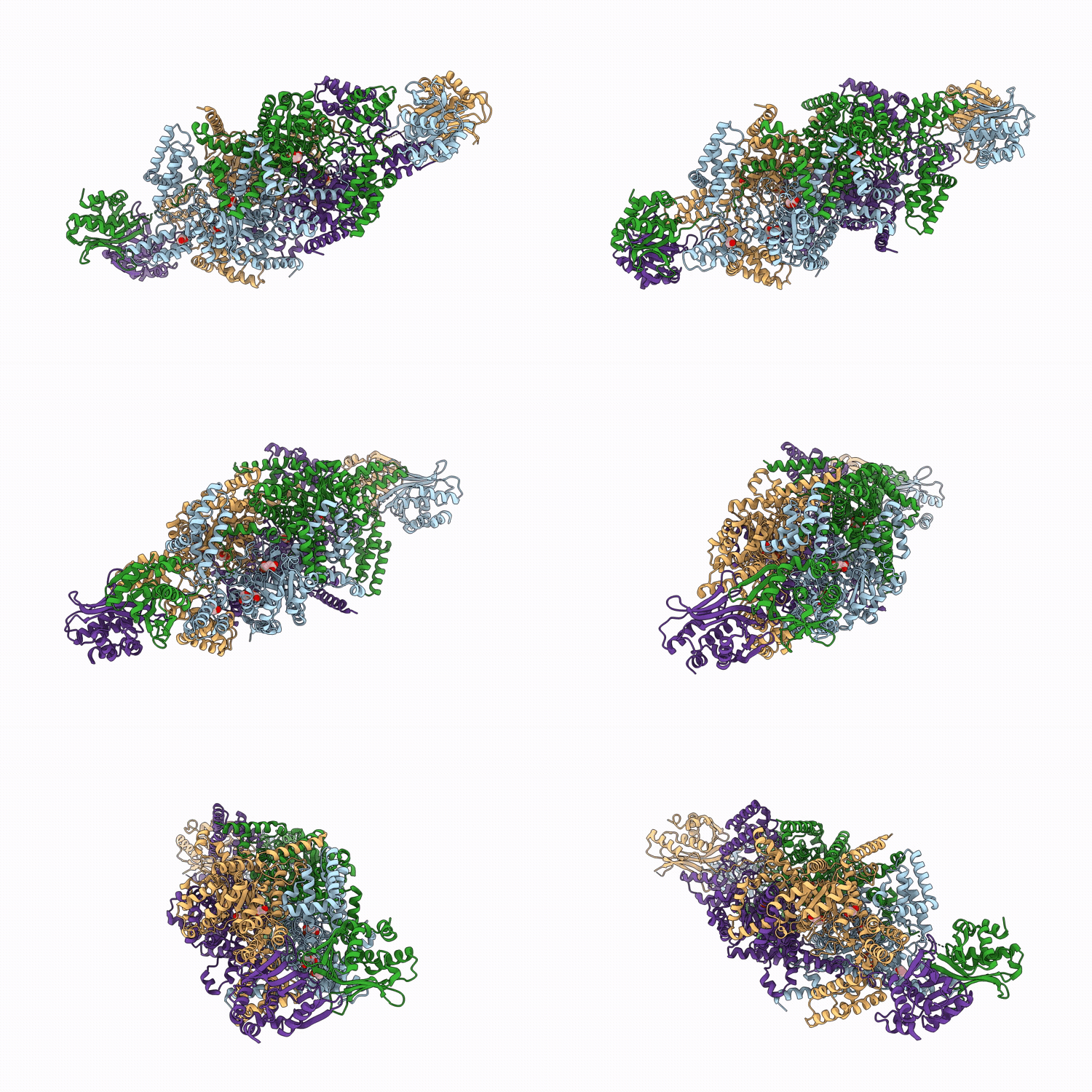
9OBO image
Deposition Date 2025-04-22
Release Date 2026-03-18
Last Version Date 2026-05-06
Entry Detail
PDB ID:
9OBO
Keywords:
Title:
Crystal structure of Mycobacterium tuberculosis isocitrate lyase 2 fixed in the apo form with disulfide bonds
Biological Source:
Source Organism(s):
Expression System(s):
Method Details:
Experimental Method:
Resolution:
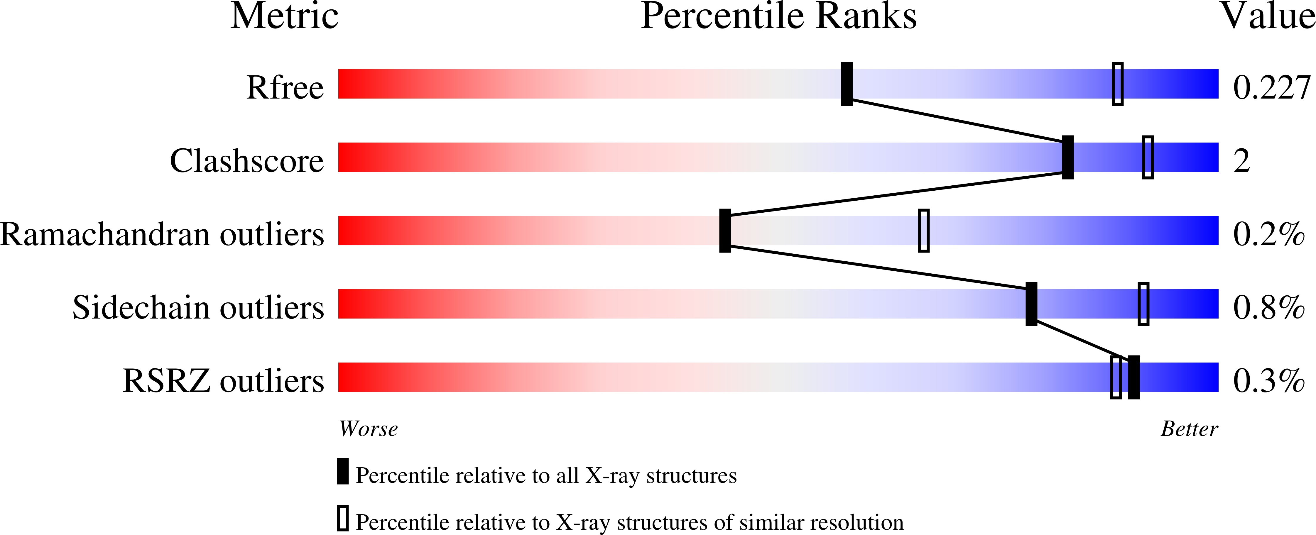
2.60 Å
R-Value Free:
0.22
R-Value Work:
0.17
Space Group:
P 1 21 1
Macromolecular Entities
Structures with similar UniProt ID
Protein Blast
Polymer Type:polypeptide(L)
Molecule:Isocitrate lyase 2
Gene (Uniprot):icl2
Mutagens:Q635C, V708C, V734C
Chain IDs:A, B, C, D
Chain Length:766
Number of Molecules:4
Biological Source:Mycobacterium tuberculosis CDC1551
Primary Citation
Structural basis of allosteric activation of Mycobacterium tuberculosis isocitrate lyase 2.
Commun Biol 9 ? ? (2026)
PMID: 41803459 DOI: 10.1038/s42003-026-09821-6

Abstact

Mycobacterium tuberculosis isocitrate lyase 2 (ICL2) is an allosterically regulated enzyme required for growth on non-glycolytic carbon substrates during infection. Although acetyl-CoA and its analogues are known to activate ICL2, the molecular basis of this regulation has remained unclear. Here, we combine protein NMR, crystallography, molecular dynamics, and mutagenesis to show that two structural features unique to ICL2, the C-terminal domain and a helical substructure in the N-terminal catalytic domain, govern its allostery. Acetyl-CoA binding promotes dimerisation of the C-terminal domain and disrupts its contacts with the helical substructure to trigger conformational changes that activate the enzyme. Together, these findings reveal how a non-substrate metabolite drives isocitrate lyase activation, uncovering the allosteric mechanism that controls M. tuberculosis metabolism and informs new therapeutic strategies.

Legend

Protein

Chemical

Disease

Primary Citation of related structures
Feedback Form
Name
Email
Institute
Feedback