
Search Count: 21
 |
Organism: Homo sapiens, Mus musculus
Method: ELECTRON MICROSCOPY Release Date: 2026-03-25 Classification: SIGNALING PROTEIN/Immune System |
 |
Organism: Homo sapiens
Method: ELECTRON MICROSCOPY Resolution:4.20 Å Release Date: 2026-03-25 Classification: SIGNALING PROTEIN/Immune System |
 |
Organism: Homo sapiens, Mus musculus
Method: ELECTRON MICROSCOPY Release Date: 2026-03-25 Classification: SIGNALING PROTEIN/Immune System |
 |
Organism: Homo sapiens, Mus musculus
Method: ELECTRON MICROSCOPY Release Date: 2026-03-25 Classification: SIGNALING PROTEIN/Imuune System |
 |
Organism: Homo sapiens, Mus musculus
Method: ELECTRON MICROSCOPY Release Date: 2026-03-25 Classification: SIGNALING PROTEIN/Immune System |
 |
Organism: Homo sapiens, Mus musculus
Method: ELECTRON MICROSCOPY Release Date: 2026-03-25 Classification: SIGNALING PROTEIN/Immune System |
 |
Organism: Homo sapiens
Method: ELECTRON MICROSCOPY Resolution:4.55 Å Release Date: 2026-03-25 Classification: SIGNALING PROTEIN/Immune System |
 |
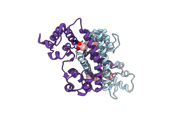
Sk5A-Matured Glycine/Glutamate In Complex With Glun1-Glun2B, Full Refinement
Organism: Homo sapiens, Mus musculus
Method: ELECTRON MICROSCOPY Release Date: 2026-03-25 Classification: SIGNALING PROTEIN/Immune System |
 |
Organism: Homo sapiens, Mus musculus
Method: ELECTRON MICROSCOPY Release Date: 2026-03-25 Classification: SIGNALING PROTEIN/Immune System |
 |
Organism: Homo sapiens, Mus musculus
Method: ELECTRON MICROSCOPY Release Date: 2026-03-25 Classification: SIGNALING PROTEIN/Immune System |
 |
Organism: Homo sapiens, Mus musculus
Method: ELECTRON MICROSCOPY Release Date: 2026-03-25 Classification: SIGNALING PROTEIN/Immune System |
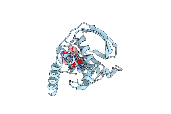 |
Organism: Escherichia coli
Method: X-RAY DIFFRACTION Resolution:1.40 Å Release Date: 2023-12-27 Classification: HYDROLASE Ligands: VLK, ZN |
 |
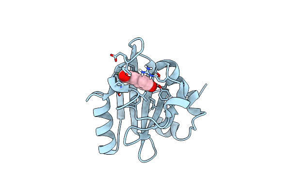
Organism: Homo sapiens
Method: SOLUTION NMR Release Date: 2023-05-31 Classification: SUGAR BINDING PROTEIN |
 |
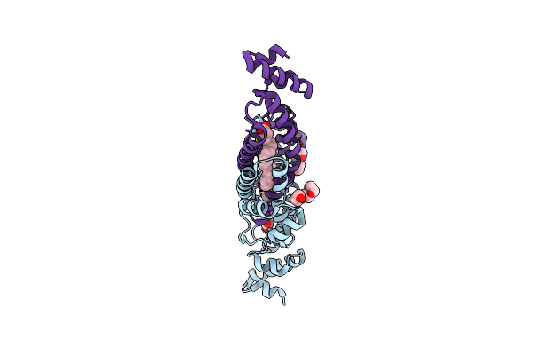
Neutron And X-Ray Structural Studies Of Short Hydrogen Bonds In Photoactive Yellow Protein (Pyp)
Organism: Halorhodospira halophila
Method: X-RAY DIFFRACTION Resolution:2.50 Å Release Date: 2008-03-18 Classification: SIGNALING PROTEIN Ligands: HC4, DOD |
 |
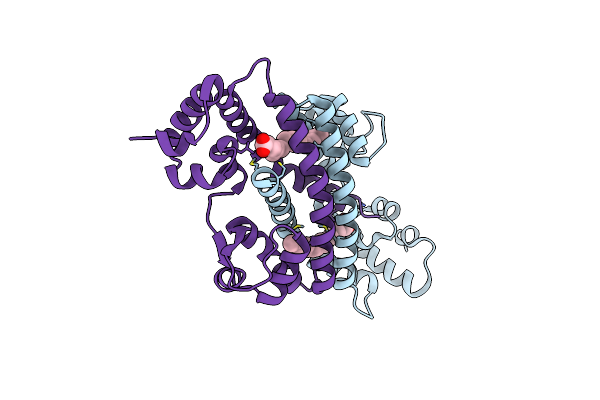
Structure Of Rv1264N, The Regulatory Domain Of The Mycobacterial Adenylyl Cylcase Rv1264, At Ph 6.0
Organism: Mycobacterium tuberculosis
Method: X-RAY DIFFRACTION Resolution:1.60 Å Release Date: 2006-11-07 Classification: LYASE Ligands: OLA, 1PE |
 |
Structure Of Rv1264N, The Regulatory Domain Of The Mycobacterial Adenylyl Cylcase Rv1264, At Ph 8.5
Organism: Mycobacterium tuberculosis
Method: X-RAY DIFFRACTION Resolution:2.35 Å Release Date: 2006-11-07 Classification: LYASE Ligands: OLA |
 |
Structure Of Rv1264N, The Regulatory Domain Of The Mycobacterial Adenylyl Cylcase Rv1264, At Ph 5.3
Organism: Mycobacterium tuberculosis
Method: X-RAY DIFFRACTION Resolution:2.68 Å Release Date: 2006-11-07 Classification: LYASE Ligands: OLA |
 |
Structure Of Rv1264N, The Regulatory Domain Of The Mycobacterial Adenylyl Cylcase Rv1264, With A Salt Precipitant
Organism: Mycobacterium tuberculosis
Method: X-RAY DIFFRACTION Resolution:2.28 Å Release Date: 2006-11-07 Classification: LYASE Ligands: CL, OLA |
 |
Organism: Mycobacterium tuberculosis
Method: X-RAY DIFFRACTION Resolution:2.30 Å Release Date: 2005-05-24 Classification: LYASE Ligands: CA, 1PE |
 |
Organism: Mycobacterium tuberculosis
Method: X-RAY DIFFRACTION Resolution:3.30 Å Release Date: 2005-05-24 Classification: LYASE Ligands: SO4, 1PE, GOL |