

Search Count: 56
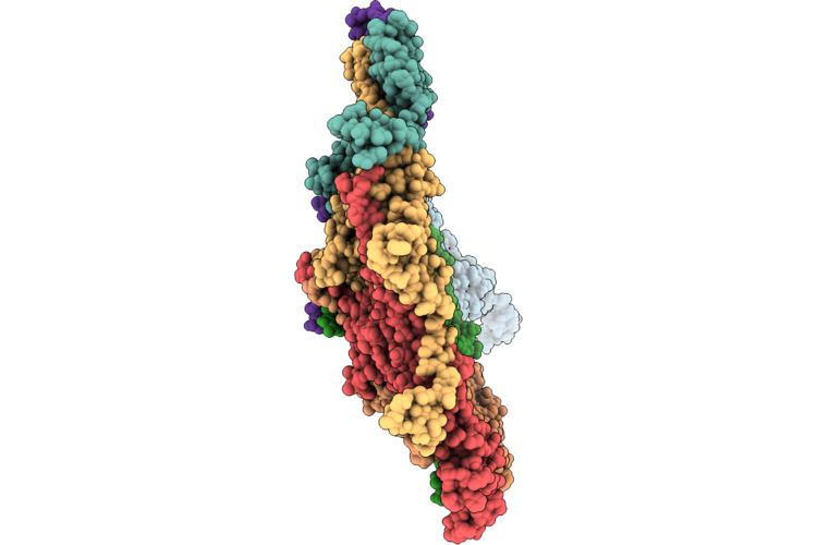 |
Capsid Of The Oenococcus Oeni Phage Oe33Pa
Organism: Oenococcus phage phioe33pa
Method: ELECTRON MICROSCOPY Release Date: 2026-06-17 Classification: VIRAL PROTEIN |
Organism: Oenococcus phage phioe33pa
Method: ELECTRON MICROSCOPY
Release Date: 2026-06-17
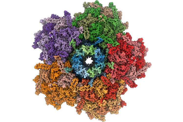 |
Capsid-Connector Assembly Of The Phage Oe33Pa
Organism: Oenococcus phage phioe33pa
Method: ELECTRON MICROSCOPY Release Date: 2026-06-17 Classification: VIRAL PROTEIN |
Organism: Oenococcus phage phioe33pa
Method: ELECTRON MICROSCOPY
Release Date: 2026-06-17
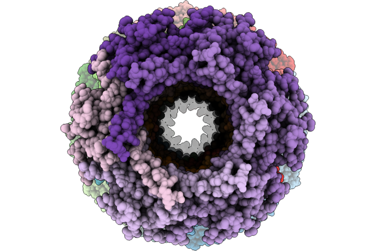 |
Connector And Major Tail Protein Of The Phage Oe33Pa
Organism: Oenococcus phage phioe33pa
Method: ELECTRON MICROSCOPY Release Date: 2026-06-17 Classification: VIRAL PROTEIN Ligands: CA |
Organism: Oenococcus phage phioe33pa
Method: ELECTRON MICROSCOPY
Release Date: 2026-06-17
Ligands: CA
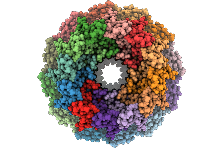 |
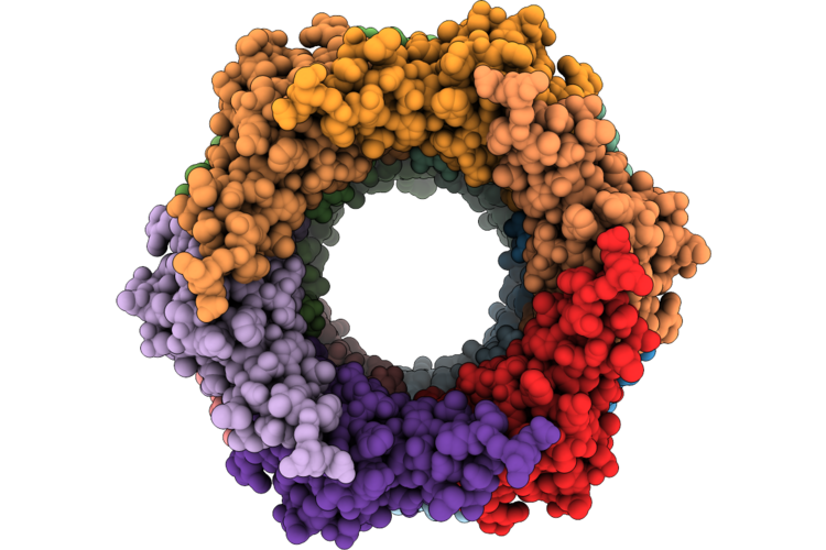
Portal-Adaptor Asssembly Of The Phage Oe33Pa
Organism: Oenococcus phage phioe33pa
Method: ELECTRON MICROSCOPY Release Date: 2026-06-17 Classification: VIRAL PROTEIN |
Organism: Oenococcus phage phioe33pa
Method: ELECTRON MICROSCOPY
Release Date: 2026-06-17
 |
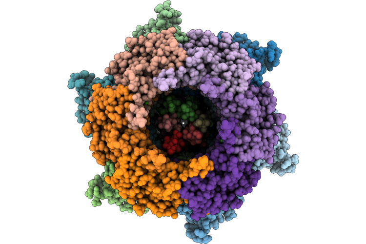
Stopper And Tail Terminator Assembly Of The Phage Oe33Pa
Organism: Oenococcus phage phioe33pa
Method: ELECTRON MICROSCOPY Release Date: 2026-06-17 Classification: VIRAL PROTEIN |
Organism: Oenococcus phage phioe33pa
Method: ELECTRON MICROSCOPY
Release Date: 2026-06-17
 |
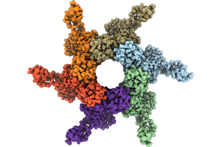
Adhesion Device (C3 Symmetrized Reconstruction) Of The Phage Oe33Pa
Organism: Oenococcus phage phioe33pa
Method: ELECTRON MICROSCOPY Release Date: 2026-06-17 Classification: VIRAL PROTEIN Ligands: CA |
Organism: Oenococcus phage phioe33pa
Method: ELECTRON MICROSCOPY
Release Date: 2026-06-17
Ligands: CA
 |
C6 Symmetrized Reconstruction Of The Adhesion Device Of The Phage Oe33Pa
Organism: Oenococcus phage phioe33pa
Method: ELECTRON MICROSCOPY Release Date: 2026-06-17 Classification: VIRAL PROTEIN |
Organism: Oenococcus phage phioe33pa
Method: ELECTRON MICROSCOPY
Release Date: 2026-06-17
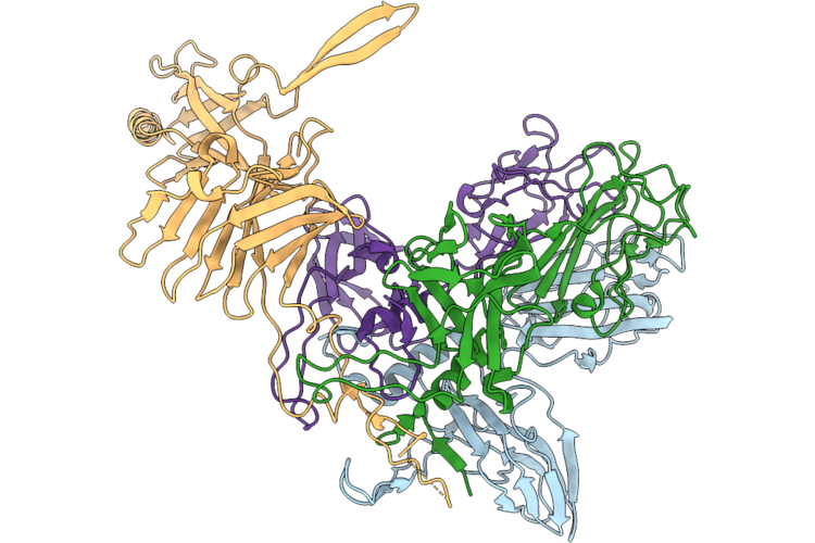 |
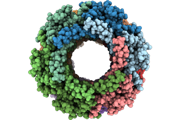
Rbp Trimer Bound To A Monomer Of The Distal Tail Protein Of The Phage Oe33Pa
Organism: Oenococcus phage phioe33pa
Method: ELECTRON MICROSCOPY Release Date: 2026-06-17 Classification: VIRAL PROTEIN |
Organism: Oenococcus phage phioe33pa
Method: ELECTRON MICROSCOPY
Release Date: 2026-06-17
 |
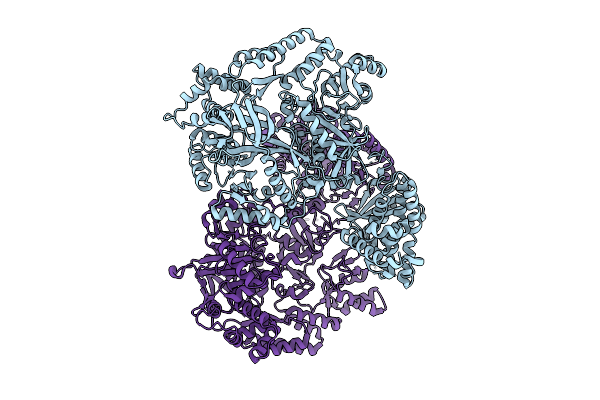
Part Of The Tail Tube Of The Phage Oe33Pa
Organism: Oenococcus phage phioe33pa
Method: ELECTRON MICROSCOPY Release Date: 2026-06-17 Classification: VIRAL PROTEIN Ligands: CA |
Organism: Oenococcus phage phioe33pa
Method: ELECTRON MICROSCOPY
Release Date: 2026-06-17
Ligands: CA
 |
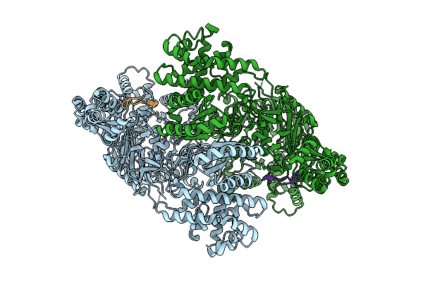
Structure Of Madb, A Class I Dehydrates From Clostridium Maddingley In The Apo State
Organism: Clostridium sp. maddingley mbc34-26
Method: ELECTRON MICROSCOPY Release Date: 2025-07-16 Classification: BIOSYNTHETIC PROTEIN |
Organism: Clostridium sp. maddingley mbc34-26
Method: ELECTRON MICROSCOPY
Release Date: 2025-07-16
 |
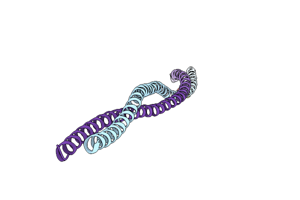
Structure Of Madb, A Class I Dehydrates From Clostridium Maddingley, In Complex With Its Substrate
Organism: Clostridium sp. maddingley mbc34-26
Method: ELECTRON MICROSCOPY Release Date: 2025-07-16 Classification: BIOSYNTHETIC PROTEIN |
Organism: Clostridium sp. maddingley mbc34-26
Method: ELECTRON MICROSCOPY
Release Date: 2025-07-16
 |
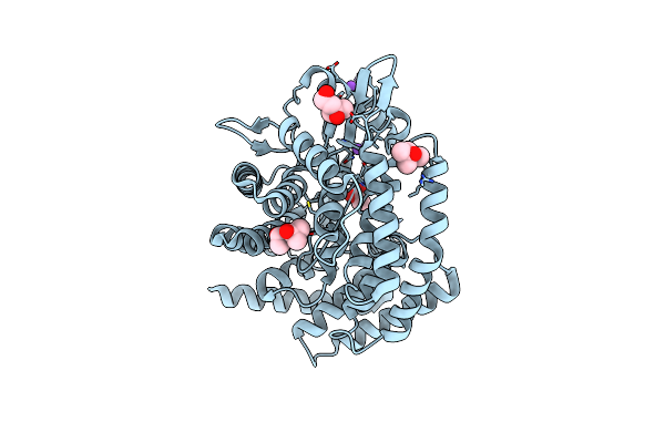
Crystal Structure Hr1 Domain Of Rho-Associated Coiled-Coil Protein Kinases (Rock-Hr1)
Organism: Homo sapiens
Method: X-RAY DIFFRACTION Resolution:2.20 Å Release Date: 2023-06-21 Classification: SIGNALING PROTEIN Ligands: DTT |
Organism: Homo sapiens
Method: X-RAY DIFFRACTION
Release Date: 2023-06-21
Ligands: DTT
 |
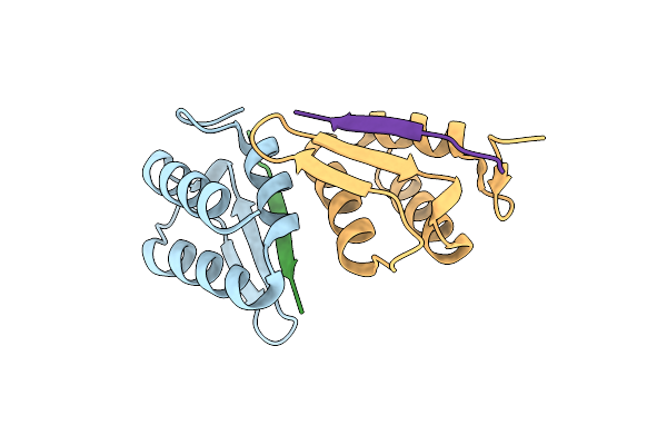
The Structure Of Madc From Clostridium Maddingley Reveals New Insights Into Class I Lanthipeptide Cyclases
Organism: Clostridium
Method: X-RAY DIFFRACTION Resolution:1.70 Å Release Date: 2023-02-22 Classification: BIOSYNTHETIC PROTEIN Ligands: ZN, NA, MPD, ACT, CL |
Organism: Clostridium
Method: X-RAY DIFFRACTION
Release Date: 2023-02-22
Ligands: ZN, NA, MPD, ACT, CL
 |
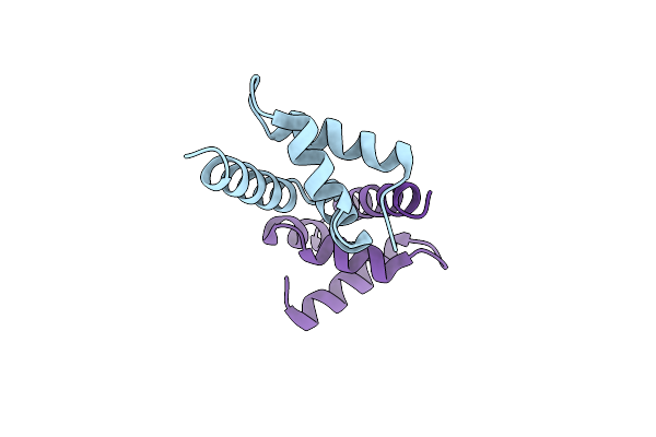
Taf14 Et Domain In Complex With C-Terminal Tail Of Taf2
Organism: Saccharomyces
Method: X-RAY DIFFRACTION Resolution:1.66 Å Release Date: 2022-08-17 Classification: TRANSCRIPTION |
Organism: Saccharomyces
Method: X-RAY DIFFRACTION
Release Date: 2022-08-17
 |
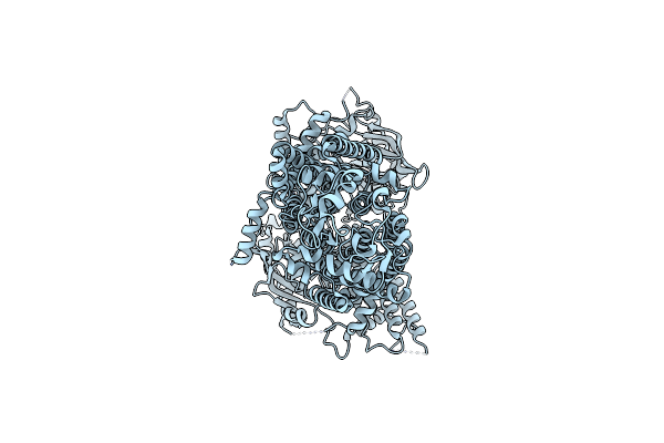
Mademoiselle Domain 2 Of Rrm4 From Ustilago Maydis
Organism: Ustilago maydis
Method: X-RAY DIFFRACTION Resolution:2.60 Å Release Date: 2022-06-15 Classification: RNA BINDING PROTEIN |
Organism: Ustilago maydis
Method: X-RAY DIFFRACTION
Release Date: 2022-06-15
 |
Cryo-Em Structure Of Pdr5 From Saccharomyces Cerevisiae In Inward-Facing Conformation Without Nucleotides
Organism: Saccharomyces cerevisiae (strain atcc 204508 / s288c)
Method: ELECTRON MICROSCOPY Release Date: 2021-11-10 Classification: TRANSPORT PROTEIN |
Organism: Saccharomyces cerevisiae (strain atcc 204508 / s288c)
Method: ELECTRON MICROSCOPY
Release Date: 2021-11-10
 |
Cryo-Em Structure Of Pdr5 From Saccharomyces Cerevisiae In Inward-Facing Conformation With Adp/Atp
Organism: Saccharomyces cerevisiae (strain atcc 204508 / s288c)
Method: ELECTRON MICROSCOPY Release Date: 2021-11-10 Classification: TRANSPORT PROTEIN Ligands: ATP, ADP |
Organism: Saccharomyces cerevisiae (strain atcc 204508 / s288c)
Method: ELECTRON MICROSCOPY
Release Date: 2021-11-10
Ligands: ATP, ADP
 |
Cryo-Em Structure Of Pdr5 From Saccharomyces Cerevisiae In Inward-Facing Conformation With Adp/Atp And Rhodamine 6G
Organism: Saccharomyces cerevisiae (strain atcc 204508 / s288c)
Method: ELECTRON MICROSCOPY Release Date: 2021-11-10 Classification: TRANSPORT PROTEIN Ligands: ATP, ADP, RHQ |
Organism: Saccharomyces cerevisiae (strain atcc 204508 / s288c)
Method: ELECTRON MICROSCOPY
Release Date: 2021-11-10
Ligands: ATP, ADP, RHQ
 |
Cryo-Em Structure Of Pdr5 From Saccharomyces Cerevisiae In Outward-Facing Conformation With Adp-Orthovanadate/Atp
Organism: Saccharomyces cerevisiae (strain atcc 204508 / s288c)
Method: ELECTRON MICROSCOPY Release Date: 2021-11-10 Classification: TRANSPORT PROTEIN Ligands: ATP, MG, AOV |
Organism: Saccharomyces cerevisiae (strain atcc 204508 / s288c)
Method: ELECTRON MICROSCOPY
Release Date: 2021-11-10
Ligands: ATP, MG, AOV
 |
Structure Of A Opubc Mutant With Bound Glycine Betaine
Organism: Bacillus subtilis
Method: X-RAY DIFFRACTION Resolution:1.42 Å Release Date: 2018-11-21 Classification: TRANSPORT PROTEIN Ligands: BET |
Organism: Bacillus subtilis
Method: X-RAY DIFFRACTION
Release Date: 2018-11-21
Ligands: BET