

Search Count: 48
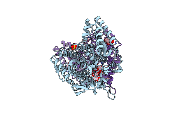 |
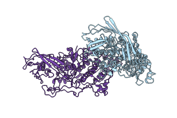
Structure Of Urethanase Umg-Sp2
Organism: Uncultured bacterium
Method: X-RAY DIFFRACTION Resolution:2.59 Å Release Date: 2025-05-21 Classification: HYDROLASE Ligands: GOL, SO4 |
Organism: Uncultured bacterium
Method: X-RAY DIFFRACTION
Release Date: 2025-05-21
Ligands: GOL, SO4
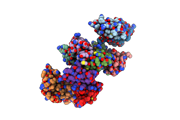 |
Flig Interacting Domain Of Spflhf (Fid)
Organism: Shewanella putrefaciens cn-32
Method: X-RAY DIFFRACTION Resolution:3.01 Å Release Date: 2025-03-26 Classification: SIGNALING PROTEIN |
Organism: Shewanella putrefaciens cn-32
Method: X-RAY DIFFRACTION
Release Date: 2025-03-26
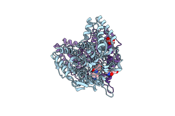 |
Crystal Structure Of A Novel Pu Plastic Degradation Urethanase Umg-Sp2 Mutant From Uncultured Bacterium In Complex With Ligand
Organism: Metagenome
Method: X-RAY DIFFRACTION Resolution:2.40 Å Release Date: 2025-01-15 Classification: HYDROLASE Ligands: A1D5H, GOL |
Organism: Metagenome
Method: X-RAY DIFFRACTION
Release Date: 2025-01-15
Ligands: A1D5H, GOL
 |
Crystal Structure Of A Novel Pu Plastic Degradation Urethanase Umg-Sp2 From Uncultured Bacterium
Organism: Uncultured bacterium
Method: X-RAY DIFFRACTION Resolution:2.16 Å Release Date: 2024-09-18 Classification: HYDROLASE Ligands: GOL, SO4, PMS |
Organism: Uncultured bacterium
Method: X-RAY DIFFRACTION
Release Date: 2024-09-18
Ligands: GOL, SO4, PMS
 |
Urethanase Umg-Sp1 Without Inhibitor Or Substrate Displays Flexible Active Site Loops
Organism: Metagenome
Method: X-RAY DIFFRACTION Resolution:2.67 Å Release Date: 2024-07-24 Classification: HYDROLASE |
Organism: Metagenome
Method: X-RAY DIFFRACTION
Release Date: 2024-07-24
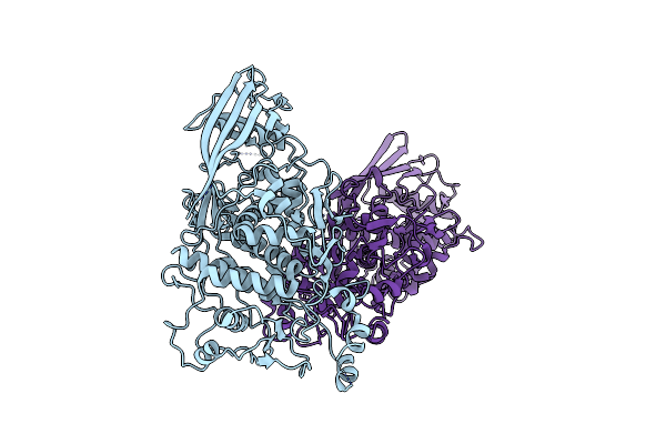 |
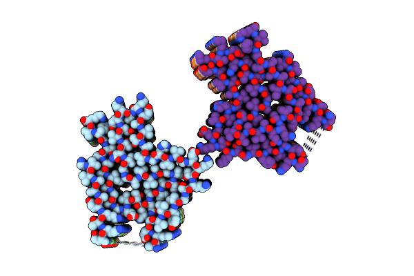
Capsid Structure Of The L-A Helper Virus From Native Viral Communities
Organism: Saccharomyces cerevisiae virus l-a
Method: ELECTRON MICROSCOPY Release Date: 2024-05-22 Classification: VIRUS |
Organism: Saccharomyces cerevisiae virus l-a
Method: ELECTRON MICROSCOPY
Release Date: 2024-05-22
 |
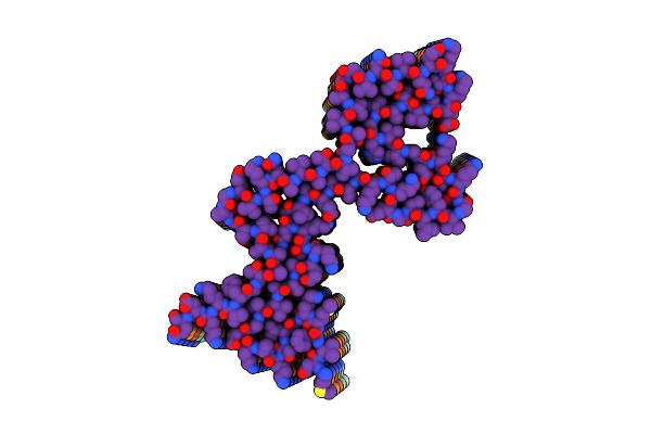
Capsid Structure Of The L-A Helper Virus From Native Viral Communities
Organism: Saccharomyces cerevisiae
Method: ELECTRON MICROSCOPY Release Date: 2023-12-20 Classification: VIRUS |
Organism: Saccharomyces cerevisiae
Method: ELECTRON MICROSCOPY
Release Date: 2023-12-20
 |
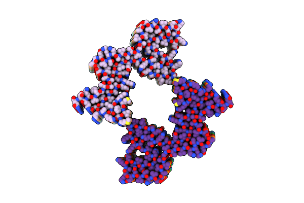
Lipidic Alpha-Synuclein Fibril - Polymorph L2B
Organism: Homo sapiens
Method: ELECTRON MICROSCOPY Release Date: 2022-10-12 Classification: PROTEIN FIBRIL |
Organism: Homo sapiens
Method: ELECTRON MICROSCOPY
Release Date: 2022-10-12
 |
Lipidic Alpha-Synuclein Fibril - Polymorph L1A
Organism: Homo sapiens
Method: ELECTRON MICROSCOPY Release Date: 2022-10-12 Classification: PROTEIN FIBRIL |
Organism: Homo sapiens
Method: ELECTRON MICROSCOPY
Release Date: 2022-10-12
 |
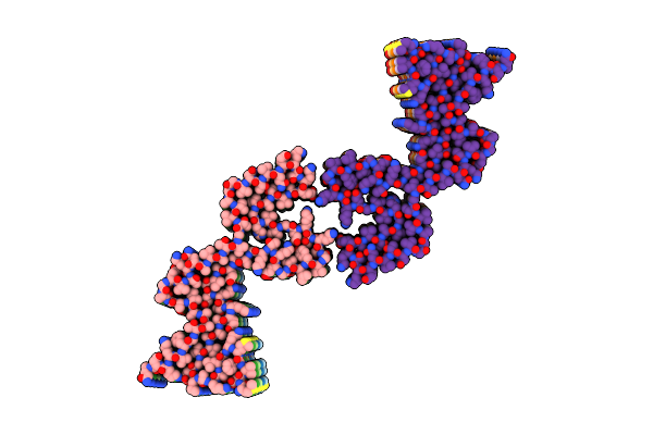
Lipidic Alpha-Synuclein Fibril - Polymorph L1B
Organism: Homo sapiens
Method: ELECTRON MICROSCOPY Release Date: 2022-10-12 Classification: PROTEIN FIBRIL |
Organism: Homo sapiens
Method: ELECTRON MICROSCOPY
Release Date: 2022-10-12
 |
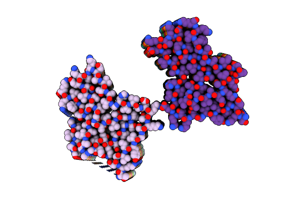
Lipidic Alpha-Synuclein Fibril - Polymorph L1C
Organism: Homo sapiens
Method: ELECTRON MICROSCOPY Release Date: 2022-10-12 Classification: PROTEIN FIBRIL |
Organism: Homo sapiens
Method: ELECTRON MICROSCOPY
Release Date: 2022-10-12
 |
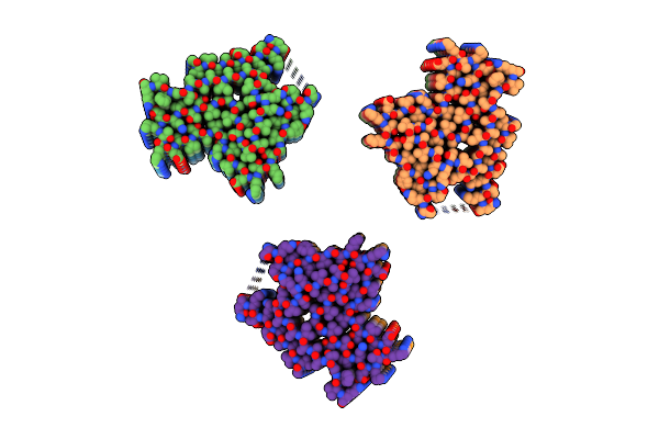
Lipidic Alpha-Synuclein Fibril - Polymorph L3A
Organism: Homo sapiens
Method: ELECTRON MICROSCOPY Release Date: 2022-10-12 Classification: PROTEIN FIBRIL |
Organism: Homo sapiens
Method: ELECTRON MICROSCOPY
Release Date: 2022-10-12
 |
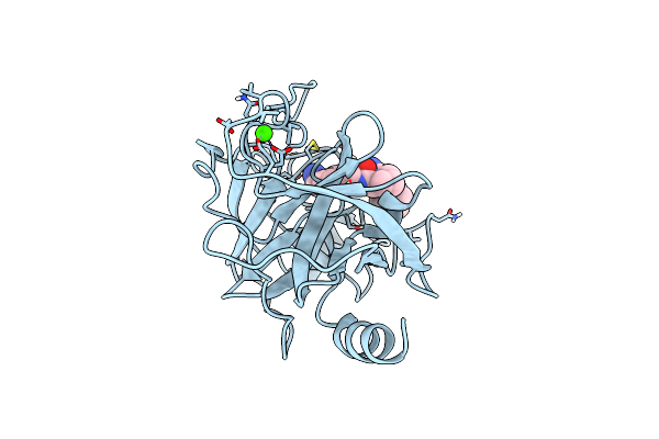
Lipidic Alpha-Synuclein Fibril - Polymorph L2A
Organism: Homo sapiens
Method: ELECTRON MICROSCOPY Release Date: 2022-08-17 Classification: PROTEIN FIBRIL |
Organism: Homo sapiens
Method: ELECTRON MICROSCOPY
Release Date: 2022-08-17
 |
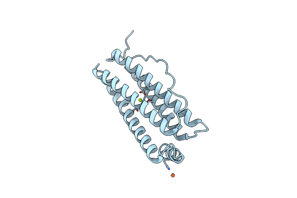
Cationic Trypsin In Complex With A D-Phe-Pro-Benzylamine Derivative
Organism: Bos taurus
Method: X-RAY DIFFRACTION Resolution:1.45 Å Release Date: 2021-03-31 Classification: HYDROLASE Ligands: CA, K73 |
Organism: Bos taurus
Method: X-RAY DIFFRACTION
Release Date: 2021-03-31
Ligands: CA, K73
 |
Molecular Structure Of Mouse Apoferritin Resolved At 2.7 Angstroms With The Glacios Cryo-Microscope
Organism: Mus musculus
Method: ELECTRON MICROSCOPY Resolution:2.73 Å Release Date: 2020-05-13 Classification: METAL BINDING PROTEIN Ligands: FE, MG |
Organism: Mus musculus
Method: ELECTRON MICROSCOPY
Release Date: 2020-05-13
Ligands: FE, MG
 |
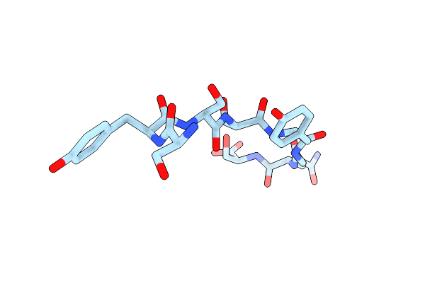
Sysgys From Low-Complexity Domain Of Fus, Residues 37-42
Organism: Homo sapiens
Method: X-RAY DIFFRACTION Resolution:1.10 Å Release Date: 2018-04-04 Classification: PROTEIN FIBRIL Ligands: HOH |
Organism: Homo sapiens
Method: X-RAY DIFFRACTION
Release Date: 2018-04-04
Ligands: HOH
 |
Syssygqs From Low-Complexity Domain Of Fus, Residues 54-61
Organism: Homo sapiens
Method: X-RAY DIFFRACTION Resolution:1.10 Å Release Date: 2018-04-04 Classification: PROTEIN FIBRIL |
Organism: Homo sapiens
Method: X-RAY DIFFRACTION
Release Date: 2018-04-04
 |
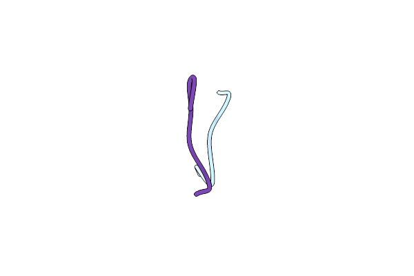
Gyngfg From Low-Complexity Domain Of Hnrnpa1, Residues 243-248
Organism: Homo sapiens
Method: X-RAY DIFFRACTION Resolution:1.10 Å Release Date: 2018-04-04 Classification: PROTEIN FIBRIL |
Organism: Homo sapiens
Method: X-RAY DIFFRACTION
Release Date: 2018-04-04
 |
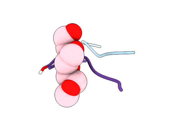
Gfgnfgts From Low-Complexity/Fg Repeat Domain Of Nup98, Residues 116-123
Organism: Homo sapiens
Method: ELECTRON CRYSTALLOGRAPHY Resolution:0.90 Å Release Date: 2018-04-04 Classification: PROTEIN FIBRIL |
Organism: Homo sapiens
Method: ELECTRON CRYSTALLOGRAPHY
Release Date: 2018-04-04
 |
Stggyg From Low-Complexity Domain Of Fus, Residues 77-82
Organism: Homo sapiens
Method: ELECTRON CRYSTALLOGRAPHY Resolution:1.10 Å Release Date: 2018-04-04 Classification: PROTEIN FIBRIL Ligands: TOE |
Organism: Homo sapiens
Method: ELECTRON CRYSTALLOGRAPHY
Release Date: 2018-04-04
Ligands: TOE