

Search Count: 20
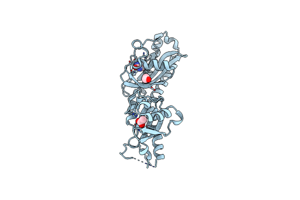 |
Ligand Binding Domain Of Roseburia Intestinalis L1-82 Uracil Chemoreceptor (Dcache) In Complex With Uracil And Acetate
Organism: Roseburia intestinalis l1-82
Method: X-RAY DIFFRACTION Resolution:1.46 Å Release Date: 2025-03-12 Classification: SIGNALING PROTEIN Ligands: URA, ACT, EDO |
Organism: Roseburia intestinalis l1-82
Method: X-RAY DIFFRACTION
Release Date: 2025-03-12
Ligands: URA, ACT, EDO
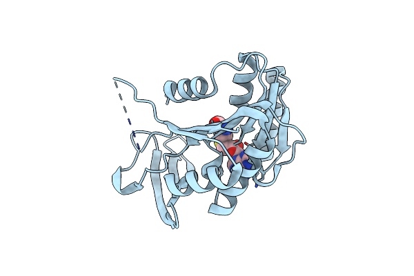 |
Xenorhabdin Methyltransferase Xrdm With Sah
Organism: Xenorhabdus doucetiae frm16 = dsm 17909
Method: X-RAY DIFFRACTION Resolution:2.10 Å Release Date: 2024-10-16 Classification: TRANSFERASE Ligands: SAH |
Organism: Xenorhabdus doucetiae frm16 = dsm 17909
Method: X-RAY DIFFRACTION
Release Date: 2024-10-16
Ligands: SAH
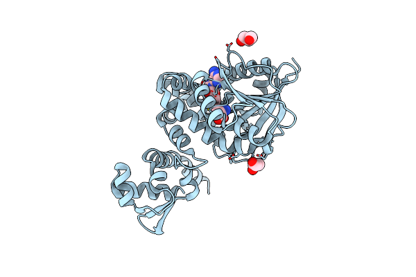 |
Holomycin Methyltransferase Dtpm With Sah
Organism: Xenorhabdus doucetiae frm16 = dsm 17909
Method: X-RAY DIFFRACTION Resolution:1.75 Å Release Date: 2024-10-16 Classification: TRANSFERASE Ligands: SAH, GOL |
Organism: Xenorhabdus doucetiae frm16 = dsm 17909
Method: X-RAY DIFFRACTION
Release Date: 2024-10-16
Ligands: SAH, GOL
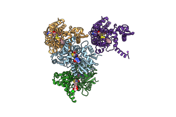 |
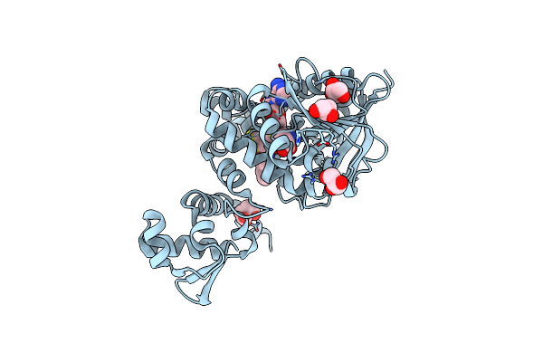
Holomycin Methyltransferase Dtpm With Sah And Xrd-271
Organism: Xenorhabdus doucetiae frm16 = dsm 17909
Method: X-RAY DIFFRACTION Resolution:2.20 Å Release Date: 2024-10-16 Classification: TRANSFERASE Ligands: SAH, YRH, CL, GOL, NA |
Organism: Xenorhabdus doucetiae frm16 = dsm 17909
Method: X-RAY DIFFRACTION
Release Date: 2024-10-16
Ligands: SAH, YRH, CL, GOL, NA
 |
Holomycin Methyltransferase Dtpm With Sah And Xrd-271Me
Organism: Xenorhabdus doucetiae frm16 = dsm 17909
Method: X-RAY DIFFRACTION Resolution:1.75 Å Release Date: 2024-10-16 Classification: TRANSFERASE Ligands: SAH, YRD, GOL, MLT |
Organism: Xenorhabdus doucetiae frm16 = dsm 17909
Method: X-RAY DIFFRACTION
Release Date: 2024-10-16
Ligands: SAH, YRD, GOL, MLT
 |
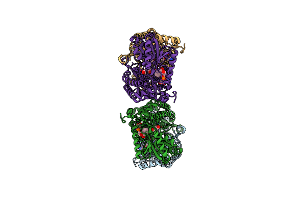
Succinic Semialdehyde Dehydrogenase From E. Coli With Bound Nad+
Organism: Escherichia coli k-12
Method: X-RAY DIFFRACTION Resolution:1.70 Å Release Date: 2024-10-02 Classification: OXIDOREDUCTASE Ligands: NAD |
Organism: Escherichia coli k-12
Method: X-RAY DIFFRACTION
Release Date: 2024-10-02
Ligands: NAD
 |
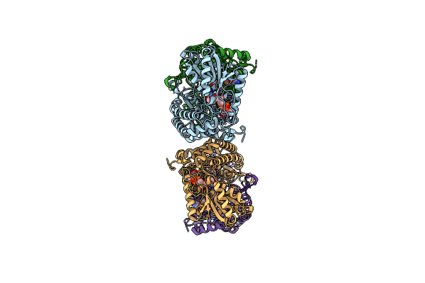
Succinic Semialdehyde Dehydrogenase From E. Coli With Bound Nad+ And Succinic Semialdehyde
Organism: Escherichia coli k-12
Method: X-RAY DIFFRACTION Resolution:2.30 Å Release Date: 2024-10-02 Classification: OXIDOREDUCTASE Ligands: NAD, SSN |
Organism: Escherichia coli k-12
Method: X-RAY DIFFRACTION
Release Date: 2024-10-02
Ligands: NAD, SSN
 |
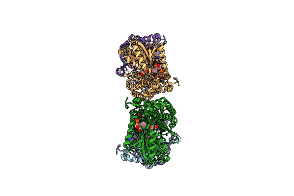
Succinic Semialdehyde Dehydrogenase From E. Coli With Q262R Substitution And Bound Nad+
Organism: Escherichia coli k-12
Method: X-RAY DIFFRACTION Resolution:1.90 Å Release Date: 2024-10-02 Classification: OXIDOREDUCTASE Ligands: NAD |
Organism: Escherichia coli k-12
Method: X-RAY DIFFRACTION
Release Date: 2024-10-02
Ligands: NAD
 |
Succinic Semialdehyde Dehydrogenase From E. Coli With Q262R Substitution And Bound Nad+, Succinic Semialdehyde
Organism: Escherichia coli k-12
Method: X-RAY DIFFRACTION Resolution:1.80 Å Release Date: 2024-10-02 Classification: OXIDOREDUCTASE Ligands: NAD, SSN |
Organism: Escherichia coli k-12
Method: X-RAY DIFFRACTION
Release Date: 2024-10-02
Ligands: NAD, SSN
 |
Crystal Structure Of Citrate Synthase From Methylophaga Sulfidovorans
Organism: Methylophaga sulfidovorans
Method: X-RAY DIFFRACTION Resolution:3.20 Å Release Date: 2024-07-31 Classification: TRANSFERASE |
Organism: Methylophaga sulfidovorans
Method: X-RAY DIFFRACTION
Release Date: 2024-07-31
 |
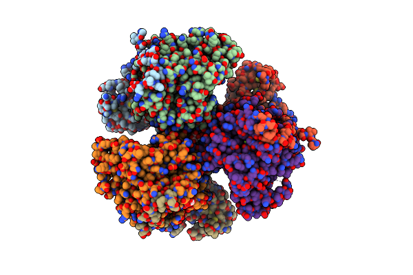
Structure Of The Non-Mitochondrial Citrate Synthase From Ananas Comosus
Organism: Ananas comosus
Method: ELECTRON MICROSCOPY Release Date: 2024-07-24 Classification: TRANSFERASE |
Organism: Ananas comosus
Method: ELECTRON MICROSCOPY
Release Date: 2024-07-24
 |
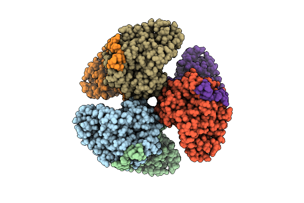
Citrate-Bound Hexamer Of Synechococcus Elongatus Citrate Synthase
Organism: Synechococcus elongatus pcc 7942 = fachb-805
Method: X-RAY DIFFRACTION Resolution:2.71 Å Release Date: 2024-04-24 Classification: TRANSFERASE Ligands: SO4, CIT, MG |
Organism: Synechococcus elongatus pcc 7942 = fachb-805
Method: X-RAY DIFFRACTION
Release Date: 2024-04-24
Ligands: SO4, CIT, MG
 |
Structure Of Hexameric Subcomplexes (Truncation Delta2-6) Of The Fractal Citrate Synthase From Synechococcus Elongatus Pcc7942
Organism: Synechococcus elongatus pcc 7942 = fachb-805
Method: ELECTRON MICROSCOPY Release Date: 2024-02-28 Classification: TRANSFERASE |
Organism: Synechococcus elongatus pcc 7942 = fachb-805
Method: ELECTRON MICROSCOPY
Release Date: 2024-02-28
 |
Pseudoatomic Model Of A Second-Order Sierpinski Triangle Formed By The Citrate Synthase From Synechococcus Elongatus
Organism: Synechococcus elongatus pcc 7942 = fachb-805
Method: ELECTRON MICROSCOPY Release Date: 2024-02-28 Classification: TRANSFERASE |
Organism: Synechococcus elongatus pcc 7942 = fachb-805
Method: ELECTRON MICROSCOPY
Release Date: 2024-02-28
 |
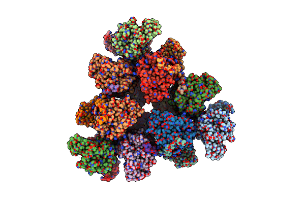
Structure Of A First Order Sierpinski Triangle Formed By The H369R Mutant Of The Citrate Synthase From Synechococcus Elongatus
Organism: Synechococcus elongatus pcc 7942 = fachb-805
Method: ELECTRON MICROSCOPY Release Date: 2024-02-28 Classification: TRANSFERASE |
Organism: Synechococcus elongatus pcc 7942 = fachb-805
Method: ELECTRON MICROSCOPY
Release Date: 2024-02-28
 |
Structure Of A First Level Sierpinski Triangle Formed By A Citrate Synthase
Organism: Synechococcus elongatus pcc 7942 = fachb-805
Method: ELECTRON MICROSCOPY Release Date: 2024-02-21 Classification: TRANSFERASE |
Organism: Synechococcus elongatus pcc 7942 = fachb-805
Method: ELECTRON MICROSCOPY
Release Date: 2024-02-21
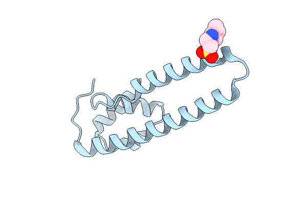 |
Structure Of The Fluorescence Recovery-Like Protein Frpl From Pseudomonas Borbori
Organism: Pseudomonas borbori
Method: X-RAY DIFFRACTION Resolution:1.80 Å Release Date: 2023-04-05 Classification: PROTEIN BINDING Ligands: NHE |
Organism: Pseudomonas borbori
Method: X-RAY DIFFRACTION
Release Date: 2023-04-05
Ligands: NHE
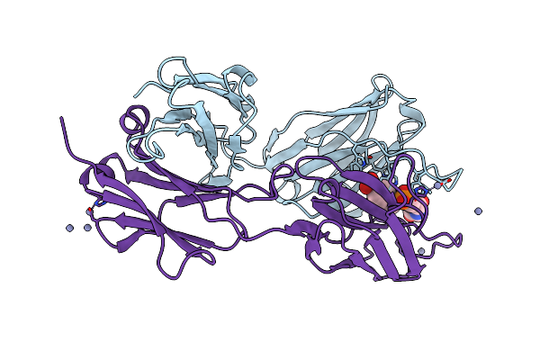 |
Structure Of A Catalytic Antibody Igg2A Fab Fragment (D2.3)
Organism: Mus musculus
Method: X-RAY DIFFRACTION Resolution:1.90 Å Release Date: 1997-10-15 Classification: CATALYTIC ANTIBODY Ligands: ZN, PNB |
Organism: Mus musculus
Method: X-RAY DIFFRACTION
Release Date: 1997-10-15
Ligands: ZN, PNB
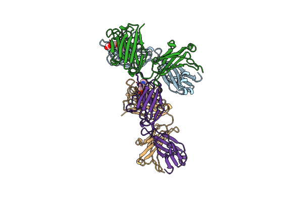 |
Structure Of A Catalytic Antibody Igg2A Fab Fragment (D2.4)
Organism: Mus musculus
Method: X-RAY DIFFRACTION Resolution:3.10 Å Release Date: 1997-10-15 Classification: CATALYTIC ANTIBODY Ligands: PNB |
Organism: Mus musculus
Method: X-RAY DIFFRACTION
Release Date: 1997-10-15
Ligands: PNB
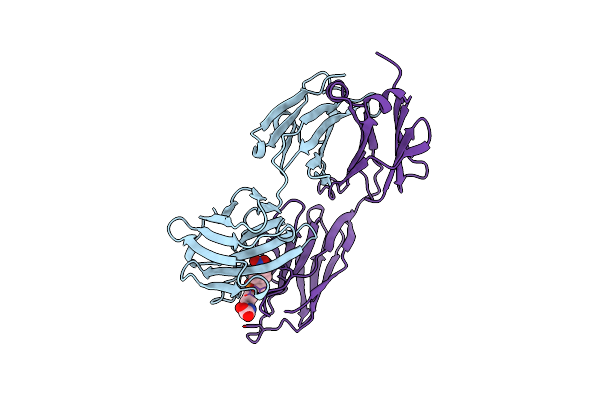 |
Structure Of A Catalytic Antibody, Igg2A Fab Fragment (D2.5)
Organism: Mus musculus
Method: X-RAY DIFFRACTION Resolution:2.20 Å Release Date: 1997-10-15 Classification: CATALYTIC ANTIBODY Ligands: PNB |
Organism: Mus musculus
Method: X-RAY DIFFRACTION
Release Date: 1997-10-15
Ligands: PNB