

Search Count: 17
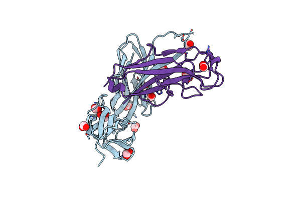 |
Crystal Structure Of The E. Coli Type 1 Pilus Assembly Inhibitor Fimi Bound To Fimc
Organism: Escherichia coli (strain k12)
Method: X-RAY DIFFRACTION Resolution:1.75 Å Release Date: 2021-12-08 Classification: STRUCTURAL PROTEIN Ligands: EDO, FMT |
Organism: Escherichia coli (strain k12)
Method: X-RAY DIFFRACTION
Release Date: 2021-12-08
Ligands: EDO, FMT
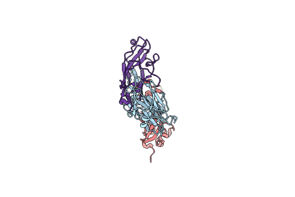 |
Crystal Structure Of The Ternary Complex Of The E. Coli Type 1 Pilus Proteins Fimc, Fimi And The N-Terminal Domain Of Fimd
Organism: Escherichia coli (strain k12)
Method: X-RAY DIFFRACTION Resolution:1.70 Å Release Date: 2021-12-08 Classification: STRUCTURAL PROTEIN Ligands: EDO |
Organism: Escherichia coli (strain k12)
Method: X-RAY DIFFRACTION
Release Date: 2021-12-08
Ligands: EDO
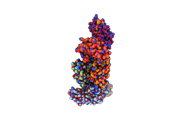 |
Crystal Structure Of The Ternary Complex Between The Type 1 Pilus Proteins Fimc, Fimi And Fima From E. Coli
Organism: Escherichia coli (strain k12)
Method: X-RAY DIFFRACTION Resolution:2.80 Å Release Date: 2020-10-07 Classification: STRUCTURAL PROTEIN Ligands: PEG, EDO |
Organism: Escherichia coli (strain k12)
Method: X-RAY DIFFRACTION
Release Date: 2020-10-07
Ligands: PEG, EDO
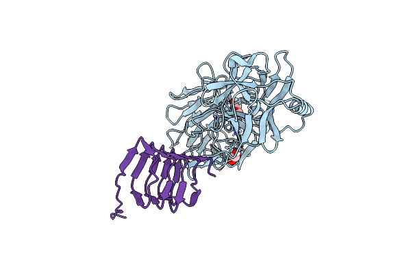 |
Crystal Structure Of The Bont/A2 Receptor-Binding Domain In Complex With The Luminal Domain Of Its Neuronal Receptor Sv2C
Organism: Clostridium botulinum, Homo sapiens
Method: X-RAY DIFFRACTION Resolution:2.30 Å Release Date: 2017-03-15 Classification: HYDROLASE Ligands: EDO, 2PE |
Organism: Clostridium botulinum, Homo sapiens
Method: X-RAY DIFFRACTION
Release Date: 2017-03-15
Ligands: EDO, 2PE
 |
Crystal Structure Of The E. Coli Type 1 Pilus Subunit Fimg (Engineered Variant With Substitution Q134E; N-Terminal Fimg Residues 1-12 Truncated) In Complex With The Donor Strand Peptide Dsf_T4R-T6R-D13N
Organism: Escherichia coli k-12
Method: X-RAY DIFFRACTION Resolution:1.50 Å Release Date: 2016-07-06 Classification: CELL ADHESION Ligands: CO |
Organism: Escherichia coli k-12
Method: X-RAY DIFFRACTION
Release Date: 2016-07-06
Ligands: CO
 |
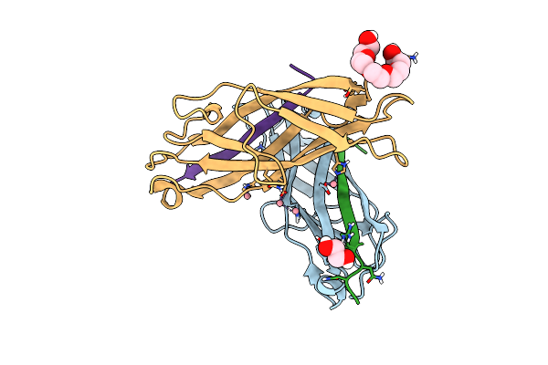
Crystal Structure Of The E. Coli Type 1 Pilus Subunit Fimg (Engineered Variant With Substitution Q134E; N-Terminal Fimg Residues 1-12 Truncated) In Complex With The Donor Strand Peptide Dsf_Sririrgyvr
Organism: Escherichia coli k-12
Method: X-RAY DIFFRACTION Resolution:1.00 Å Release Date: 2016-07-06 Classification: CELL ADHESION Ligands: CO, EDO |
Organism: Escherichia coli k-12
Method: X-RAY DIFFRACTION
Release Date: 2016-07-06
Ligands: CO, EDO
 |
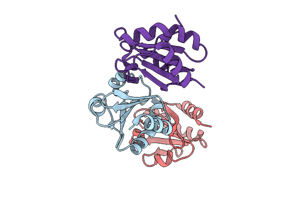
Crystal Structure Of The E. Coli Type 1 Pilus Subunit Fimg (Engineered Variant With Substitutions Q134E And S138E; N-Terminal Fimg Residues 1-12 Truncated) In Complex With The Donor Strand Peptide Dsf_T4R-T6R-D13N
Organism: Escherichia coli (strain k12), Escherichia coli k-12
Method: X-RAY DIFFRACTION Resolution:1.30 Å Release Date: 2016-07-06 Classification: CELL ADHESION Ligands: CO, EDO, 1PE |
Organism: Escherichia coli (strain k12), Escherichia coli k-12
Method: X-RAY DIFFRACTION
Release Date: 2016-07-06
Ligands: CO, EDO, 1PE
 |
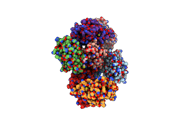
Structure Of Proline-Free E. Coli Thioredoxin
Organism: Escherichia coli k-12
Method: X-RAY DIFFRACTION Resolution:1.65 Å Release Date: 2015-06-24 Classification: OXIDOREDUCTASE |
Organism: Escherichia coli k-12
Method: X-RAY DIFFRACTION
Release Date: 2015-06-24
 |
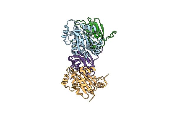
Crystal Structure Of The Mixed Disulfide Complex Of Thioredoxin-Like Tlpas(C110S) And Copper Chaperone Scois(C74S)
Organism: Bradyrhizobium diazoefficiens usda 110
Method: X-RAY DIFFRACTION Resolution:2.20 Å Release Date: 2014-10-01 Classification: OXIDODREDUCTASE/COPPER BINDING PROTEIN Ligands: SCN, PEG, NA |
Organism: Bradyrhizobium diazoefficiens usda 110
Method: X-RAY DIFFRACTION
Release Date: 2014-10-01
Ligands: SCN, PEG, NA
 |
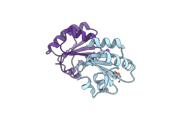
Crystal Structure Of The Mixed Disulfide Intermediate Between Thioredoxin-Like Tlpas(C110S) And Subunit Ii Of Cytochrome C Oxidase Coxbpd (C233S)
Organism: Bradyrhizobium diazoefficiens, Bradyrhizobium diazoefficiens (strain jcm 10833 / iam 13628 / nbrc 14792 / usda 110)
Method: X-RAY DIFFRACTION Resolution:2.00 Å Release Date: 2014-10-01 Classification: PROTEIN BINDING |
 |
E. Coli Thioredoxin Variant With Pro76 As Single Proline Residue
Organism: Escherichia coli
Method: X-RAY DIFFRACTION Resolution:1.40 Å Release Date: 2013-05-29 Classification: OXIDOREDUCTASE Ligands: CU, NA |
Organism: Escherichia coli
Method: X-RAY DIFFRACTION
Release Date: 2013-05-29
Ligands: CU, NA
 |
E. Coli Thioredoxin Variant With (4S)-Fluoropro76 As Single Proline Residue
Organism: Escherichia coli
Method: X-RAY DIFFRACTION Resolution:1.55 Å Release Date: 2013-05-29 Classification: OXIDOREDUCTASE Ligands: CU |
Organism: Escherichia coli
Method: X-RAY DIFFRACTION
Release Date: 2013-05-29
Ligands: CU
 |
E. Coli Thioredoxin Variant With (4R)-Fluoropro76 As Single Proline Residue
Organism: Escherichia coli
Method: X-RAY DIFFRACTION Resolution:1.10 Å Release Date: 2013-05-29 Classification: OXIDOREDUCTASE Ligands: CU |
Organism: Escherichia coli
Method: X-RAY DIFFRACTION
Release Date: 2013-05-29
Ligands: CU
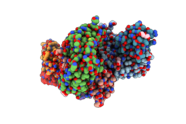 |
Structure Of The Major Type 1 Pilus Subunit Fima Bound To The Fimc Chaperone
Organism: Escherichia coli
Method: X-RAY DIFFRACTION Resolution:3.20 Å Release Date: 2012-05-30 Classification: STRUCTURAL PROTEIN/CHAPERONE Ligands: PEG, EDO, TRS |
Organism: Escherichia coli
Method: X-RAY DIFFRACTION
Release Date: 2012-05-30
Ligands: PEG, EDO, TRS
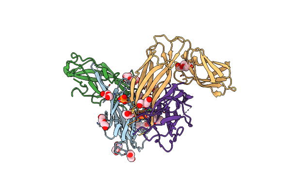 |
Structure Of The Major Type 1 Pilus Subunit Fima Bound To The Fimc (2.5 A Resolution)
Organism: Escherichia coli
Method: X-RAY DIFFRACTION Resolution:2.50 Å Release Date: 2012-05-30 Classification: STRUCTURAL PROTEIN/CHAPERONE Ligands: PEG, PG4, PO4, PGE |
Organism: Escherichia coli
Method: X-RAY DIFFRACTION
Release Date: 2012-05-30
Ligands: PEG, PG4, PO4, PGE
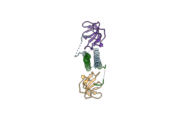 |
Eb1C/Eb3C Heterodimer In Complex With The Cap-Gly Domain Of P150Glued
Organism: Homo sapiens
Method: X-RAY DIFFRACTION Resolution:2.30 Å Release Date: 2012-01-25 Classification: PROTEIN BINDING |
Organism: Homo sapiens
Method: X-RAY DIFFRACTION
Release Date: 2012-01-25
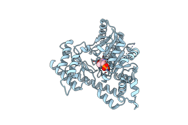 |
High-Resolution Structure Of Native Malus Domestica Acc Synthase
Organism: Malus domestica
Method: X-RAY DIFFRACTION Resolution:1.35 Å Release Date: 2010-12-15 Classification: LYASE Ligands: PLR |
Organism: Malus domestica
Method: X-RAY DIFFRACTION
Release Date: 2010-12-15
Ligands: PLR