

Search Count: 22
 |
Crystal Structure Of Schistosoma Mansoni Hdac8 In Complex With A Formate Molecule In The Active Site
Organism: Schistosoma mansoni
Method: X-RAY DIFFRACTION Resolution:2.85 Å Release Date: 2023-05-31 Classification: HYDROLASE Ligands: ZN, K, CL, FMT, PEG |
Organism: Schistosoma mansoni
Method: X-RAY DIFFRACTION
Release Date: 2023-05-31
Ligands: ZN, K, CL, FMT, PEG
 |
Crystal Structure Of Schistosoma Mansoni Hdac8 With Dmso Bound In The Active Site
Organism: Schistosoma mansoni
Method: X-RAY DIFFRACTION Resolution:1.90 Å Release Date: 2022-08-31 Classification: HYDROLASE Ligands: ZN, K, GOL, DMS |
Organism: Schistosoma mansoni
Method: X-RAY DIFFRACTION
Release Date: 2022-08-31
Ligands: ZN, K, GOL, DMS
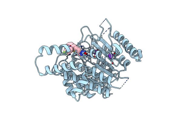 |
Crystal Structure Of Schistosoma Mansoni Hdac8 In Complex With A Tricyclic Thieno[3,2-B]Indole Capped Hydroxamate-Based Inhibitor, Chlorine Derivative
Organism: Schistosoma mansoni
Method: X-RAY DIFFRACTION Resolution:2.20 Å Release Date: 2022-07-27 Classification: HYDROLASE Ligands: ZN, K, 4UI |
Organism: Schistosoma mansoni
Method: X-RAY DIFFRACTION
Release Date: 2022-07-27
Ligands: ZN, K, 4UI
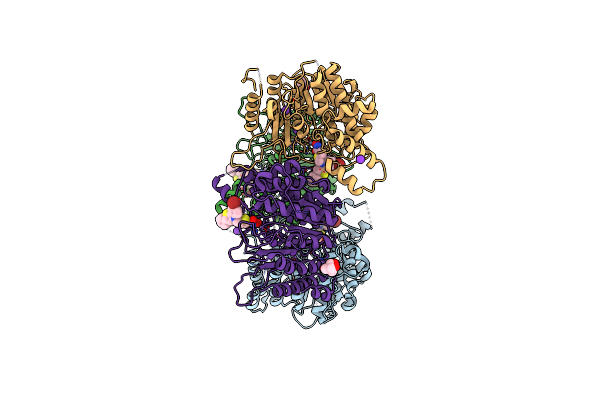 |
Tetartohedrally Twinned Crystal Structure Of Schistosoma Mansoni Hdac8 In Complex With A Tricyclic Thieno[3,2-B]Indole Capped Hydroxamate-Based Inhibitor, Bromine Derivative
Organism: Schistosoma mansoni
Method: X-RAY DIFFRACTION Resolution:2.30 Å Release Date: 2022-07-20 Classification: HYDROLASE Ligands: ZN, K, 4VX, 144, CL |
Organism: Schistosoma mansoni
Method: X-RAY DIFFRACTION
Release Date: 2022-07-20
Ligands: ZN, K, 4VX, 144, CL
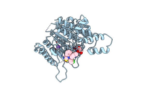 |
Crystal Structure Of Schistosoma Mansoni Hdac8 In Complex With A 3-Chlorophenyl-Spiroindoline Capped Hydroxamate-Based Inhibitor, Bound To A Novel Site
Organism: Schistosoma mansoni
Method: X-RAY DIFFRACTION Resolution:1.80 Å Release Date: 2022-07-20 Classification: HYDROLASE Ligands: ZN, K, CL, TLA, 4WB |
Organism: Schistosoma mansoni
Method: X-RAY DIFFRACTION
Release Date: 2022-07-20
Ligands: ZN, K, CL, TLA, 4WB
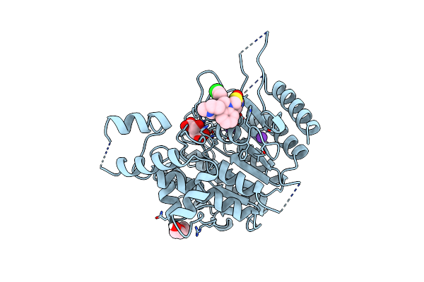 |
Crystal Structure Of Schistosoma Mansoni Hdac8 In Complex With A 4-Chlorophenyl-Spiroindoline Capped Hydroxamate-Based Inhibitor, Bound To A Novel Site
Organism: Schistosoma mansoni
Method: X-RAY DIFFRACTION Resolution:2.25 Å Release Date: 2022-07-20 Classification: HYDROLASE Ligands: ZN, K, TLA, 4XI, PEG |
Organism: Schistosoma mansoni
Method: X-RAY DIFFRACTION
Release Date: 2022-07-20
Ligands: ZN, K, TLA, 4XI, PEG
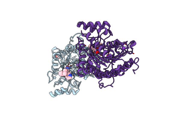 |
Crystal Structure Of Danio Rerio Histone Deacetylase 6 Catalytic Domain 2 (Cd2) Complexed With Nf2376
Organism: Danio rerio
Method: X-RAY DIFFRACTION Resolution:2.04 Å Release Date: 2020-12-02 Classification: HYDROLASE Ligands: ZN, K, QQD, EDO |
Organism: Danio rerio
Method: X-RAY DIFFRACTION
Release Date: 2020-12-02
Ligands: ZN, K, QQD, EDO
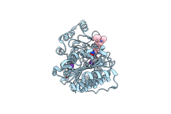 |
Crystal Structure Of Danio Rerio Histone Deacetylase 6 Catalytic Domain 2 (Cd2) Complexed With Nf2657
Organism: Danio rerio
Method: X-RAY DIFFRACTION Resolution:2.09 Å Release Date: 2020-12-02 Classification: HYDROLASE Ligands: ZN, K, QQG |
Organism: Danio rerio
Method: X-RAY DIFFRACTION
Release Date: 2020-12-02
Ligands: ZN, K, QQG
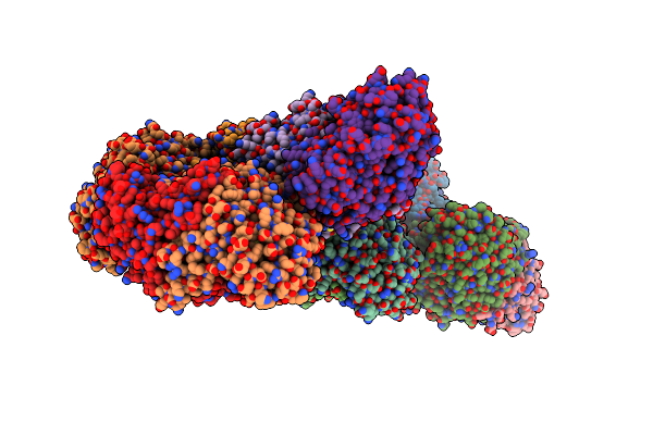 |
Methanococcus Jannaschii Serine Hydroxymethyl-Transferase In Complex With Plp
Organism: Methanocaldococcus jannaschii
Method: X-RAY DIFFRACTION Resolution:3.00 Å Release Date: 2014-07-30 Classification: TRANSFERASE Ligands: PLP |
Organism: Methanocaldococcus jannaschii
Method: X-RAY DIFFRACTION
Release Date: 2014-07-30
Ligands: PLP
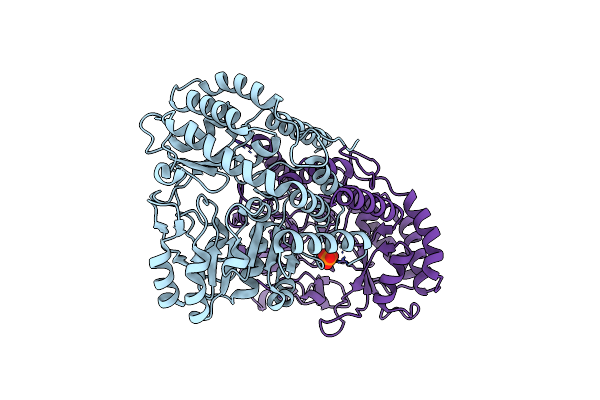 |
Methanococcus Jannaschii Serine Hydroxymethyl-Transferase, Apo Form
Organism: Methanocaldococcus jannaschii
Method: X-RAY DIFFRACTION Resolution:2.83 Å Release Date: 2014-04-16 Classification: TRANSFERASE Ligands: PO4 |
Organism: Methanocaldococcus jannaschii
Method: X-RAY DIFFRACTION
Release Date: 2014-04-16
Ligands: PO4
 |
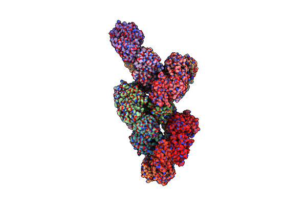
Crystal Structure Of Schistosoma Mansoni Peroxiredoxin I C48S Mutant With One Decamer In The Asu
Organism: Schistosoma mansoni
Method: X-RAY DIFFRACTION Resolution:2.49 Å Release Date: 2013-09-11 Classification: OXIDOREDUCTASE Ligands: SO4, PEG |
Organism: Schistosoma mansoni
Method: X-RAY DIFFRACTION
Release Date: 2013-09-11
Ligands: SO4, PEG
 |
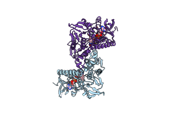
Crystal Structure Of Schistosoma Mansoni Peroxiredoxin 1 C48P Mutant Form With Four Decamers In The Asymmetric Unit
Organism: Schistosoma mansoni
Method: X-RAY DIFFRACTION Resolution:3.52 Å Release Date: 2013-09-11 Classification: OXIDOREDUCTASE |
Organism: Schistosoma mansoni
Method: X-RAY DIFFRACTION
Release Date: 2013-09-11
 |
Crystal Structure Of Plasmodium Falciparum Oxidised Thioredoxin Reductase At 2.9 Angstrom
Organism: Plasmodium falciparum
Method: X-RAY DIFFRACTION Resolution:2.90 Å Release Date: 2012-08-29 Classification: OXIDOREDUCTASE Ligands: FAD |
Organism: Plasmodium falciparum
Method: X-RAY DIFFRACTION
Release Date: 2012-08-29
Ligands: FAD
 |
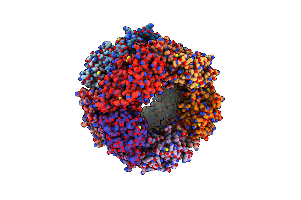
Crystal Structure Of Decameric Form Of Peroxiredoxin I From Schistosoma Mansoni
Organism: Schistosoma mansoni
Method: X-RAY DIFFRACTION Resolution:3.00 Å Release Date: 2012-03-21 Classification: OXIDOREDUCTASE |
Organism: Schistosoma mansoni
Method: X-RAY DIFFRACTION
Release Date: 2012-03-21
 |
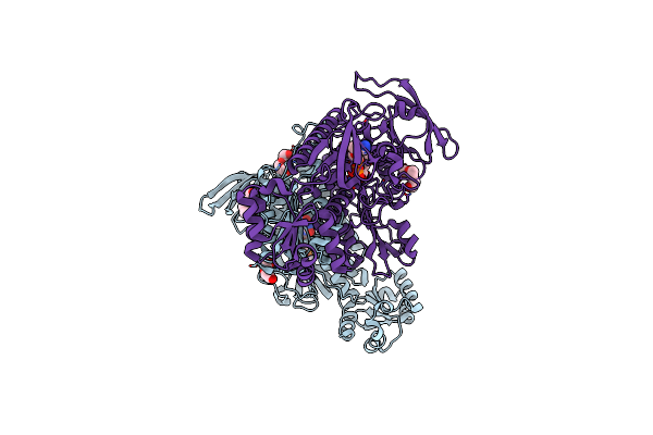
Crystal Structure Of High Molecular Weight (Hmw) Form Of Peroxiredoxin I From Schistosoma Mansoni
Organism: Schistosoma mansoni
Method: X-RAY DIFFRACTION Resolution:3.00 Å Release Date: 2012-03-21 Classification: OXIDOREDUCTASE |
Organism: Schistosoma mansoni
Method: X-RAY DIFFRACTION
Release Date: 2012-03-21
 |
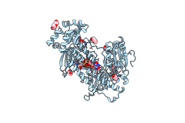
Thioredoxin Glutathione Reductase From Schistosoma Mansoni With The Reduced C-Terminal End
Organism: Schistosoma mansoni
Method: X-RAY DIFFRACTION Resolution:3.10 Å Release Date: 2010-07-21 Classification: OXIDOREDUCTASE/FLAVOPROTEIN Ligands: FAD, PGE, PEG |
Organism: Schistosoma mansoni
Method: X-RAY DIFFRACTION
Release Date: 2010-07-21
Ligands: FAD, PGE, PEG
 |
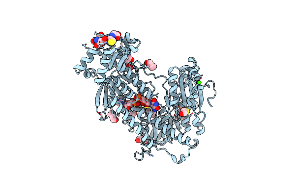
Oxidized Thioredoxin Glutathione Reductase From Schistosoma Mansoni
Organism: Schistosoma mansoni
Method: X-RAY DIFFRACTION Resolution:1.90 Å Release Date: 2010-07-21 Classification: OXIDOREDUCTASE Ligands: FAD, PEG, GOL, PG4 |
Organism: Schistosoma mansoni
Method: X-RAY DIFFRACTION
Release Date: 2010-07-21
Ligands: FAD, PEG, GOL, PG4
 |
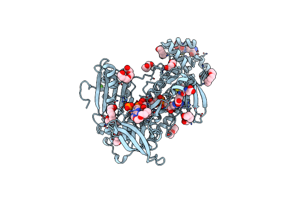
Thioredoxin Glutathione Reductase From Schistosoma Mansoni In Complex With Gsh
Organism: Schistosoma mansoni
Method: X-RAY DIFFRACTION Resolution:1.90 Å Release Date: 2010-07-21 Classification: OXIDOREDUCTASE Ligands: FAD, GSH, BME, PGE, PG4, CA, PEG |
Organism: Schistosoma mansoni
Method: X-RAY DIFFRACTION
Release Date: 2010-07-21
Ligands: FAD, GSH, BME, PGE, PG4, CA, PEG
 |
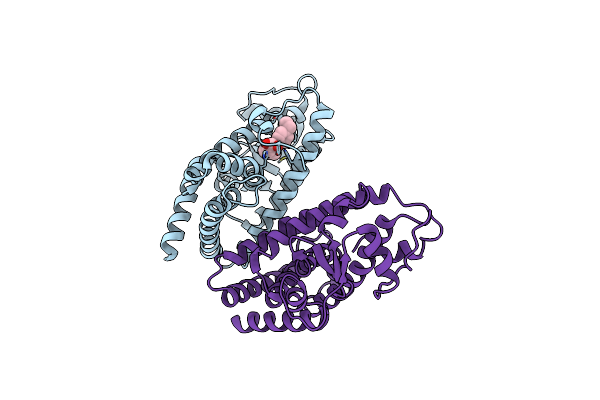
Thioredoxin Glutathione Reductase From Schistosoma Mansoni In Complex With Nadph
Organism: Schistosoma mansoni
Method: X-RAY DIFFRACTION Resolution:2.30 Å Release Date: 2010-07-21 Classification: OXIDOREDUCTASE Ligands: FAD, NDP, GSH, CA, PEG, PG4 |
Organism: Schistosoma mansoni
Method: X-RAY DIFFRACTION
Release Date: 2010-07-21
Ligands: FAD, NDP, GSH, CA, PEG, PG4
 |
Crystal Structure Of The Complex Between Ppar-Gamma And The Agonist Lt248 (Clofibric Acid Analogue)
Organism: Homo sapiens
Method: X-RAY DIFFRACTION Resolution:2.65 Å Release Date: 2008-12-30 Classification: TRANSCRIPTION Ligands: GRR |
Organism: Homo sapiens
Method: X-RAY DIFFRACTION
Release Date: 2008-12-30
Ligands: GRR