

Search Count: 78
 |
Chitinase Tfec From Yersinia Pseudotuberculosis
Organism: Yersinia pseudotuberculosis serotype o:3 (strain ypiii)
Method: X-RAY DIFFRACTION Resolution:1.90 Å Release Date: 2025-11-05 Classification: HYDROLASE Ligands: SRT |
Organism: Yersinia pseudotuberculosis serotype o:3 (strain ypiii)
Method: X-RAY DIFFRACTION
Release Date: 2025-11-05
Ligands: SRT
 |
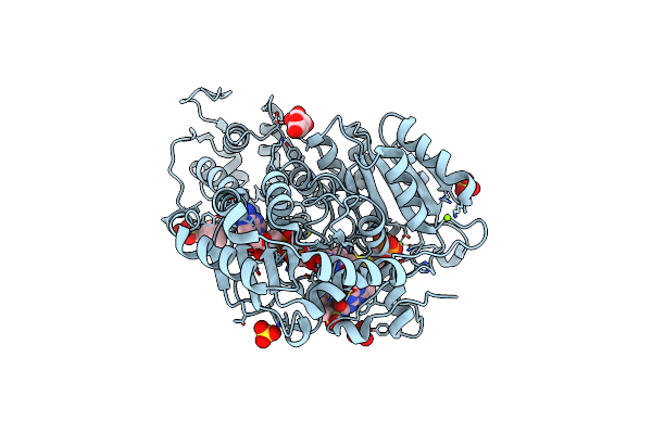
Kras In Complex With Darpin 784_F5
Organism: Homo sapiens, Synthetic construct
Method: X-RAY DIFFRACTION Resolution:2.00 Å Release Date: 2025-06-25 Classification: SIGNALING PROTEIN Ligands: EDO, MG, GNP, 1PE, PGE, SRT |
Organism: Homo sapiens, Synthetic construct
Method: X-RAY DIFFRACTION
Release Date: 2025-06-25
Ligands: EDO, MG, GNP, 1PE, PGE, SRT
 |
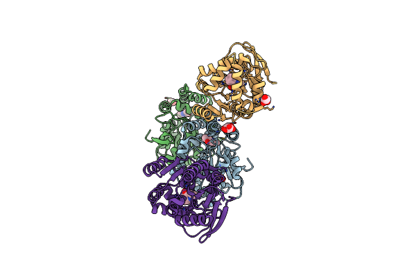
Crystal Structure Of Wild-Type Arabidopsis Thaliana Acetohydroxyacid Synthase In Complex With Commercial Herbicide Triasulfuron
Organism: Arabidopsis thaliana
Method: X-RAY DIFFRACTION Resolution:2.40 Å Release Date: 2024-10-23 Classification: TRANSFERASE Ligands: MG, FAD, NHE, A1AUE, SRT, AUJ, SO4 |
Organism: Arabidopsis thaliana
Method: X-RAY DIFFRACTION
Release Date: 2024-10-23
Ligands: MG, FAD, NHE, A1AUE, SRT, AUJ, SO4
 |
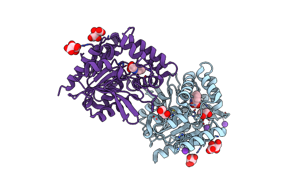
Crystal Structure Of The Cofactor-Devoid 1-H-3-Hydroxy-4- Oxoquinaldine 2,4-Dioxygenase (Hod) S101A Variant Complexed With 2-Methyl-Quinolin-4(1H)-One Under Normoxyc Conditions
Organism: Paenarthrobacter nitroguajacolicus
Method: X-RAY DIFFRACTION Resolution:2.00 Å Release Date: 2024-01-17 Classification: OXIDOREDUCTASE Ligands: VFH, GOL, SRT |
Organism: Paenarthrobacter nitroguajacolicus
Method: X-RAY DIFFRACTION
Release Date: 2024-01-17
Ligands: VFH, GOL, SRT
 |
Crystal Structure Of The Cofactor-Devoid 1-H-3-Hydroxy-4- Oxoquinaldine 2,4-Dioxygenase (Hod) H251A Variant Complexed With N-Acetylanthranilate As Result Of In Crystallo Turnover Of Its Natural Substrate 1-H-3-Hydroxy-4- Oxoquinaldine Under Hyperoxic Conditions
Organism: Paenarthrobacter nitroguajacolicus
Method: X-RAY DIFFRACTION Resolution:2.00 Å Release Date: 2024-01-17 Classification: OXIDOREDUCTASE Ligands: ZZ8, SRT, GOL, K |
Organism: Paenarthrobacter nitroguajacolicus
Method: X-RAY DIFFRACTION
Release Date: 2024-01-17
Ligands: ZZ8, SRT, GOL, K
 |
Crystal Structure Of Human Trim7 Pryspry Domain Bound To (2-(1-Oxoisoindolin-2-Yl)-3-Phenylpropanoyl)-L-Glutamine
Organism: Homo sapiens
Method: X-RAY DIFFRACTION Resolution:1.60 Å Release Date: 2024-01-17 Classification: LIGASE Ligands: Y3C, EDO, SRT |
Organism: Homo sapiens
Method: X-RAY DIFFRACTION
Release Date: 2024-01-17
Ligands: Y3C, EDO, SRT
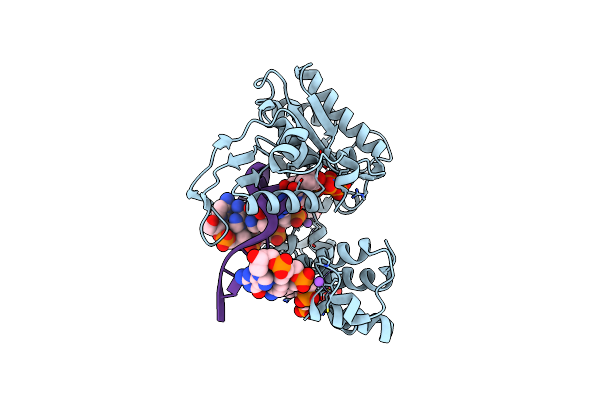 |
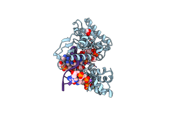
Dna Polymerase Lambda, Ttp:At Mg2+ Reaction State Ternary Complex, 120 Sec
Organism: Homo sapiens, Synthetic construct
Method: X-RAY DIFFRACTION Resolution:1.89 Å Release Date: 2022-07-06 Classification: REPLICATION Ligands: PPV, TTP, MG, NA, EDO, SRT |
Organism: Homo sapiens, Synthetic construct
Method: X-RAY DIFFRACTION
Release Date: 2022-07-06
Ligands: PPV, TTP, MG, NA, EDO, SRT
 |
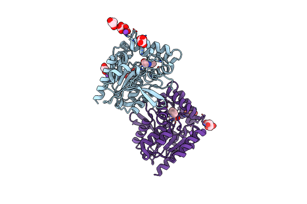
Dna Polymerase Lambda, Ttp:At Mg2+ Product State Ternary Complex, 60 Min
Organism: Homo sapiens, Synthetic construct
Method: X-RAY DIFFRACTION Resolution:1.65 Å Release Date: 2022-07-06 Classification: REPLICATION Ligands: SRT, NA, MG, PPV, EDO |
Organism: Homo sapiens, Synthetic construct
Method: X-RAY DIFFRACTION
Release Date: 2022-07-06
Ligands: SRT, NA, MG, PPV, EDO
 |
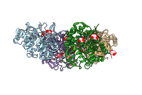
Crystal Structure Of The Cofactor-Devoid 1-H-3-Hydroxy-4- Oxoquinaldine 2,4-Dioxygenase (Hod) Catalytically Inactive H251A Variant Complexed With 2-Methyl-Quinolin-4(1H)-One Under Normoxic Conditions
Organism: Paenarthrobacter nitroguajacolicus
Method: X-RAY DIFFRACTION Resolution:2.00 Å Release Date: 2022-06-01 Classification: OXIDOREDUCTASE Ligands: VFH, K, GOL, SRT, NA |
Organism: Paenarthrobacter nitroguajacolicus
Method: X-RAY DIFFRACTION
Release Date: 2022-06-01
Ligands: VFH, K, GOL, SRT, NA
 |
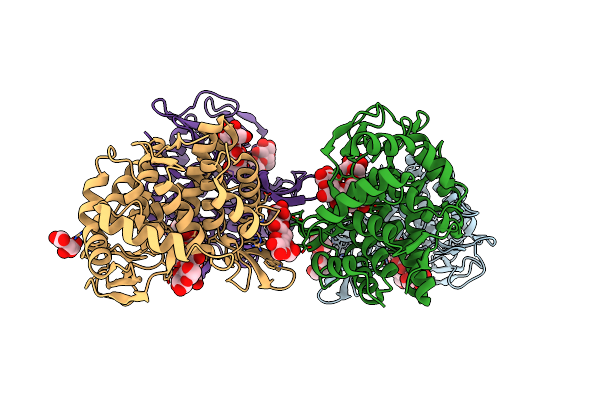
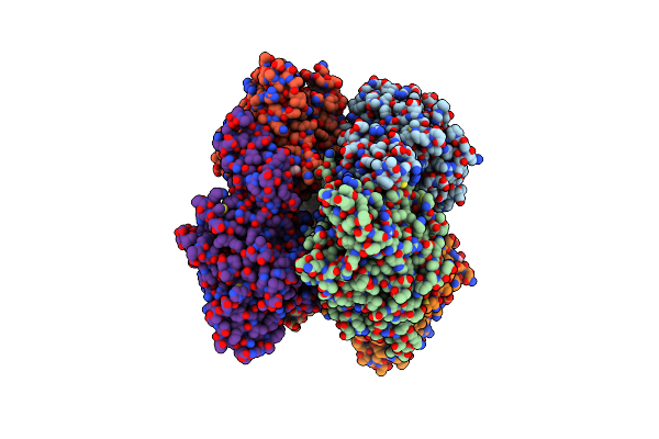
Structure Of The Bacterial Cellulose Synthase Subunit Z From Enterobacter Sp. Cjf-002
Organism: Enterobacter sp. cjf-002
Method: X-RAY DIFFRACTION Resolution:1.93 Å Release Date: 2022-02-09 Classification: HYDROLASE Ligands: GOL, SRT |
Organism: Enterobacter sp. cjf-002
Method: X-RAY DIFFRACTION
Release Date: 2022-02-09
Ligands: GOL, SRT
 |
Structure Of The Bacterial Cellulose Synthase Subunit Z In Complex With Cellooligosaccharides From Enterobacter Sp. Cjf-002
Organism: Enterobacter sp. cjf-002
Method: X-RAY DIFFRACTION Resolution:1.30 Å Release Date: 2022-02-09 Classification: HYDROLASE Ligands: SRT |
Organism: Enterobacter sp. cjf-002
Method: X-RAY DIFFRACTION
Release Date: 2022-02-09
Ligands: SRT
 |
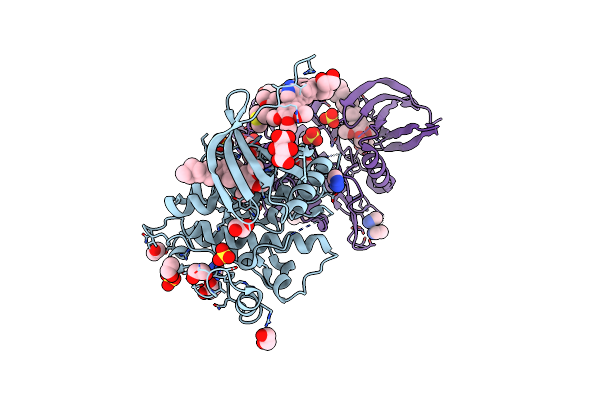
Crystal Structure Of Phthalate Dioxygenase From Comamonas Testosteroni Kf1
Organism: Comamonas testosteroni (strain dsm 14576 / kf-1)
Method: X-RAY DIFFRACTION Resolution:2.11 Å Release Date: 2021-12-15 Classification: OXIDOREDUCTASE Ligands: FE2, FES, TLA, SRT, EDO |
Organism: Comamonas testosteroni (strain dsm 14576 / kf-1)
Method: X-RAY DIFFRACTION
Release Date: 2021-12-15
Ligands: FE2, FES, TLA, SRT, EDO
 |
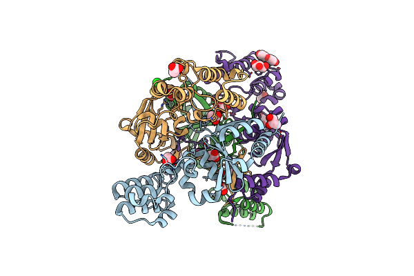
Xchem Group Deposition -- Crystal Structure Of Human Acvr1 In Complex With Fm010913A
Organism: Homo sapiens
Method: X-RAY DIFFRACTION Resolution:1.30 Å Release Date: 2021-06-23 Classification: TRANSFERASE Ligands: LU8, EDO, DMS, SRT, HUH, SO4 |
Organism: Homo sapiens
Method: X-RAY DIFFRACTION
Release Date: 2021-06-23
Ligands: LU8, EDO, DMS, SRT, HUH, SO4
 |
The Structure Of A Sensor Domain Of A Histidine Kinase (Vxra) From Vibrio Cholerae O1 Biovar Eltor Str. N16961, 2Nd Form
Organism: Vibrio cholerae serotype o1 (strain atcc 39315 / el tor inaba n16961)
Method: X-RAY DIFFRACTION Resolution:2.25 Å Release Date: 2020-10-14 Classification: SIGNALING PROTEIN Ligands: GOL, ACT, CL, PEG, SRT |
Organism: Vibrio cholerae serotype o1 (strain atcc 39315 / el tor inaba n16961)
Method: X-RAY DIFFRACTION
Release Date: 2020-10-14
Ligands: GOL, ACT, CL, PEG, SRT
 |
The Crystal Structure Of A Beta-Lactamase From Escherichia Coli Cft073
Organism: Escherichia coli o6:h1 (strain cft073 / atcc 700928 / upec)
Method: X-RAY DIFFRACTION Resolution:1.90 Å Release Date: 2020-06-17 Classification: HYDROLASE Ligands: GOL, SO4, SRT |
Organism: Escherichia coli o6:h1 (strain cft073 / atcc 700928 / upec)
Method: X-RAY DIFFRACTION
Release Date: 2020-06-17
Ligands: GOL, SO4, SRT
 |
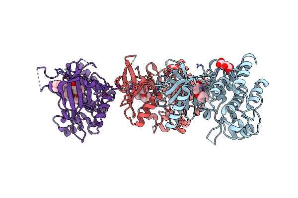
Structure Of Fima Type-1 (Fima1) Prepilin Of The Type V Major Fimbrium
Organism: Porphyromonas gingivalis atcc 33277
Method: X-RAY DIFFRACTION Resolution:2.10 Å Release Date: 2020-04-15 Classification: CELL ADHESION Ligands: GOL, SO4, SRT |
Organism: Porphyromonas gingivalis atcc 33277
Method: X-RAY DIFFRACTION
Release Date: 2020-04-15
Ligands: GOL, SO4, SRT
 |
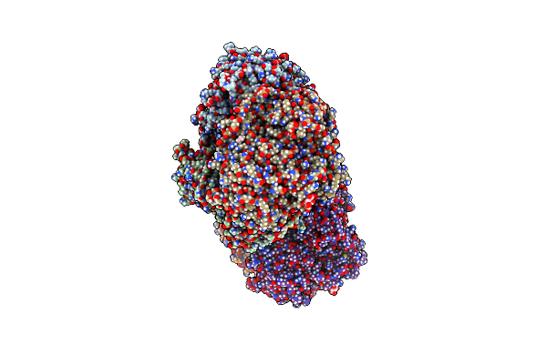
Trk-A In Complex With Ligand
Organism: Homo sapiens
Method: X-RAY DIFFRACTION Resolution:3.00 Å Release Date: 2020-02-26 Classification: TRANSFERASE Ligands: ZN, OOY, SRT |
Organism: Homo sapiens
Method: X-RAY DIFFRACTION
Release Date: 2020-02-26
Ligands: ZN, OOY, SRT
 |
Bacteroides Salyersiae Gh164 Beta-Mannosidase
Organism: Bacteroides salyersiae
Method: X-RAY DIFFRACTION Resolution:1.91 Å Release Date: 2020-01-01 Classification: HYDROLASE Ligands: EDO, SRT, CL, TLA, PG4, TAR |
Organism: Bacteroides salyersiae
Method: X-RAY DIFFRACTION
Release Date: 2020-01-01
Ligands: EDO, SRT, CL, TLA, PG4, TAR
 |
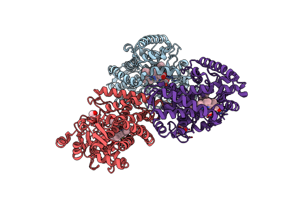
Crystal Structure Of Ppk2 In Complex With Phosphonic Acid Inhibitor
Organism: Cytophaga hutchinsonii (strain atcc 33406 / ncimb 9469)
Method: X-RAY DIFFRACTION Resolution:2.20 Å Release Date: 2019-01-16 Classification: transferase/transferase inhibitor Ligands: BOY, GOL, SRT |
Organism: Cytophaga hutchinsonii (strain atcc 33406 / ncimb 9469)
Method: X-RAY DIFFRACTION
Release Date: 2019-01-16
Ligands: BOY, GOL, SRT
 |
Cytochrome P450-153 From Pseudomonas Sp. 19-Rlim
Organism: Pseudomonas sp. 19-rlim
Method: X-RAY DIFFRACTION Resolution:1.80 Å Release Date: 2018-12-12 Classification: OXIDOREDUCTASE Ligands: HEM, SRT, PGE, GOL |
Organism: Pseudomonas sp. 19-rlim
Method: X-RAY DIFFRACTION
Release Date: 2018-12-12
Ligands: HEM, SRT, PGE, GOL