

Search Count: 39
 |
Smad4 Mh2 Domain (Residues 314-552) Bound By Nanobody A1 (Nba1)
Organism: Homo sapiens, Lama glama
Method: X-RAY DIFFRACTION Resolution:2.10 Å Release Date: 2026-09-30 Classification: TRANSCRIPTION Ligands: SO4, EDO |
Organism: Homo sapiens, Lama glama
Method: X-RAY DIFFRACTION
Release Date: 2026-09-30
Ligands: SO4, EDO
 |
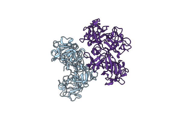
Smad4 Mh2 Domain (Residues 314-552) Bound By Nanobody A7 (Nba7)
Organism: Homo sapiens, Lama glama
Method: X-RAY DIFFRACTION Resolution:3.04 Å Release Date: 2026-09-30 Classification: TRANSCRIPTION Ligands: ACT, PEG, EDO |
Organism: Homo sapiens, Lama glama
Method: X-RAY DIFFRACTION
Release Date: 2026-09-30
Ligands: ACT, PEG, EDO
 |
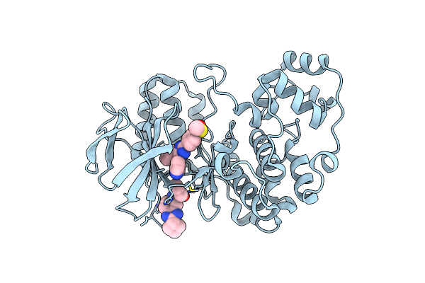
Full-Length Crystal Structure Of Human Fascin 1
Organism: Homo sapiens
Method: X-RAY DIFFRACTION Resolution:2.20 Å Release Date: 2025-06-25 Classification: PROTEIN BINDING |
Organism: Homo sapiens
Method: X-RAY DIFFRACTION
Release Date: 2025-06-25
 |
Crystal Structure Of P38Alpha C162S In Complex With Sb20358 And Cas 2094667-81-7 (Behind Catalytic Site; Y35 In), P 21 21 21
Organism: Mus musculus
Method: X-RAY DIFFRACTION Resolution:2.25 Å Release Date: 2023-06-14 Classification: ONCOPROTEIN Ligands: IIM, 87B |
Organism: Mus musculus
Method: X-RAY DIFFRACTION
Release Date: 2023-06-14
Ligands: IIM, 87B
 |
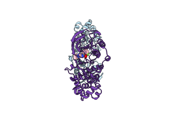
Crystal Structure Of P38Alpha C162S In Complex With Atpgs And Cas 2094667-81-7 (In Catalytic Site, Y35 Out), P 1 21 1
Organism: Mus musculus
Method: X-RAY DIFFRACTION Resolution:2.60 Å Release Date: 2023-06-14 Classification: ONCOPROTEIN Ligands: 87B, ATP |
Organism: Mus musculus
Method: X-RAY DIFFRACTION
Release Date: 2023-06-14
Ligands: 87B, ATP
 |
Crystal Structure Of P38Alpha C162S In Complex With Cas2094511-69-8, P 1 21 1
Organism: Mus musculus
Method: X-RAY DIFFRACTION Resolution:2.15 Å Release Date: 2023-05-17 Classification: ONCOPROTEIN Ligands: 8DI |
Organism: Mus musculus
Method: X-RAY DIFFRACTION
Release Date: 2023-05-17
Ligands: 8DI
 |
Crystal Structure Of The Zebrafish Foxh1 Bound To The Tgtggatt Site
Organism: Danio rerio
Method: X-RAY DIFFRACTION Resolution:0.98 Å Release Date: 2022-11-16 Classification: DNA BINDING PROTEIN Ligands: K |
Organism: Danio rerio
Method: X-RAY DIFFRACTION
Release Date: 2022-11-16
Ligands: K
 |
Crystal Structure Of The Zebrafish Foxh1 Bound To The Tgtgtatt Site
Organism: Danio rerio
Method: X-RAY DIFFRACTION Resolution:1.18 Å Release Date: 2022-11-16 Classification: DNA BINDING PROTEIN Ligands: K |
Organism: Danio rerio
Method: X-RAY DIFFRACTION
Release Date: 2022-11-16
Ligands: K
 |
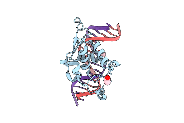
Crystal Structure Of The Human Foxh1 Bound To The Tgtggatt Site
Organism: Homo sapiens
Method: X-RAY DIFFRACTION Resolution:1.47 Å Release Date: 2022-11-16 Classification: DNA BINDING PROTEIN Ligands: K |
Organism: Homo sapiens
Method: X-RAY DIFFRACTION
Release Date: 2022-11-16
Ligands: K
 |
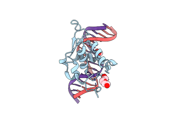
Crystal Structure Of The Zebrafish Foxh1 Bound To The Tgtttatt Site
Organism: Danio rerio
Method: X-RAY DIFFRACTION Resolution:2.17 Å Release Date: 2022-11-16 Classification: DNA BINDING PROTEIN Ligands: ACT |
Organism: Danio rerio
Method: X-RAY DIFFRACTION
Release Date: 2022-11-16
Ligands: ACT
 |
Crystal Structure Of The Zebrafish Foxh1 Bound To The Tgtttact Site (Fkh Motif Gtaaaca)
Organism: Danio rerio
Method: X-RAY DIFFRACTION Resolution:2.13 Å Release Date: 2022-11-16 Classification: DNA BINDING PROTEIN Ligands: ACT, PGE |
Organism: Danio rerio
Method: X-RAY DIFFRACTION
Release Date: 2022-11-16
Ligands: ACT, PGE
 |
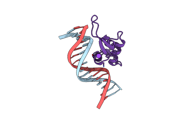
Crystal Structure Of The Human Foxa2 Bound To The Tgtttact Site (Forkhead Motif Gtaaaca)
Organism: Homo sapiens
Method: X-RAY DIFFRACTION Resolution:1.99 Å Release Date: 2022-11-16 Classification: DNA BINDING PROTEIN Ligands: K, SO4 |
Organism: Homo sapiens
Method: X-RAY DIFFRACTION
Release Date: 2022-11-16
Ligands: K, SO4
 |
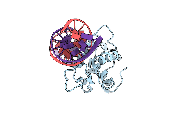
Crystal Structure Of The Human Foxa2 Bound To The Tgtttatt Site (Forkhead Motif Ataaaca)
Organism: Homo sapiens
Method: X-RAY DIFFRACTION Resolution:2.18 Å Release Date: 2022-11-16 Classification: DNA BINDING PROTEIN Ligands: K |
Organism: Homo sapiens
Method: X-RAY DIFFRACTION
Release Date: 2022-11-16
Ligands: K
 |
Crystal Structure Of The Xenopus Foxh1 Bound To The Tgtggatt Site
Organism: Xenopus laevis
Method: X-RAY DIFFRACTION Resolution:2.82 Å Release Date: 2022-11-16 Classification: DNA BINDING PROTEIN |
Organism: Xenopus laevis
Method: X-RAY DIFFRACTION
Release Date: 2022-11-16
 |
Crystal Structure Of Thioredoxin T From Drosophila Melanogaster
Organism: Drosophila melanogaster
Method: X-RAY DIFFRACTION Resolution:2.33 Å Release Date: 2020-10-21 Classification: NUCLEAR PROTEIN Ligands: ZN |
Organism: Drosophila melanogaster
Method: X-RAY DIFFRACTION
Release Date: 2020-10-21
Ligands: ZN
 |
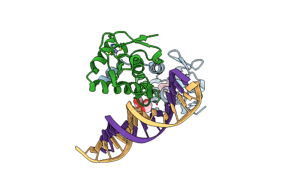
Crystal Structure Of The Germline-Specific Thioredoxin Protein Deadhead (Thioredoxin-1) From Drospohila Melanogaster, P43212
Organism: Drosophila melanogaster
Method: X-RAY DIFFRACTION Resolution:1.95 Å Release Date: 2020-10-21 Classification: OXIDOREDUCTASE Ligands: SO4, NA |
Organism: Drosophila melanogaster
Method: X-RAY DIFFRACTION
Release Date: 2020-10-21
Ligands: SO4, NA
 |
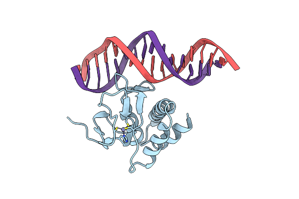
Crystal Structure Of The Smad3-Smad5 Mh1 Domain Chimera Bound To The Ggcgc Site
Organism: Homo sapiens
Method: X-RAY DIFFRACTION Resolution:2.33 Å Release Date: 2020-08-26 Classification: SIGNALING PROTEIN Ligands: ACT, EDO, ZN, PGE |
Organism: Homo sapiens
Method: X-RAY DIFFRACTION
Release Date: 2020-08-26
Ligands: ACT, EDO, ZN, PGE
 |
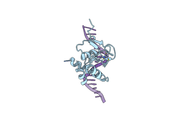
Crystal Structure Of The Mh1 Domain Of Smad5-Smad3 Chimera Construct Bound To The Ggcgc Site
Organism: Homo sapiens
Method: X-RAY DIFFRACTION Resolution:1.78 Å Release Date: 2019-11-20 Classification: TRANSCRIPTION Ligands: ZN |
Organism: Homo sapiens
Method: X-RAY DIFFRACTION
Release Date: 2019-11-20
Ligands: ZN
 |
Crystal Structure Of The Ggct Site-Bound Mh1 Domain Of Smad5 Containing A Gggs Insertion In The Loop1
Organism: Homo sapiens
Method: X-RAY DIFFRACTION Resolution:2.92 Å Release Date: 2019-11-20 Classification: TRANSCRIPTION Ligands: ZN |
Organism: Homo sapiens
Method: X-RAY DIFFRACTION
Release Date: 2019-11-20
Ligands: ZN
 |
Crystal Structure Of Smad5-Mh1 Bound To The Ggcgc Site.
Organism: Homo sapiens
Method: X-RAY DIFFRACTION Resolution:2.31 Å Release Date: 2019-10-02 Classification: TRANSCRIPTION Ligands: ZN |
Organism: Homo sapiens
Method: X-RAY DIFFRACTION
Release Date: 2019-10-02
Ligands: ZN