

Search Count: 16
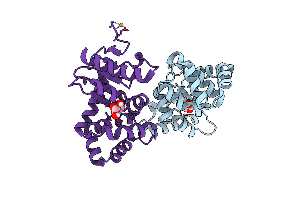 |
Crystal Structure Of E.Coli Dsba Mutant E24A/K58A
Organism: Escherichia coli k-12
Method: X-RAY DIFFRACTION Resolution:1.47 Å Release Date: 2023-02-15 Classification: OXIDOREDUCTASE Ligands: GOL, CU |
Organism: Escherichia coli k-12
Method: X-RAY DIFFRACTION
Release Date: 2023-02-15
Ligands: GOL, CU
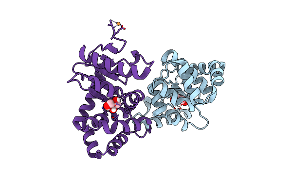 |
Crystal Structure Of E.Coli Dsba Mutant K58A
Organism: Escherichia coli k-12
Method: X-RAY DIFFRACTION Resolution:1.62 Å Release Date: 2023-02-15 Classification: OXIDOREDUCTASE Ligands: GOL, CU |
Organism: Escherichia coli k-12
Method: X-RAY DIFFRACTION
Release Date: 2023-02-15
Ligands: GOL, CU
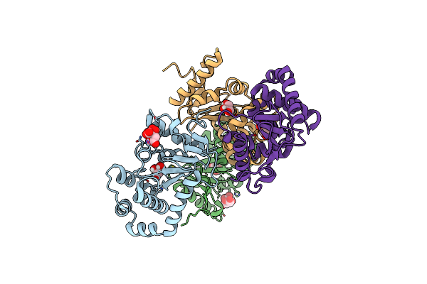 |
Crystal Structure Of E.Coli Dsba Mutant E24A/E37A/K58A
Organism: Escherichia coli k-12
Method: X-RAY DIFFRACTION Resolution:2.30 Å Release Date: 2023-02-15 Classification: OXIDOREDUCTASE Ligands: GOL, FLC |
Organism: Escherichia coli k-12
Method: X-RAY DIFFRACTION
Release Date: 2023-02-15
Ligands: GOL, FLC
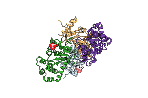 |
Crystal Structure Of E.Coli Dsba Mutant E37A
Organism: Escherichia coli k-12
Method: X-RAY DIFFRACTION Resolution:2.13 Å Release Date: 2023-02-15 Classification: OXIDOREDUCTASE Ligands: FLC |
Organism: Escherichia coli k-12
Method: X-RAY DIFFRACTION
Release Date: 2023-02-15
Ligands: FLC
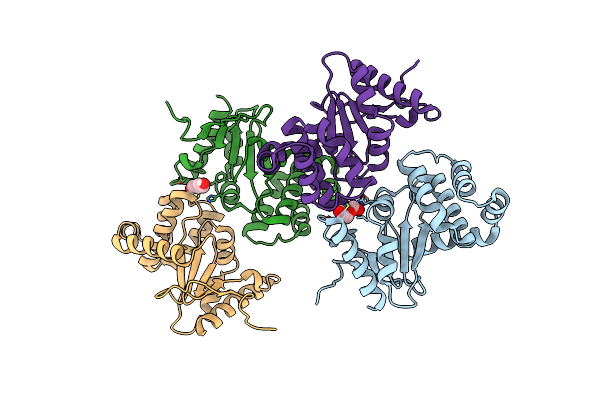 |
Crystal Structure Of E.Coli Dsba Mutant E24A
Organism: Escherichia coli k-12
Method: X-RAY DIFFRACTION Resolution:2.29 Å Release Date: 2023-02-15 Classification: OXIDOREDUCTASE Ligands: PEG |
Organism: Escherichia coli k-12
Method: X-RAY DIFFRACTION
Release Date: 2023-02-15
Ligands: PEG
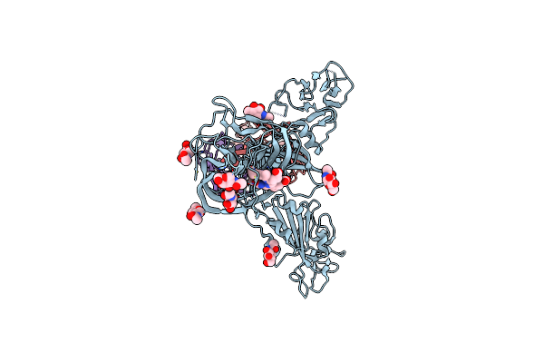 |
Cryoem Structure Of Sars-Cov-2 Spike Monomer In Complex With Neutralising Antibody P008_60
Organism: Severe acute respiratory syndrome coronavirus 2, Homo sapiens
Method: ELECTRON MICROSCOPY Release Date: 2022-08-17 Classification: VIRAL PROTEIN Ligands: NAG, 3Q9 |
Organism: Severe acute respiratory syndrome coronavirus 2, Homo sapiens
Method: ELECTRON MICROSCOPY
Release Date: 2022-08-17
Ligands: NAG, 3Q9
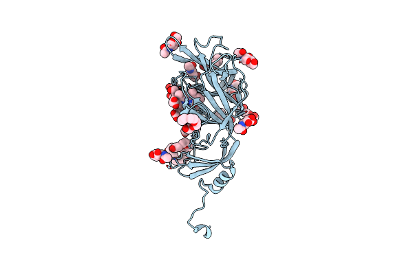 |
Crystal Structure Of Sars-Cov-2 Spike Protein N-Terminal Domain In Complex With Biliverdin
Organism: Severe acute respiratory syndrome coronavirus 2
Method: X-RAY DIFFRACTION Resolution:1.82 Å Release Date: 2021-04-28 Classification: VIRAL PROTEIN Ligands: BLA, NAG, PG4, PEG, PGE, 1PE |
Organism: Severe acute respiratory syndrome coronavirus 2
Method: X-RAY DIFFRACTION
Release Date: 2021-04-28
Ligands: BLA, NAG, PG4, PEG, PGE, 1PE
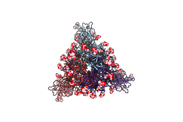 |
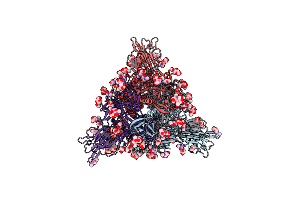
Trimeric Sars-Cov-2 Spike Ectodomain In Complex With Biliverdin (Closed Conformation)
Organism: Severe acute respiratory syndrome coronavirus 2
Method: ELECTRON MICROSCOPY Release Date: 2021-04-28 Classification: VIRAL PROTEIN Ligands: BLA, NAG |
Organism: Severe acute respiratory syndrome coronavirus 2
Method: ELECTRON MICROSCOPY
Release Date: 2021-04-28
Ligands: BLA, NAG
 |
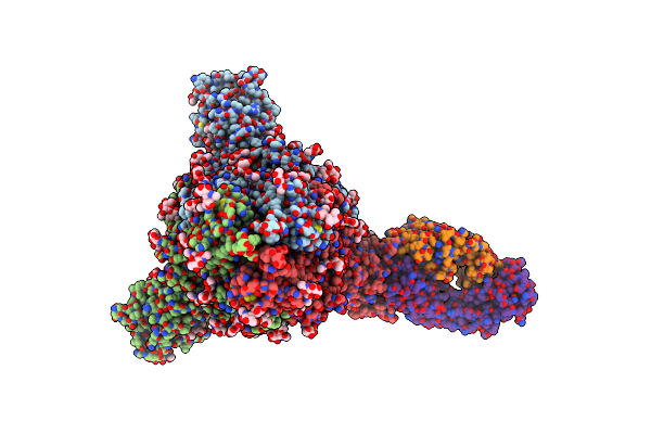
Trimeric Sars-Cov-2 Spike Ectodomain In Complex With Biliverdin (One Rbd Erect)
Organism: Severe acute respiratory syndrome coronavirus 2
Method: ELECTRON MICROSCOPY Release Date: 2021-04-28 Classification: VIRAL PROTEIN Ligands: BLA, NAG |
Organism: Severe acute respiratory syndrome coronavirus 2
Method: ELECTRON MICROSCOPY
Release Date: 2021-04-28
Ligands: BLA, NAG
 |
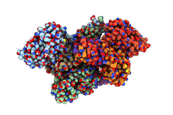
Trimeric Sars-Cov-2 Spike Ectodomain Bound To P008_056 Fab
Organism: Severe acute respiratory syndrome coronavirus 2, Homo sapiens
Method: ELECTRON MICROSCOPY Release Date: 2021-04-28 Classification: VIRAL PROTEIN Ligands: BLA, NAG |
Organism: Severe acute respiratory syndrome coronavirus 2, Homo sapiens
Method: ELECTRON MICROSCOPY
Release Date: 2021-04-28
Ligands: BLA, NAG
 |
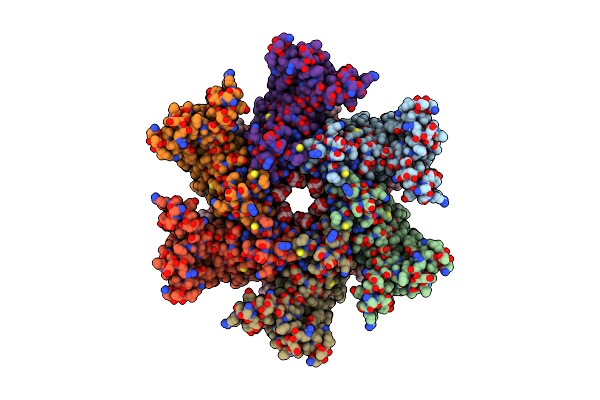
Crystal Structure Of Salmonella Enterica Typhimurium Bcfh
Organism: Salmonella typhimurium
Method: X-RAY DIFFRACTION Resolution:2.31 Å Release Date: 2021-04-21 Classification: OXIDOREDUCTASE Ligands: MG, EDO, PGE |
Organism: Salmonella typhimurium
Method: X-RAY DIFFRACTION
Release Date: 2021-04-21
Ligands: MG, EDO, PGE
 |
Cryoem Structure Of Serinc From Drosophila Melanogaster
Organism: Drosophila melanogaster
Method: ELECTRON MICROSCOPY Release Date: 2020-01-01 Classification: MEMBRANE PROTEIN Ligands: LMN, P5S, CDL |
Organism: Drosophila melanogaster
Method: ELECTRON MICROSCOPY
Release Date: 2020-01-01
Ligands: LMN, P5S, CDL
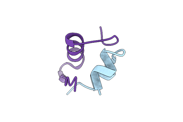 |
Insulin Glargine Crystal Structure 1
Organism: Homo sapiens
Method: X-RAY DIFFRACTION Resolution:1.66 Å Release Date: 2014-02-12 Classification: HORMONE |
Organism: Homo sapiens
Method: X-RAY DIFFRACTION
Release Date: 2014-02-12
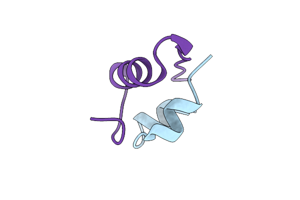 |
Insulin Glargine Crystal Structure 2
Organism: Homo sapiens
Method: X-RAY DIFFRACTION Resolution:1.80 Å Release Date: 2014-02-12 Classification: HORMONE |
Organism: Homo sapiens
Method: X-RAY DIFFRACTION
Release Date: 2014-02-12
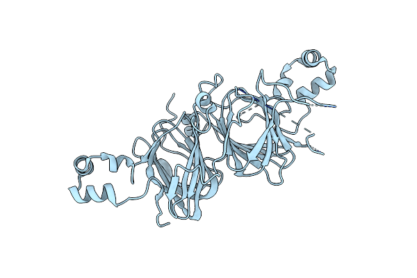 |
Structure Of Amaranth 11S Proglobulin Seed Storage Protein From Amaranthus Hypochondriacus L.
Organism: Amaranthus hypochondriacus
Method: X-RAY DIFFRACTION Resolution:2.28 Å Release Date: 2012-01-11 Classification: PLANT PROTEIN |
Organism: Amaranthus hypochondriacus
Method: X-RAY DIFFRACTION
Release Date: 2012-01-11
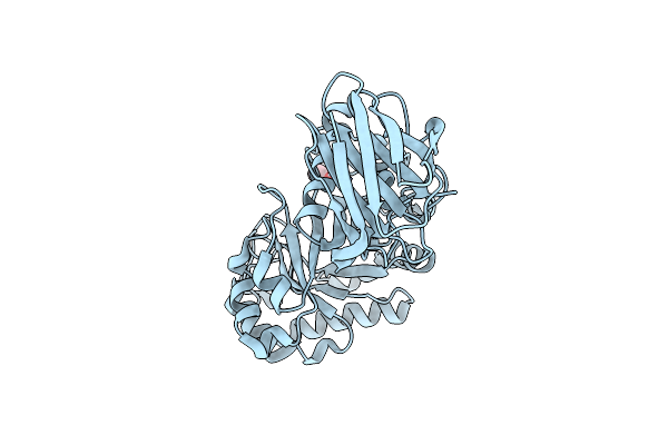 |
The First Crystallographic Structure Of A Xylanase From Glycosyl Hydrolase Family 5: Implications For Catalysis
Organism: Erwinia chrysanthemi
Method: X-RAY DIFFRACTION Resolution:1.42 Å Release Date: 2003-09-16 Classification: HYDROLASE Ligands: ACT |
Organism: Erwinia chrysanthemi
Method: X-RAY DIFFRACTION
Release Date: 2003-09-16
Ligands: ACT