

Search Count: 292
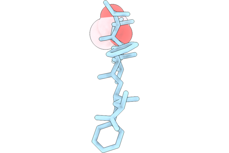 |
Structure Of Peptide Segment Klvffa From Amyloid-Beta Determined By Liquid Cell Microed At Room Temperature
Organism: Homo sapiens
Method: ELECTRON CRYSTALLOGRAPHY Resolution:1.00 Å Release Date: 2026-08-26 Classification: PROTEIN FIBRIL Ligands: ACT |
Organism: Homo sapiens
Method: ELECTRON CRYSTALLOGRAPHY
Release Date: 2026-08-26
Ligands: ACT
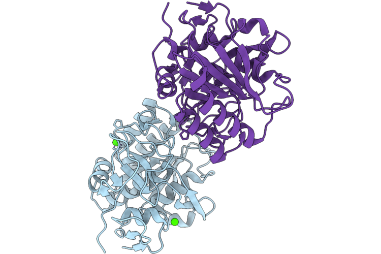 |
Microed Structure Of Proteinase K From Microcrystals Frozen By Traditional Cryoem Methods
Organism: Parengyodontium album
Method: ELECTRON CRYSTALLOGRAPHY Resolution:2.20 Å Release Date: 2026-07-01 Classification: HYDROLASE Ligands: CA |
Organism: Parengyodontium album
Method: ELECTRON CRYSTALLOGRAPHY
Release Date: 2026-07-01
Ligands: CA
 |
Microed Structure Of Proteinase K From Spray-Frozen Microcrystals
Organism: Parengyodontium album
Method: ELECTRON CRYSTALLOGRAPHY Resolution:2.50 Å Release Date: 2026-07-01 Classification: HYDROLASE |
Organism: Parengyodontium album
Method: ELECTRON CRYSTALLOGRAPHY
Release Date: 2026-07-01
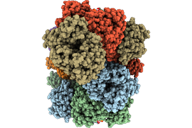 |
Cryoem Structure Of Adhe Spirosome From Clostridium Thermocellum Uncovered By Visual Proteomics.
Organism: Acetivibrio thermocellus dsm 1313
Method: ELECTRON MICROSCOPY Release Date: 2026-06-24 Classification: PROTEIN FIBRIL Ligands: FE, NAD |
Organism: Acetivibrio thermocellus dsm 1313
Method: ELECTRON MICROSCOPY
Release Date: 2026-06-24
Ligands: FE, NAD
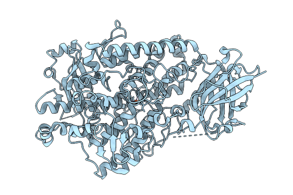 |
Rt Xfel Structure Of Soybean Lipoxygenase-1 In Small Unit-Cell
Organism: Glycine max
Method: X-RAY DIFFRACTION Resolution:1.95 Å Release Date: 2026-04-08 Classification: OXIDOREDUCTASE Ligands: FE |
Organism: Glycine max
Method: X-RAY DIFFRACTION
Release Date: 2026-04-08
Ligands: FE
 |
Rt Xfel Structure Of Soybean Lipoxygenase-1 In Large Unit-Cell
Organism: Glycine max
Method: X-RAY DIFFRACTION Resolution:1.95 Å Release Date: 2026-04-08 Classification: OXIDOREDUCTASE Ligands: FE |
Organism: Glycine max
Method: X-RAY DIFFRACTION
Release Date: 2026-04-08
Ligands: FE
 |
Structure Of Lysozyme-N,N',N"-Triacetylchitotriose Complex Determined Using Reyes For Automated Microed
Organism: Gallus gallus
Method: ELECTRON CRYSTALLOGRAPHY Resolution:2.20 Å Release Date: 2025-12-10 Classification: HYDROLASE Ligands: CL, NA |
Organism: Gallus gallus
Method: ELECTRON CRYSTALLOGRAPHY
Release Date: 2025-12-10
Ligands: CL, NA
 |
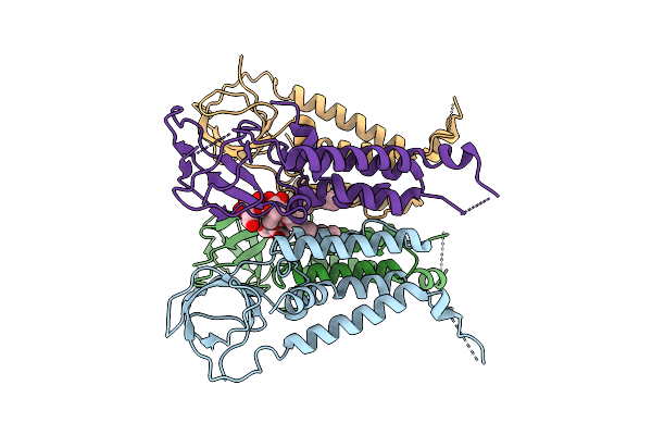
Crystal Structure Of Krasg13C In Complex With Nucleotide-Based Covalent Inhibitor 7B
Organism: Homo sapiens
Method: X-RAY DIFFRACTION Resolution:1.85 Å Release Date: 2025-11-12 Classification: HYDROLASE Ligands: A1I08, MG |
Organism: Homo sapiens
Method: X-RAY DIFFRACTION
Release Date: 2025-11-12
Ligands: A1I08, MG
 |
Cryo-Em Structure Of Kwaa Tetramer With C2 Symmetry
Organism: Escherichia coli
Method: ELECTRON MICROSCOPY Resolution:3.69 Å Release Date: 2025-11-05 Classification: IMMUNE SYSTEM Ligands: AV0 |
Organism: Escherichia coli
Method: ELECTRON MICROSCOPY
Release Date: 2025-11-05
Ligands: AV0
 |
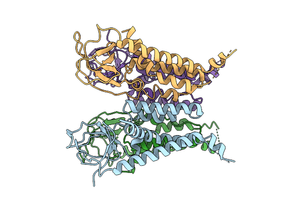
Cryoem Structure Of Alpha-Synuclein Fibril Induced By Psychosine
Organism: Homo sapiens
Method: ELECTRON MICROSCOPY Resolution:2.72 Å Release Date: 2025-10-08 Classification: PROTEIN FIBRIL |
Organism: Homo sapiens
Method: ELECTRON MICROSCOPY
Release Date: 2025-10-08
 |
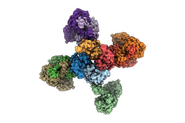
Cryo-Em Structure Of Kwaa With C4 Symmetry
Organism: Escherichia coli
Method: ELECTRON MICROSCOPY Release Date: 2025-08-06 Classification: IMMUNE SYSTEM |
Organism: Escherichia coli
Method: ELECTRON MICROSCOPY
Release Date: 2025-08-06
 |
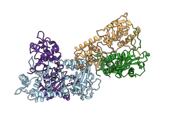
Cryo-Em Structure Of Kwaa-Kwab Complex
Organism: Escherichia coli
Method: ELECTRON MICROSCOPY Resolution:3.75 Å Release Date: 2025-08-06 Classification: IMMUNE SYSTEM |
Organism: Escherichia coli
Method: ELECTRON MICROSCOPY
Release Date: 2025-08-06
 |
Crystal Structure Of Kwab
Organism: Escherichia coli
Method: X-RAY DIFFRACTION Resolution:3.92 Å Release Date: 2025-08-06 Classification: IMMUNE SYSTEM |
Organism: Escherichia coli
Method: X-RAY DIFFRACTION
Release Date: 2025-08-06
 |
Crystal Structure Of Kwab Dimer
Organism: Escherichia coli
Method: X-RAY DIFFRACTION Resolution:3.60 Å Release Date: 2025-08-06 Classification: ANTIVIRAL PROTEIN |
Organism: Escherichia coli
Method: X-RAY DIFFRACTION
Release Date: 2025-08-06
 |
Cryo-Em Structure Of Local Kwaa-Kwab Complex
Organism: Escherichia coli
Method: ELECTRON MICROSCOPY Resolution:3.86 Å Release Date: 2025-08-06 Classification: ANTIVIRAL PROTEIN |
Organism: Escherichia coli
Method: ELECTRON MICROSCOPY
Release Date: 2025-08-06
 |
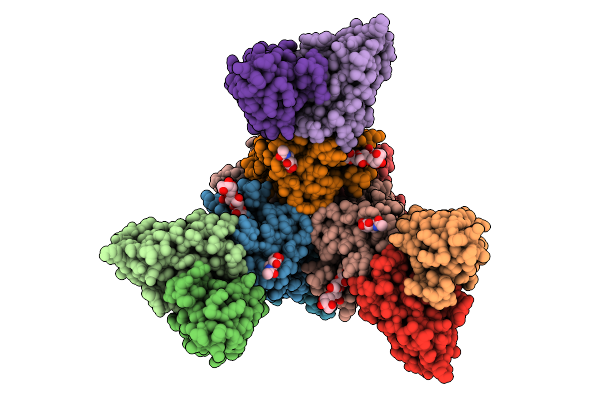
Junv Gp1, Gp2, Ssp And Cr1-28 Fab Complex In A Pseudotyped Virus Membrane
Organism: Mammarenavirus juninense, Homo sapiens
Method: ELECTRON MICROSCOPY Release Date: 2025-07-23 Classification: VIRAL PROTEIN/IMMUNE SYSTEM Ligands: MYR, NAG |
Organism: Mammarenavirus juninense, Homo sapiens
Method: ELECTRON MICROSCOPY
Release Date: 2025-07-23
Ligands: MYR, NAG
 |
Junv Gp1, Gp2, Ssp Complex With Neutralizing Antibody In A Pseudotyped Virus Membrane
Organism: Mammarenavirus juninense, Mus musculus
Method: ELECTRON MICROSCOPY Resolution:3.80 Å Release Date: 2025-07-23 Classification: VIRAL PROTEIN Ligands: MYR, NAG |
Organism: Mammarenavirus juninense, Mus musculus
Method: ELECTRON MICROSCOPY
Release Date: 2025-07-23
Ligands: MYR, NAG
 |
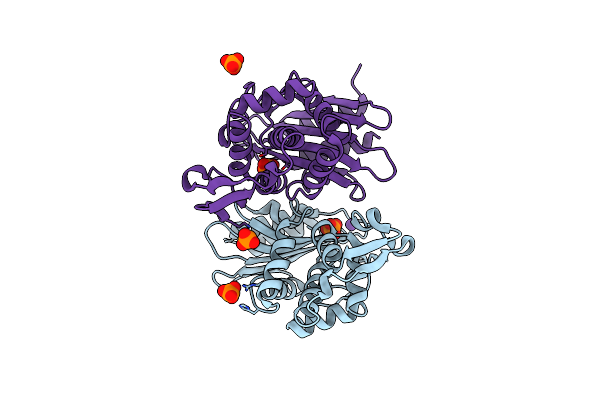
Serial Synchrotron X-Ray Diffraction Structure Of The Apo Form Of Papain
Organism: Carica papaya
Method: X-RAY DIFFRACTION Resolution:1.80 Å Release Date: 2025-06-18 Classification: HYDROLASE |
Organism: Carica papaya
Method: X-RAY DIFFRACTION
Release Date: 2025-06-18
 |
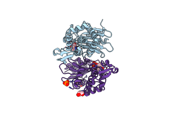
X-Ray Diffraction Structure Of Apo-Form Ctx-M-14 Beta-Lactamase
Organism: Escherichia coli
Method: X-RAY DIFFRACTION Resolution:1.50 Å Release Date: 2025-06-18 Classification: HYDROLASE Ligands: PO4, K |
Organism: Escherichia coli
Method: X-RAY DIFFRACTION
Release Date: 2025-06-18
Ligands: PO4, K
 |
X-Ray Diffraction Structure Of Ctx-M-14 Beta-Lactamase Co-Crystallized With Avibactam
Organism: Escherichia coli
Method: X-RAY DIFFRACTION Resolution:1.60 Å Release Date: 2025-06-18 Classification: HYDROLASE Ligands: NXL, GOL, PO4 |
Organism: Escherichia coli
Method: X-RAY DIFFRACTION
Release Date: 2025-06-18
Ligands: NXL, GOL, PO4