

Search Count: 23
 |
Cryoem Map Of The Large Glutamate Dehydrogenase Composed Of 180 Kda Subunits From Mycobacterium Smegmatis Obtained In The Presence Of Nad+ And L-Glutamate. Open Tetramer.
Organism: Mycolicibacterium smegmatis
Method: ELECTRON MICROSCOPY Release Date: 2026-01-14 Classification: OXIDOREDUCTASE Ligands: NAD |
Organism: Mycolicibacterium smegmatis
Method: ELECTRON MICROSCOPY
Release Date: 2026-01-14
Ligands: NAD
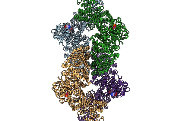 |
Cryoem Map Of The Large Glutamate Dehydrogenase Composed Of 180 Kda Subunits From Mycobacterium Smegmatis Obtained In The Presence Of Nad+ And L-Glutamate. Closed1 Tetramer.
Organism: Mycolicibacterium smegmatis
Method: ELECTRON MICROSCOPY Release Date: 2026-01-14 Classification: OXIDOREDUCTASE Ligands: NAD |
Organism: Mycolicibacterium smegmatis
Method: ELECTRON MICROSCOPY
Release Date: 2026-01-14
Ligands: NAD
 |
Cryoem Map Of The Large Glutamate Dehydrogenase Composed Of 180 Kda Subunits From Mycobacterium Smegmatis Obtained In The Presence Of Nad+ And L-Glutamate. Closed2 Tetramer
Organism: Mycolicibacterium smegmatis
Method: ELECTRON MICROSCOPY Release Date: 2026-01-14 Classification: OXIDOREDUCTASE Ligands: NAD |
Organism: Mycolicibacterium smegmatis
Method: ELECTRON MICROSCOPY
Release Date: 2026-01-14
Ligands: NAD
 |
Cryoem Map Of The Large Glutamate Dehydrogenase Composed Of 180 Kda Subunits From Mycobacterium Smegmatis Obtained In The Presence Of Nad+ And L-Glutamate. Empty Monomer.
Organism: Mycolicibacterium smegmatis
Method: ELECTRON MICROSCOPY Release Date: 2026-01-14 Classification: OXIDOREDUCTASE |
Organism: Mycolicibacterium smegmatis
Method: ELECTRON MICROSCOPY
Release Date: 2026-01-14
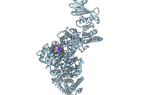 |
Cryoem Map Of The Large Glutamate Dehydrogenase Composed Of 180 Kda Subunits From Mycobacterium Smegmatis Obtained In The Presence Of Nad+ And L-Glutamate. Cofactor-Monomer.
Organism: Mycolicibacterium smegmatis
Method: ELECTRON MICROSCOPY Release Date: 2026-01-14 Classification: OXIDOREDUCTASE Ligands: NAD |
Organism: Mycolicibacterium smegmatis
Method: ELECTRON MICROSCOPY
Release Date: 2026-01-14
Ligands: NAD
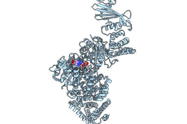 |
Cryoem Map Of The Large Glutamate Dehydrogenase Composed Of 180 Kda Subunits From Mycobacterium Smegmatis Obtained In The Presence Of Nad+ And L-Glutamate. Cofactor/Ligand-Monomer
Organism: Mycolicibacterium smegmatis
Method: ELECTRON MICROSCOPY Release Date: 2026-01-14 Classification: OXIDOREDUCTASE Ligands: GLU, NAD |
Organism: Mycolicibacterium smegmatis
Method: ELECTRON MICROSCOPY
Release Date: 2026-01-14
Ligands: GLU, NAD
 |
Cryoem Map Of The Large Glutamate Dehydrogenase Composed Of 180 Kda Subunits From Mycobacterium Smegmatis Obtained In The Presence Of Nad+ And L-Glutamate. Total-Monomer
Organism: Mycolicibacterium smegmatis
Method: ELECTRON MICROSCOPY Release Date: 2026-01-14 Classification: OXIDOREDUCTASE Ligands: NAD |
Organism: Mycolicibacterium smegmatis
Method: ELECTRON MICROSCOPY
Release Date: 2026-01-14
Ligands: NAD
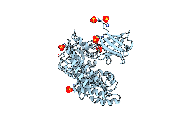 |
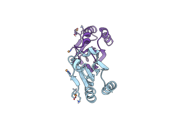
Crystal Structure Of The Protease Inhibitor U-Omp19 From Brucella Abortus Fused To Maltose-Binding Protein
Organism: Escherichia coli, Brucella abortus
Method: X-RAY DIFFRACTION Resolution:2.55 Å Release Date: 2022-04-20 Classification: MEMBRANE PROTEIN Ligands: SO4 |
Organism: Escherichia coli, Brucella abortus
Method: X-RAY DIFFRACTION
Release Date: 2022-04-20
Ligands: SO4
 |
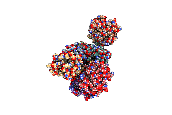
Crystal Structure Of Thioredoxin E101G Mutant
Organism: Escherichia coli
Method: X-RAY DIFFRACTION Resolution:1.31 Å Release Date: 2017-02-22 Classification: OXIDOREDUCTASE Ligands: CU |
Organism: Escherichia coli
Method: X-RAY DIFFRACTION
Release Date: 2017-02-22
Ligands: CU
 |
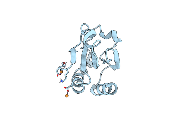
Crystal Structure Of Thioredoxin L107A Mutant
Organism: Escherichia coli o157:h7
Method: X-RAY DIFFRACTION Resolution:2.14 Å Release Date: 2017-02-22 Classification: OXIDOREDUCTASE Ligands: CU |
Organism: Escherichia coli o157:h7
Method: X-RAY DIFFRACTION
Release Date: 2017-02-22
Ligands: CU
 |
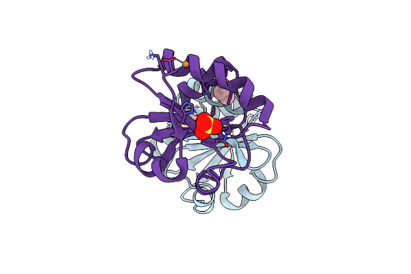
Crystal Structure Of Thioredoxin L94A Mutant
Organism: Escherichia coli
Method: X-RAY DIFFRACTION Resolution:1.20 Å Release Date: 2017-02-22 Classification: OXIDOREDUCTASE Ligands: CU |
Organism: Escherichia coli
Method: X-RAY DIFFRACTION
Release Date: 2017-02-22
Ligands: CU
 |
Crystal Structure Of Thioredoxin N106A Mutant
Organism: Escherichia coli
Method: X-RAY DIFFRACTION Resolution:1.10 Å Release Date: 2017-02-22 Classification: OXIDOREDUCTASE Ligands: CU, EOH, SO4 |
Organism: Escherichia coli
Method: X-RAY DIFFRACTION
Release Date: 2017-02-22
Ligands: CU, EOH, SO4
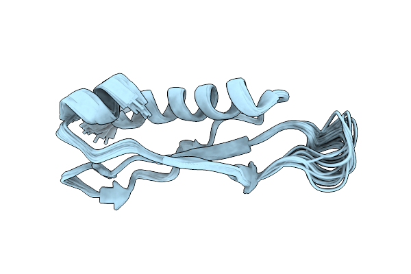 |
The Second Dsrbd Domain From A. Thaliana Dicer-Like 1
Organism: Arabidopsis thaliana
Method: SOLUTION NMR Release Date: 2013-01-23 Classification: HYDROLASE |
Organism: Arabidopsis thaliana
Method: SOLUTION NMR
Release Date: 2013-01-23
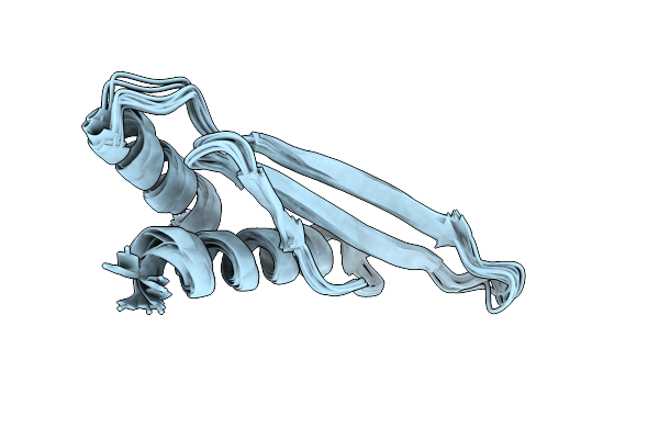 |
Solution Structure Of The Second Dsrbd Of Hyl1
Organism: Arabidopsis thaliana
Method: SOLUTION NMR Release Date: 2010-09-29 Classification: RNA BINDING PROTEIN, PLANT PROTEIN |
Organism: Arabidopsis thaliana
Method: SOLUTION NMR
Release Date: 2010-09-29
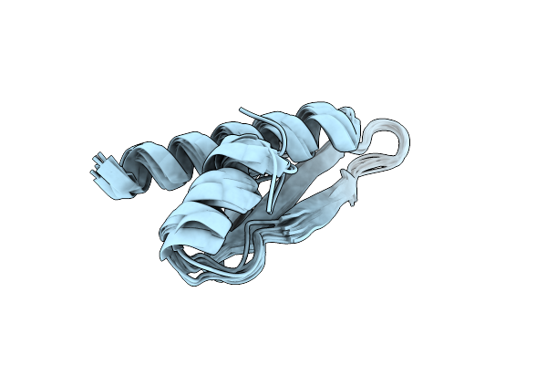 |
Backbone 1H, 13C, And 15N Chemical Shift Assignments For The First Dsrbd Of Protein Hyl1
Organism: Arabidopsis thaliana
Method: SOLUTION NMR Release Date: 2010-09-29 Classification: RNA BINDING PROTEIN, PLANT PROTEIN |
Organism: Arabidopsis thaliana
Method: SOLUTION NMR
Release Date: 2010-09-29
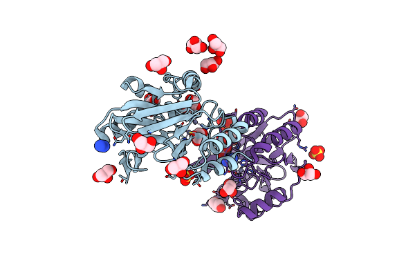 |
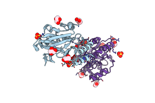
Bacillus Cereus Metallo-Beta-Lactamase (Bcii) Arg (121) Cys Mutant. Solved At Ph7 Using 20Mm Znso4 In Buffer. 1Mm Dtt Was Used As A Reducing Agent
Organism: Bacillus cereus
Method: X-RAY DIFFRACTION Resolution:2.00 Å Release Date: 2005-03-31 Classification: HYDROLASE Ligands: AZI, GOL, ZN, SO4 |
Organism: Bacillus cereus
Method: X-RAY DIFFRACTION
Release Date: 2005-03-31
Ligands: AZI, GOL, ZN, SO4
 |
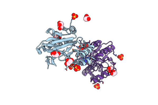
Bacillus Cereus Metallo-Beta-Lactamase (Bcii) Arg (121) Cys Mutant. Solved At Ph5 Using 20Mm Znso4 In Buffer. 1Mm Dtt Was Used As A Reducing Agent.
Organism: Bacillus cereus
Method: X-RAY DIFFRACTION Resolution:1.80 Å Release Date: 2005-03-31 Classification: HYDROLASE Ligands: GOL, ZN, SO4, AZI |
Organism: Bacillus cereus
Method: X-RAY DIFFRACTION
Release Date: 2005-03-31
Ligands: GOL, ZN, SO4, AZI
 |
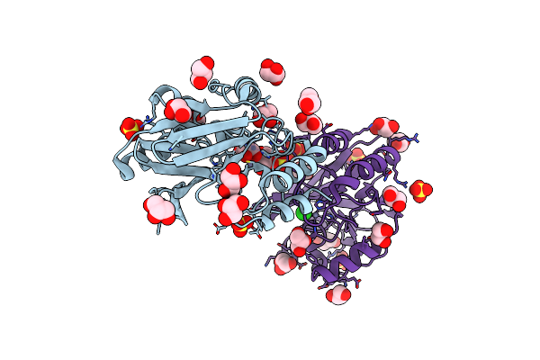
Bacillus Cereus Metallo-Beta-Lactamase (Bcii) Arg (121) Cys Mutant. Solved At Ph4.5 Using 20Mm Znso4 In Buffer. 1Mm Dtt Was Used As A Reducing Agent. Cys221 Is Oxidized.
Organism: Bacillus cereus
Method: X-RAY DIFFRACTION Resolution:2.30 Å Release Date: 2005-03-31 Classification: HYDROLASE Ligands: GOL, ZN, SO4, AZI |
Organism: Bacillus cereus
Method: X-RAY DIFFRACTION
Release Date: 2005-03-31
Ligands: GOL, ZN, SO4, AZI
 |
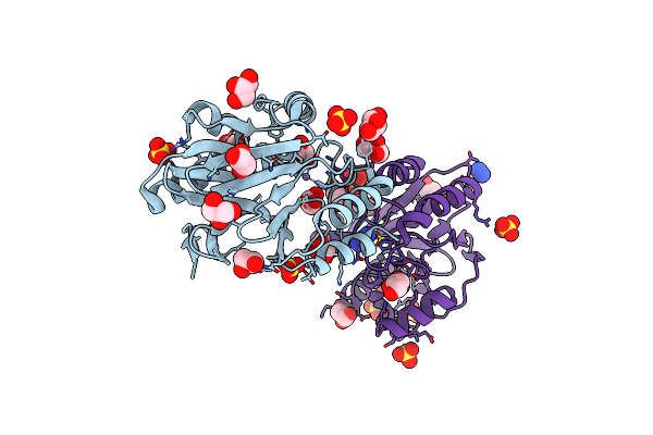
Bacillus Cereus Metallo-Beta-Lactamase (Bcii) Arg (121) Cys Mutant. Solved At Ph4.5 Using 20Mm Znso4 In The Buffer. 1Mm Dtt And 1Mm Tcep- Hcl Were Used As Reducing Agents. Cys221 Is Reduced.
Organism: Bacillus cereus
Method: X-RAY DIFFRACTION Resolution:2.40 Å Release Date: 2005-03-31 Classification: HYDROLASE Ligands: GOL, ZN, SO4, CL |
Organism: Bacillus cereus
Method: X-RAY DIFFRACTION
Release Date: 2005-03-31
Ligands: GOL, ZN, SO4, CL
 |
Bacillus Cereus Metallo-Beta-Lactamase (Bcii) Arg (121) Cys Mutant. Solved At Ph5 Using 20 Micromolar Znso4 In The Buffer. 1Mm Dtt Was Used As A Reducing Agent. Cys221 Is Oxidized.
Organism: Bacillus cereus
Method: X-RAY DIFFRACTION Resolution:2.30 Å Release Date: 2005-03-31 Classification: HYDROLASE Ligands: GOL, ZN, SO4, AZI |
Organism: Bacillus cereus
Method: X-RAY DIFFRACTION
Release Date: 2005-03-31
Ligands: GOL, ZN, SO4, AZI