

Search Count: 24
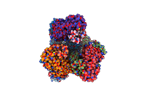 |
Ultrapotent Sars-Cov-2 Neutralizing Antibodies With Protective Efficacy Against Newly Emerged Mutational Variants
Organism: Severe acute respiratory syndrome coronavirus 2, Homo sapiens
Method: ELECTRON MICROSCOPY Release Date: 2021-09-15 Classification: VIRAL PROTEIN Ligands: NAG |
Organism: Severe acute respiratory syndrome coronavirus 2, Homo sapiens
Method: ELECTRON MICROSCOPY
Release Date: 2021-09-15
Ligands: NAG
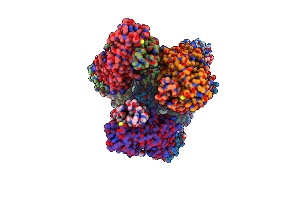 |
Ultrapotent Sars-Cov-2 Neutralizing Antibodies With Protective Efficacy Against Newly Emerged Mutational Variants
Organism: Severe acute respiratory syndrome coronavirus 2, Homo sapiens
Method: ELECTRON MICROSCOPY Release Date: 2021-09-15 Classification: VIRAL PROTEIN Ligands: NAG |
Organism: Severe acute respiratory syndrome coronavirus 2, Homo sapiens
Method: ELECTRON MICROSCOPY
Release Date: 2021-09-15
Ligands: NAG
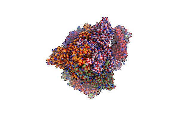 |
S-3C1-F3B Structure, All The Three Rbds Are In The Up Conformation And Each Of Them Associates With A 3C1 Fab
Organism: Severe acute respiratory syndrome coronavirus 2, Mus musculus
Method: ELECTRON MICROSCOPY Release Date: 2020-12-02 Classification: VIRAL PROTEIN |
Organism: Severe acute respiratory syndrome coronavirus 2, Mus musculus
Method: ELECTRON MICROSCOPY
Release Date: 2020-12-02
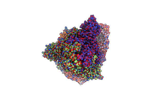 |
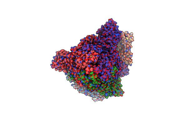
S-3C1-F3A Structure, Two Rbds Are Up And One Rbd Is Down, Each Rbd Binds With A 3C1 Fab.
Organism: Severe acute respiratory syndrome coronavirus 2, Mus musculus
Method: ELECTRON MICROSCOPY Release Date: 2020-12-02 Classification: VIRAL PROTEIN |
Organism: Severe acute respiratory syndrome coronavirus 2, Mus musculus
Method: ELECTRON MICROSCOPY
Release Date: 2020-12-02
 |
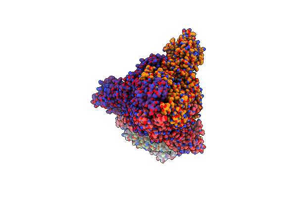
S-3C1-F2 Structure, Two Rbds Are Up And One Rbd Is Down, The Two Up Rbd Bind With A 3C1 Fab.
Organism: Severe acute respiratory syndrome coronavirus 2, Mus musculus
Method: ELECTRON MICROSCOPY Release Date: 2020-12-02 Classification: VIRAL PROTEIN |
Organism: Severe acute respiratory syndrome coronavirus 2, Mus musculus
Method: ELECTRON MICROSCOPY
Release Date: 2020-12-02
 |
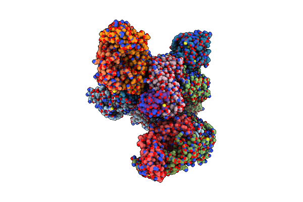
S-3C1-F1 Structure, One Rbd Is Up And Two Rbds Are Down, The Up Rbd Binds With A 3C1 Fab
Organism: Severe acute respiratory syndrome coronavirus 2, Mus musculus
Method: ELECTRON MICROSCOPY Release Date: 2020-12-02 Classification: VIRAL PROTEIN |
Organism: Severe acute respiratory syndrome coronavirus 2, Mus musculus
Method: ELECTRON MICROSCOPY
Release Date: 2020-12-02
 |
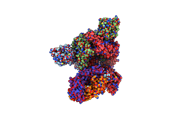
S-2H2-F3A Structure, Two Rbds Are Up And One Rbd Is Down, Each Rbd Binds With A 2H2 Fab.
Organism: Severe acute respiratory syndrome coronavirus 2, Mus musculus
Method: ELECTRON MICROSCOPY Release Date: 2020-12-02 Classification: VIRAL PROTEIN |
Organism: Severe acute respiratory syndrome coronavirus 2, Mus musculus
Method: ELECTRON MICROSCOPY
Release Date: 2020-12-02
 |
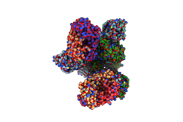
S-2H2-F1 Structure, One Rbd Is Up And Two Rbds Are Down, Only Up Rbd Binds With A 2H2 Fab
Organism: Severe acute respiratory syndrome coronavirus 2, Mus musculus
Method: ELECTRON MICROSCOPY Release Date: 2020-12-02 Classification: VIRAL PROTEIN |
Organism: Severe acute respiratory syndrome coronavirus 2, Mus musculus
Method: ELECTRON MICROSCOPY
Release Date: 2020-12-02
 |
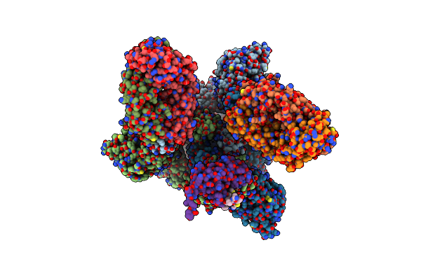
S-2H2-F2 Structure, Two Rbds Are Up And One Rbd Is Down, Each Up Rbd Binds With A 2H2 Fab.
Organism: Severe acute respiratory syndrome coronavirus 2, Mus musculus
Method: ELECTRON MICROSCOPY Release Date: 2020-12-02 Classification: VIRAL PROTEIN |
Organism: Severe acute respiratory syndrome coronavirus 2, Mus musculus
Method: ELECTRON MICROSCOPY
Release Date: 2020-12-02
 |
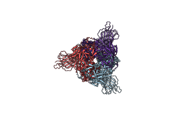
S-2H2-F3B Structure, Three Rbds Are Up And Each Rbd Binds With A 2H2 Fab.
Organism: Severe acute respiratory syndrome coronavirus 2, Mus musculus
Method: ELECTRON MICROSCOPY Release Date: 2020-12-02 Classification: VIRAL PROTEIN |
Organism: Severe acute respiratory syndrome coronavirus 2, Mus musculus
Method: ELECTRON MICROSCOPY
Release Date: 2020-12-02
 |
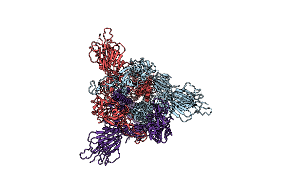
Sars-Cov2 S Protein At Close State
Organism: Severe acute respiratory syndrome coronavirus 2
Method: ELECTRON MICROSCOPY Release Date: 2020-11-25 Classification: VIRAL PROTEIN |
Organism: Severe acute respiratory syndrome coronavirus 2
Method: ELECTRON MICROSCOPY
Release Date: 2020-11-25
 |
Sars-Cov2 S Protein At Open State
Organism: Severe acute respiratory syndrome coronavirus 2
Method: ELECTRON MICROSCOPY Release Date: 2020-11-25 Classification: VIRAL PROTEIN |
Organism: Severe acute respiratory syndrome coronavirus 2
Method: ELECTRON MICROSCOPY
Release Date: 2020-11-25
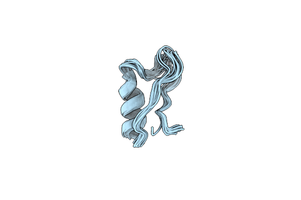 |
Ion Channel Modulation By Scorpion Haemolymph And Its Defensin Ingredients Uncovers Origin Of Neurotoxins In Telson Formed In Paleozoic Scorpion
|
Organism: Buthus sp. iy-2001
Method: SOLUTION NMR
Release Date: 2018-03-14
 |
Structure Of Pngf-Ii In C2 Space Group
Organism: Elizabethkingia meningoseptica
Method: X-RAY DIFFRACTION Resolution:1.90 Å Release Date: 2015-01-28 Classification: HYDROLASE Ligands: ZN |
Organism: Elizabethkingia meningoseptica
Method: X-RAY DIFFRACTION
Release Date: 2015-01-28
Ligands: ZN
 |
Structure Of Pngf-Ii In P21 Space Group
Organism: Elizabethkingia meningoseptica
Method: X-RAY DIFFRACTION Resolution:2.81 Å Release Date: 2015-01-28 Classification: HYDROLASE |
Organism: Elizabethkingia meningoseptica
Method: X-RAY DIFFRACTION
Release Date: 2015-01-28
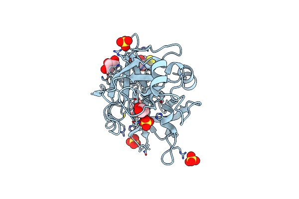 |
Crystal Structure Of The Ligand-Free Inactive Matriptase
Organism: Homo sapiens
Method: X-RAY DIFFRACTION Resolution:1.48 Å Release Date: 2013-03-06 Classification: HYDROLASE Ligands: SO4, GOL, GSH |
Organism: Homo sapiens
Method: X-RAY DIFFRACTION
Release Date: 2013-03-06
Ligands: SO4, GOL, GSH
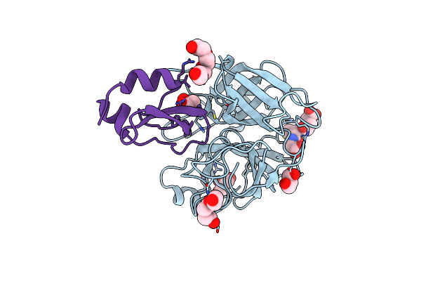 |
Crystal Structure Of The Inactive Matriptase In Complex With Its Inhibitor Hai-1
Organism: Homo sapiens
Method: X-RAY DIFFRACTION Resolution:2.29 Å Release Date: 2013-03-06 Classification: hydrolase/hydrolase inhibitor Ligands: PG4, GOL, PGE, GSH |
Organism: Homo sapiens
Method: X-RAY DIFFRACTION
Release Date: 2013-03-06
Ligands: PG4, GOL, PGE, GSH
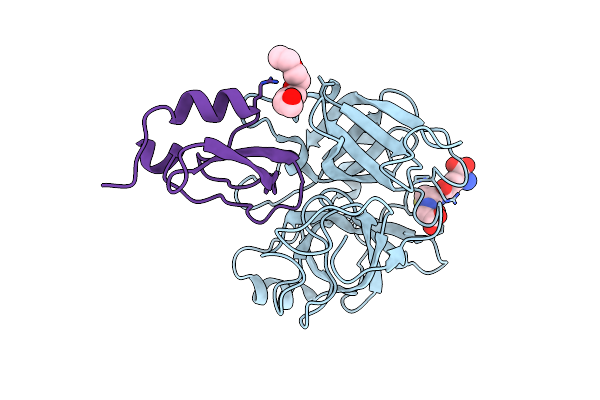 |
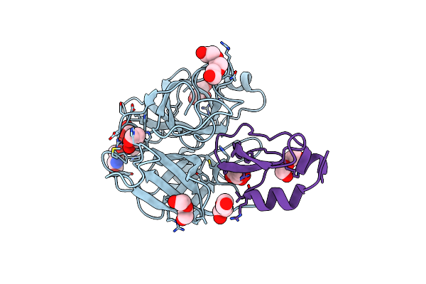
Crystal Structure Of Matriptase In Complex With Its Inhibitor Hai-1
Organism: Homo sapiens
Method: X-RAY DIFFRACTION Resolution:2.45 Å Release Date: 2013-03-06 Classification: hydrolase/hydrolase inhibitor Ligands: PG4, GSH |
Organism: Homo sapiens
Method: X-RAY DIFFRACTION
Release Date: 2013-03-06
Ligands: PG4, GSH
 |
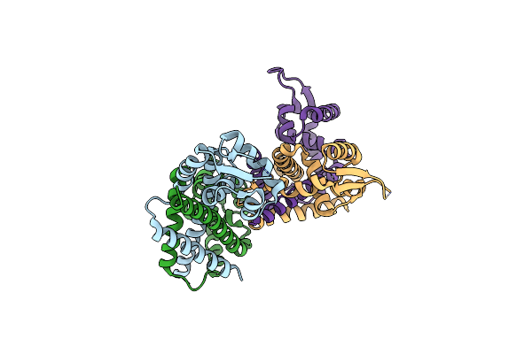
Crystal Structure Of Matriptase In Complex With Its Inhibitor Hai-1
Organism: Homo sapiens
Method: X-RAY DIFFRACTION Resolution:2.01 Å Release Date: 2013-03-06 Classification: hydrolase/hydrolase inhibitor Ligands: GSH, GOL, PEG, PGE |
Organism: Homo sapiens
Method: X-RAY DIFFRACTION
Release Date: 2013-03-06
Ligands: GSH, GOL, PEG, PGE
 |
Crystal Structure Of Abfr Of Staphylococcus Epidermidis
Organism: Staphylococcus epidermidis
Method: X-RAY DIFFRACTION Resolution:2.50 Å Release Date: 2013-01-02 Classification: TRANSCRIPTION |
Organism: Staphylococcus epidermidis
Method: X-RAY DIFFRACTION
Release Date: 2013-01-02