
Search Count: 141
All
Selected
 |
Organism: Homo sapiens, Escherichia coli str. k-12 substr. mg1655, Synthetic construct
Method: ELECTRON MICROSCOPY Resolution:3.42 Å Release Date: 2026-04-01 Classification: HYDROLASE Ligands: ZN, ATP, MG, ADP, LDZ |
 |
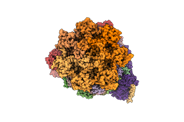
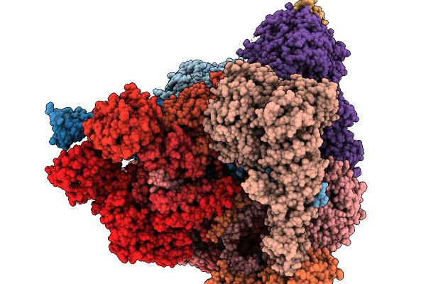
Substrate-Free Human 26S Proteasome Purified By Midnolin, 20S Proteasome, Rpts And Rpn11 Part
Organism: Homo sapiens
Method: ELECTRON MICROSCOPY Release Date: 2026-04-01 Classification: HYDROLASE Ligands: ZN, ATP, MG, ADP, LDZ |
 |
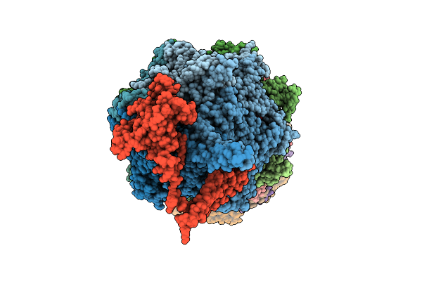
Structure Of Human 26S Proteasome Complexed With Midnolin, 19S Proteasome With Ubl Bound
Organism: Escherichia coli k-12, Homo sapiens, Purpureocillium lilacinum
Method: ELECTRON MICROSCOPY Resolution:3.65 Å Release Date: 2026-04-01 Classification: HYDROLASE Ligands: ADP, ATP, ZN, MG |
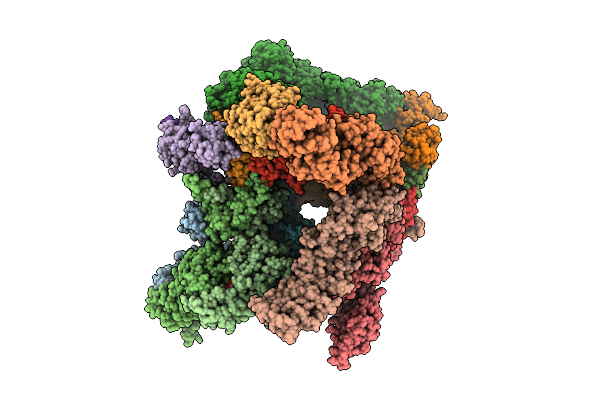 |
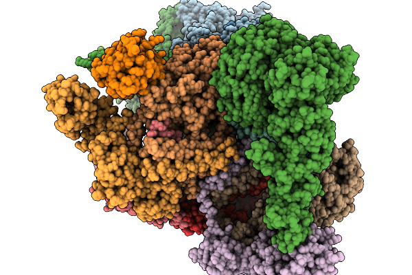
Structure Of Human 26S Proteasome Complexed With Midnolin, 19S Proteasome With Ubl And Catch Domain Resolved
Organism: Escherichia coli k-12, Homo sapiens, Pseudotamlana agarivorans
Method: ELECTRON MICROSCOPY Release Date: 2026-04-01 Classification: HYDROLASE Ligands: ADP, ATP, ZN, MG |
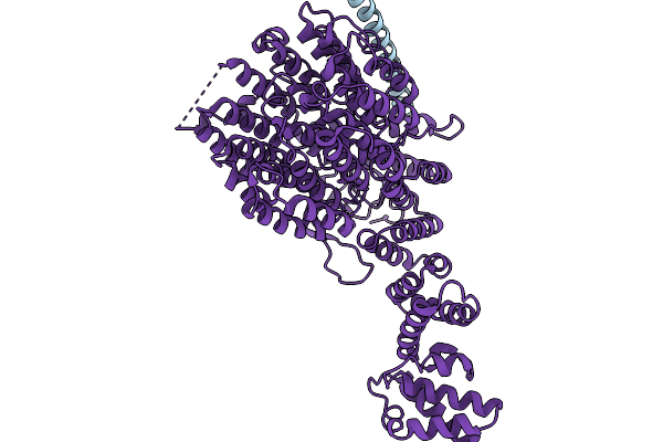 |
Focused Refinement Of Rpn1 And The C-Terminal Helix Of Midnolin In The Substrate-Engaged Human 26S Proteasome
Organism: Homo sapiens
Method: ELECTRON MICROSCOPY Release Date: 2026-03-25 Classification: HYDROLASE |
 |
Substrate-Engaged Human 26S Proteasome Bound To Midnolin With Rpt1 At Top Of Spiral Staircase
Organism: Homo sapiens
Method: ELECTRON MICROSCOPY Release Date: 2026-03-25 Classification: HYDROLASE Ligands: ZN, ATP, MG, ADP, LDZ |
 |
Substrate-Engaged Human 26S Proteasome Bound To Midnolin With Rpt5 At Top Of Spiral Staircase
Organism: Homo sapiens
Method: ELECTRON MICROSCOPY Release Date: 2026-03-25 Classification: HYDROLASE Ligands: ZN, ATP, MG, ADP, LDZ |
 |
Substrate-Engaged Human 26S Proteasome Bound To Midnolin With Rpt2 At Top Of Spiral Staircase
Organism: Homo sapiens
Method: ELECTRON MICROSCOPY Release Date: 2026-03-25 Classification: HYDROLASE Ligands: ADP, ATP, MG, LDZ, ZN |
 |
Focused Refinement Of 19S In The Substrate-Engaged Human 26S Proteasome Bound To Midnolin With Rpt6 At Top Of Spiral Staircase
Organism: Homo sapiens
Method: ELECTRON MICROSCOPY Release Date: 2026-03-25 Classification: HYDROLASE Ligands: ADP, ZN, ATP, MG |
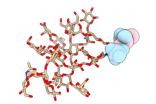 |
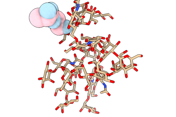
Tlp-2A, A Glycofibril Obtained From A Karst Cave From Guilin City, Guangxi Zhuang Autonomous Region, China
Organism: Unidentified
Method: ELECTRON MICROSCOPY Release Date: 2026-02-18 Classification: PROTEIN FIBRIL |
 |
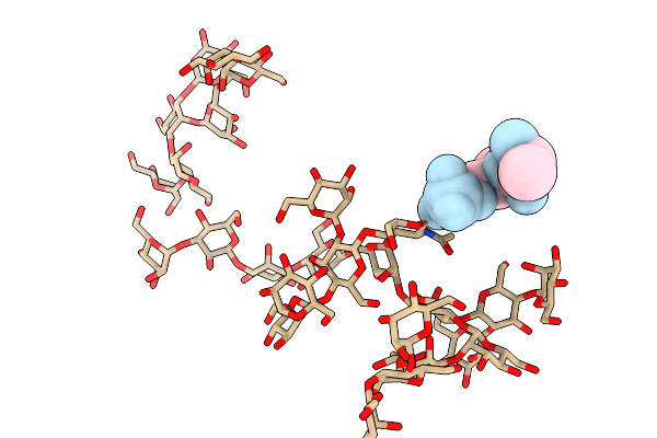
Tlp-2F, A Glycofibril Obtained From A Karst Cave From Guilin City, Guangxi Zhuang Autonomous Region, China
Organism: Unidentified
Method: ELECTRON MICROSCOPY Release Date: 2026-02-18 Classification: UNKNOWN FUNCTION |
 |
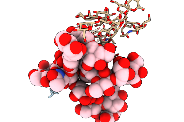
Tlp-2G, A Glycofibril Obtained From A Karst Cave From Guilin City, Guangxi Zhuang Autonomous Region, China
Organism: Unidentified
Method: ELECTRON MICROSCOPY Release Date: 2026-02-18 Classification: UNKNOWN FUNCTION Ligands: HOH |
 |
Tlp-2H, A Glycofibril Obtained From A Karst Cave From Guilin City, Guangxi Zhuang Autonomous Region, China
Organism: Unidentified
Method: ELECTRON MICROSCOPY Release Date: 2026-02-18 Classification: UNKNOWN FUNCTION |
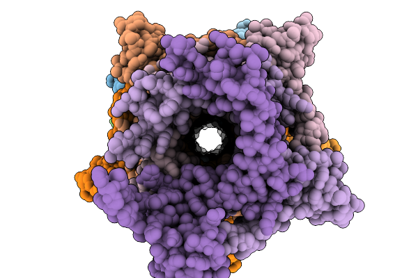 |
Pilus-Like-Alpha, A Bacteria Pilus-Like Structure Obtained From A Karst Cave From Guilin City, Guangxi Zhuang Autonomous Region, China
Organism: Unidentified
Method: ELECTRON MICROSCOPY Resolution:3.07 Å Release Date: 2026-02-18 Classification: PROTEIN FIBRIL |
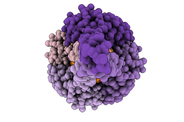 |
Pilus-Like-Beta, A Bacteria Pilus-Like Structure Obtained From A Karst Cave From Guilin City, Guangxi Zhuang Autonomous Region, China
Organism: Unidentified
Method: ELECTRON MICROSCOPY Resolution:3.37 Å Release Date: 2026-02-18 Classification: PROTEIN FIBRIL |
 |
Pilus-Like-Gamma, A Bacteria Pilus-Like Structure Obtained From A Karstcave From Guilin City, Guangxi Zhuangautonomous Region, China
Organism: Unidentified
Method: ELECTRON MICROSCOPY Release Date: 2026-02-18 Classification: PROTEIN FIBRIL |
 |
Organism: Arabidopsis thaliana
Method: ELECTRON MICROSCOPY Resolution:3.20 Å Release Date: 2025-10-29 Classification: PLANT PROTEIN Ligands: K |
 |
Organism: Arabidopsis thaliana
Method: ELECTRON MICROSCOPY Resolution:3.40 Å Release Date: 2025-10-29 Classification: PROTON TRANSPORT Ligands: K |
 |
Organism: Arabidopsis thaliana
Method: ELECTRON MICROSCOPY Resolution:3.20 Å Release Date: 2025-10-29 Classification: PROTON TRANSPORT Ligands: K |
 |
Organism: Homo sapiens
Method: X-RAY DIFFRACTION Resolution:2.49 Å Release Date: 2025-05-28 Classification: CELL CYCLE Ligands: A1D8H, EDO, MG |