

Search Count: 18
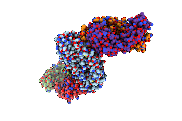 |
Pfrh5 Bound To Monoclonal Antibody R5.015 And R5.016 Fab Fragments
Organism: Plasmodium falciparum (isolate 3d7), Homo sapiens
Method: X-RAY DIFFRACTION Resolution:2.53 Å Release Date: 2022-02-09 Classification: IMMUNE SYSTEM |
Organism: Plasmodium falciparum (isolate 3d7), Homo sapiens
Method: X-RAY DIFFRACTION
Release Date: 2022-02-09
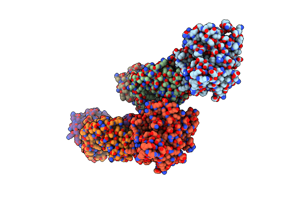 |
Pfcyrpa Bound To Monoclonal Antibody Cy.007 Fab Fragment
Organism: Plasmodium falciparum 3d7, Gallus gallus
Method: X-RAY DIFFRACTION Resolution:3.09 Å Release Date: 2022-02-09 Classification: IMMUNE SYSTEM Ligands: NH4 |
Organism: Plasmodium falciparum 3d7, Gallus gallus
Method: X-RAY DIFFRACTION
Release Date: 2022-02-09
Ligands: NH4
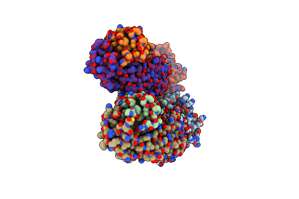 |
Pfcyrpa Bound To Monoclonal Antibody Cy.004 Fab Fragment
Organism: Plasmodium falciparum (isolate 3d7), Gallus gallus
Method: X-RAY DIFFRACTION Resolution:2.79 Å Release Date: 2022-02-09 Classification: IMMUNE SYSTEM Ligands: CA |
Organism: Plasmodium falciparum (isolate 3d7), Gallus gallus
Method: X-RAY DIFFRACTION
Release Date: 2022-02-09
Ligands: CA
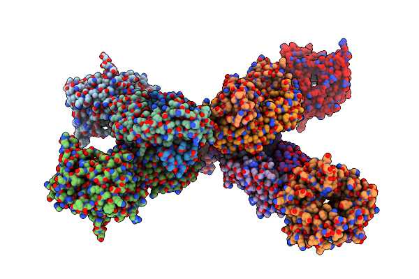 |
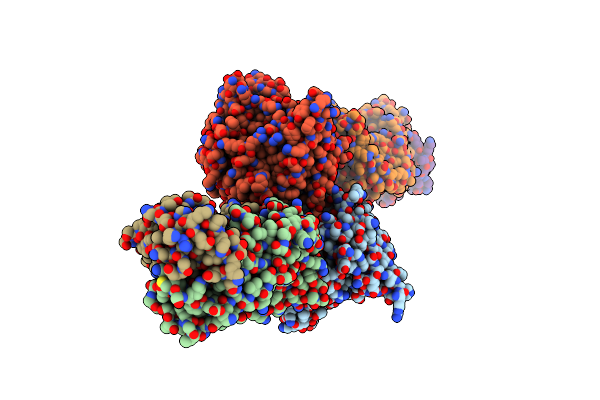
Pfcyrpa Bound To Monoclonal Antibody Cy.003 Fab Fragment
Organism: Plasmodium falciparum (isolate 3d7), Gallus gallus
Method: X-RAY DIFFRACTION Resolution:3.14 Å Release Date: 2022-02-09 Classification: IMMUNE SYSTEM |
Organism: Plasmodium falciparum (isolate 3d7), Gallus gallus
Method: X-RAY DIFFRACTION
Release Date: 2022-02-09
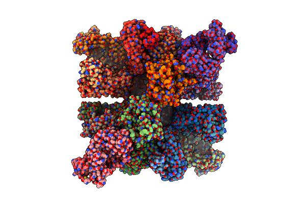 |
Pfcyrpa Bound To Fab Fragments From Monoclonal Antibodies Cy.003, Cy.004 And Cy.007
Organism: Plasmodium falciparum 3d7, Gallus gallus
Method: X-RAY DIFFRACTION Resolution:3.27 Å Release Date: 2022-02-09 Classification: IMMUNE SYSTEM Ligands: CA |
Organism: Plasmodium falciparum 3d7, Gallus gallus
Method: X-RAY DIFFRACTION
Release Date: 2022-02-09
Ligands: CA
 |
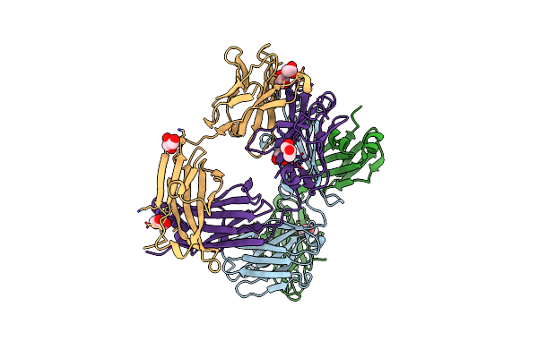
Pfcyrpa Bound To Monoclonal Antibody Cy.002 Fab Fragment
Organism: Plasmodium falciparum (isolate 3d7), Gallus gallus
Method: X-RAY DIFFRACTION Resolution:2.72 Å Release Date: 2022-02-09 Classification: IMMUNE SYSTEM |
Organism: Plasmodium falciparum (isolate 3d7), Gallus gallus
Method: X-RAY DIFFRACTION
Release Date: 2022-02-09
 |
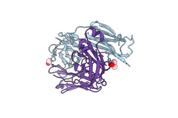
Pfrh5-Binding Monoclonal Antibody R5.004
Organism: Homo sapiens
Method: X-RAY DIFFRACTION Resolution:1.66 Å Release Date: 2019-06-26 Classification: IMMUNE SYSTEM Ligands: GOL, ACT |
Organism: Homo sapiens
Method: X-RAY DIFFRACTION
Release Date: 2019-06-26
Ligands: GOL, ACT
 |
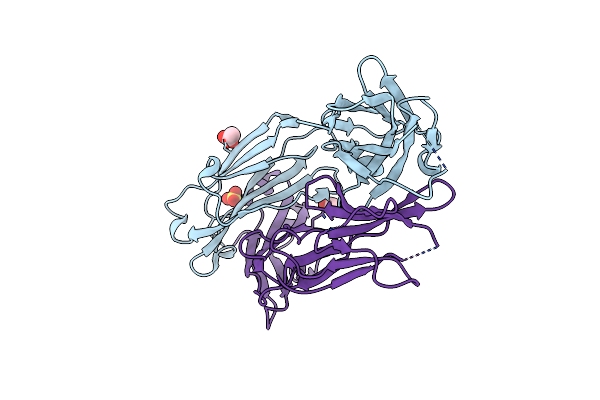
Pfrh5-Binding Monoclonal Antibody R5.011
Organism: Homo sapiens
Method: X-RAY DIFFRACTION Resolution:2.28 Å Release Date: 2019-06-26 Classification: IMMUNE SYSTEM Ligands: ACT, CAC |
Organism: Homo sapiens
Method: X-RAY DIFFRACTION
Release Date: 2019-06-26
Ligands: ACT, CAC
 |
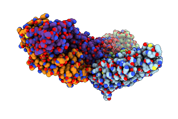
Pfrh5-Binding Monoclonal Antibody R5.016
Organism: Homo sapiens
Method: X-RAY DIFFRACTION Resolution:2.10 Å Release Date: 2019-06-26 Classification: IMMUNE SYSTEM Ligands: ACT, SO4 |
Organism: Homo sapiens
Method: X-RAY DIFFRACTION
Release Date: 2019-06-26
Ligands: ACT, SO4
 |
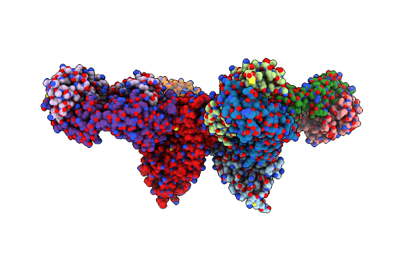
Pfrh5 Bound To Monoclonal Antibodies R5.004 And R5.016
Organism: Plasmodium falciparum (isolate 3d7), Homo sapiens
Method: X-RAY DIFFRACTION Resolution:4.01 Å Release Date: 2019-06-26 Classification: CELL ADHESION |
Organism: Plasmodium falciparum (isolate 3d7), Homo sapiens
Method: X-RAY DIFFRACTION
Release Date: 2019-06-26
 |
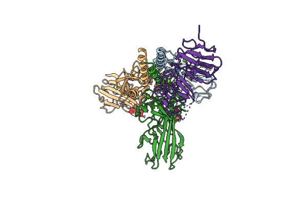
Pfrh5 Bound To Monoclonal Antibodies R5.011 And R5.016
Organism: Plasmodium falciparum, Homo sapiens
Method: X-RAY DIFFRACTION Resolution:3.58 Å Release Date: 2019-06-26 Classification: CELL ADHESION |
Organism: Plasmodium falciparum, Homo sapiens
Method: X-RAY DIFFRACTION
Release Date: 2019-06-26
 |
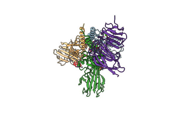
Crystal Structure Of The Metalloproteinase Enhancer Pcpe-1 Bound To The Procollagen C Propeptide Trimer (Long)
Organism: Homo sapiens
Method: X-RAY DIFFRACTION Resolution:2.78 Å Release Date: 2018-07-11 Classification: STRUCTURAL PROTEIN Ligands: CA, FLC |
Organism: Homo sapiens
Method: X-RAY DIFFRACTION
Release Date: 2018-07-11
Ligands: CA, FLC
 |
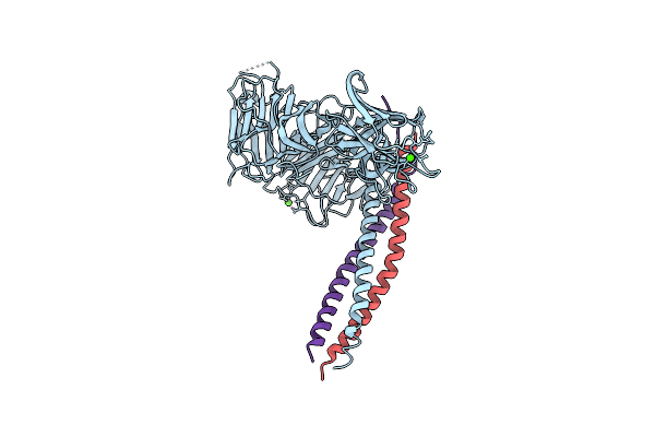
Crystal Structure Of The Metalloproteinase Enhancer Pcpe-1 Bound To The Procollagen C Propeptide Trimer (Short)
Organism: Homo sapiens
Method: X-RAY DIFFRACTION Resolution:2.70 Å Release Date: 2018-07-04 Classification: STRUCTURAL PROTEIN Ligands: CA, FLC, CL |
Organism: Homo sapiens
Method: X-RAY DIFFRACTION
Release Date: 2018-07-04
Ligands: CA, FLC, CL
 |
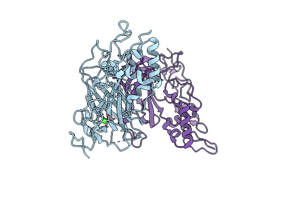
Crystal Structure Of The Heterotrimeric Integrin-Binding Region Of Laminin-111
Organism: Mus musculus
Method: X-RAY DIFFRACTION Resolution:2.13 Å Release Date: 2017-02-08 Classification: CELL ADHESION Ligands: CA |
Organism: Mus musculus
Method: X-RAY DIFFRACTION
Release Date: 2017-02-08
Ligands: CA
 |
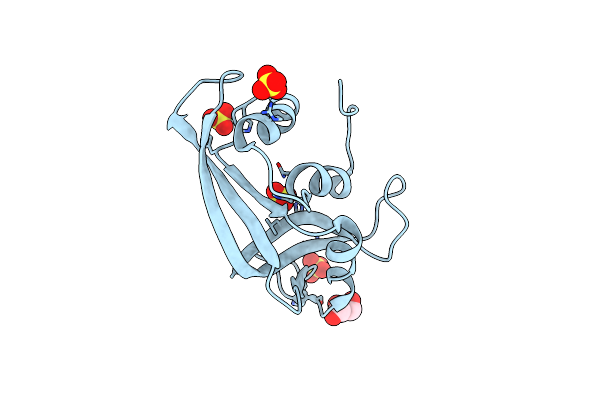
Crystal Structure Of Laminin Beta2 Le5-Lf-Le6
Organism: Rattus norvegicus
Method: X-RAY DIFFRACTION Resolution:1.85 Å Release Date: 2016-07-27 Classification: CELL ADHESION Ligands: CA, CL |
Organism: Rattus norvegicus
Method: X-RAY DIFFRACTION
Release Date: 2016-07-27
Ligands: CA, CL
 |
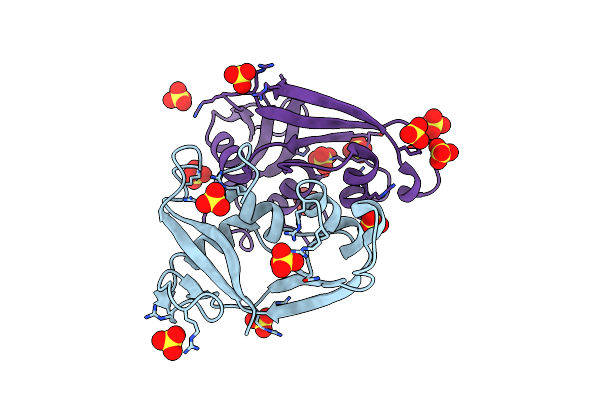
Structure Of Human Rnase 6 In Complex With Sulphate Anions
Organism: Homo sapiens
Method: X-RAY DIFFRACTION Resolution:1.72 Å Release Date: 2016-04-06 Classification: HYDROLASE Ligands: SO4, GOL |
Organism: Homo sapiens
Method: X-RAY DIFFRACTION
Release Date: 2016-04-06
Ligands: SO4, GOL
 |
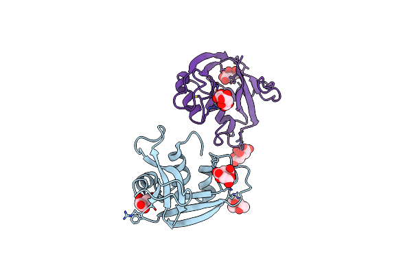
Structure Of The Human Eosinophil Cationic Protein In Complex With Sulfate Anions
Organism: Homo sapiens
Method: X-RAY DIFFRACTION Resolution:1.69 Å Release Date: 2012-06-27 Classification: HYDROLASE Ligands: SO4 |
Organism: Homo sapiens
Method: X-RAY DIFFRACTION
Release Date: 2012-06-27
Ligands: SO4
 |
Structure Of The Human Eosinophil Cationic Protein In Complex With Citrate Anions
Organism: Homo sapiens
Method: X-RAY DIFFRACTION Resolution:1.70 Å Release Date: 2012-06-27 Classification: HYDROLASE Ligands: CIT, MPD |
Organism: Homo sapiens
Method: X-RAY DIFFRACTION
Release Date: 2012-06-27
Ligands: CIT, MPD