

Search Count: 28
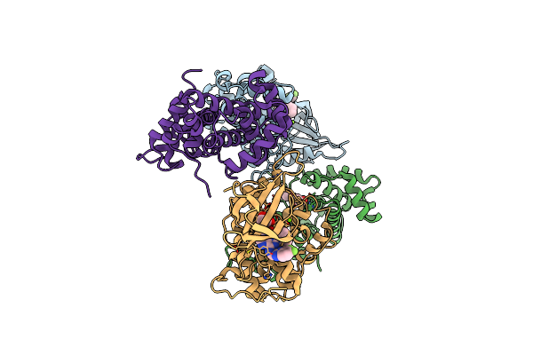 |
Crystal Structure Of Human Cdk12/Cyclink In Complex With An Inhibitor
Organism: Homo sapiens
Method: X-RAY DIFFRACTION Resolution:3.06 Å Release Date: 2017-12-27 Classification: TRANSFERASE/INHIBITOR Ligands: MG, CJM, EDO |
Organism: Homo sapiens
Method: X-RAY DIFFRACTION
Release Date: 2017-12-27
Ligands: MG, CJM, EDO
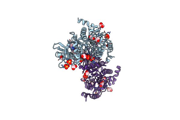 |
Crystal Structure Of The Protein Kinase Ck2 Catalytic Subunit In Complex With Pyrazolo-Pyrimidine Macrocyclic Ligand
Organism: Homo sapiens
Method: X-RAY DIFFRACTION Resolution:2.52 Å Release Date: 2017-05-17 Classification: TRANSFERASE Ligands: SO4, EDO, 8GQ |
Organism: Homo sapiens
Method: X-RAY DIFFRACTION
Release Date: 2017-05-17
Ligands: SO4, EDO, 8GQ
 |
Crystal Structure Of The Bcl6 Btb Domain In Complex With Pyrazolo-Pyrimidine Ligand
Organism: Homo sapiens
Method: X-RAY DIFFRACTION Resolution:1.72 Å Release Date: 2017-05-17 Classification: TRANSFERASE Ligands: 8HH |
Organism: Homo sapiens
Method: X-RAY DIFFRACTION
Release Date: 2017-05-17
Ligands: 8HH
 |
Crystal Structure Of The Bcl6 Btb Domain In Complex With Pyrazolo-Pyrimidine Macrocyclic Ligand
Organism: Homo sapiens
Method: X-RAY DIFFRACTION Resolution:1.81 Å Release Date: 2017-05-17 Classification: TRANSFERASE Ligands: CL, 8GQ |
Organism: Homo sapiens
Method: X-RAY DIFFRACTION
Release Date: 2017-05-17
Ligands: CL, 8GQ
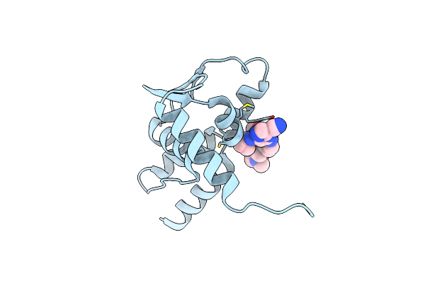 |
Crystal Structure Of The Bcl6 Btb Domain In Complex With Pyrazolo-Pyrimidine Ligand
Organism: Homo sapiens
Method: X-RAY DIFFRACTION Resolution:1.38 Å Release Date: 2017-05-17 Classification: TRANSFERASE Ligands: 8GN |
Organism: Homo sapiens
Method: X-RAY DIFFRACTION
Release Date: 2017-05-17
Ligands: 8GN
 |
Crystal Structure Of The Bcl6 Btb Domain In Complex With Pyrazolo-Pyrimidine Ligand
Organism: Homo sapiens
Method: X-RAY DIFFRACTION Resolution:1.58 Å Release Date: 2017-05-17 Classification: TRANSFERASE Ligands: CL, 8HN |
Organism: Homo sapiens
Method: X-RAY DIFFRACTION
Release Date: 2017-05-17
Ligands: CL, 8HN
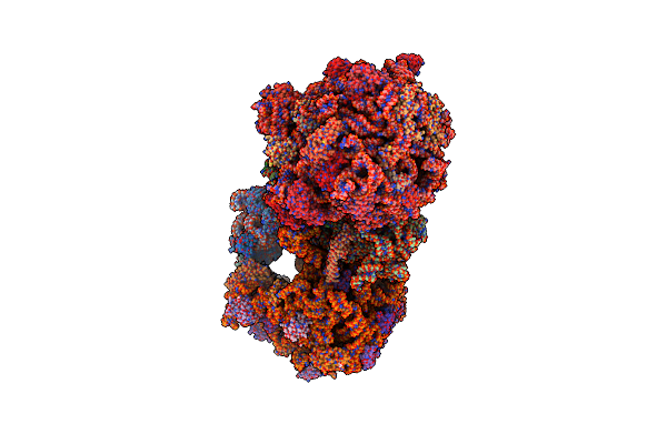 |
High-Resolution Structure Of The Escherichia Coli Ribosome
|
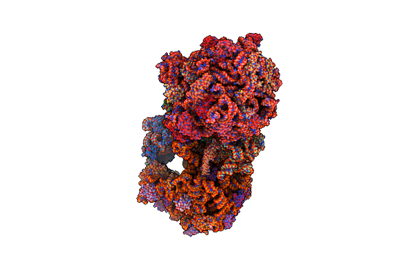 |
Structure Of The Wt E Coli Ribosome Bound To Tetracycline
Organism: Escherichia coli (strain k12)
Method: X-RAY DIFFRACTION Resolution:2.80 Å Release Date: 2016-07-27 Classification: RIBOSOME/ANTIBIOTIC Ligands: MG, PG4, MPD, PUT, TAC, ZN, PEG, EDO, PGE, SPD, 1PE, ACY, GUN, TRS |
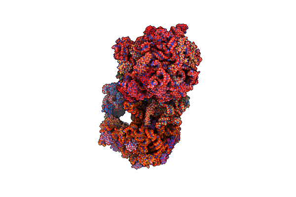 |
Structure Of The 70S E Coli Ribosome With The U1052G Mutation In The 16S Rrna Bound To Tetracycline
Organism: Escherichia coli (strain k12)
Method: X-RAY DIFFRACTION Resolution:3.00 Å Release Date: 2016-07-27 Classification: RIBOSOME/ANTIBIOTIC Ligands: MG, PG4, MPD, PUT, TAC, ZN, PEG, EDO, PGE, SPD, 1PE, ACY, GUN, TRS |
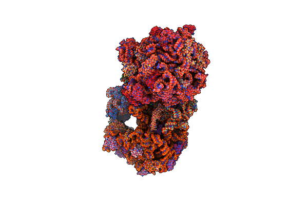 |
Structure Of The E Coli 70S Ribosome With The U1060A Mutation In 16S Rrna
Organism: Escherichia coli, Escherichia coli (strain k12)
Method: X-RAY DIFFRACTION Resolution:3.32 Å Release Date: 2016-07-06 Classification: ribosome/antibiotic Ligands: MG, PG4, MPD, PUT, ZN, PEG, EDO, PGE, SPD, 1PE, ACY, GUN, TRS |
Organism: Escherichia coli, Escherichia coli (strain k12)
Method: X-RAY DIFFRACTION
Release Date: 2016-07-06
Ligands: MG, PG4, MPD, PUT, ZN, PEG, EDO, PGE, SPD, 1PE, ACY, GUN, TRS
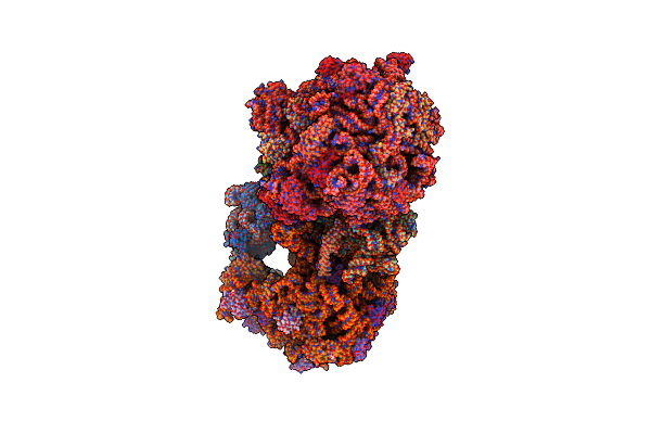 |
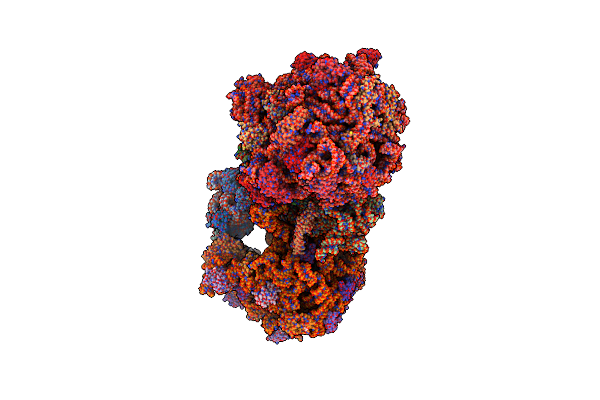
Structure Of The E Coli 70S Ribosome With The U1052G Mutation In 16S Rrna Bound To Tigecycline
Organism: Escherichia coli, Escherichia coli (strain k12), Escherichia coli o139:h28 (strain e24377a / etec)
Method: X-RAY DIFFRACTION Resolution:3.10 Å Release Date: 2016-07-06 Classification: ribosome/antibiotic Ligands: MG, PG4, MPD, PUT, T1C, ZN, PEG, EDO, PGE, SPD, 1PE, ACY, GUN, TRS |
Organism: Escherichia coli, Escherichia coli (strain k12), Escherichia coli o139:h28 (strain e24377a / etec)
Method: X-RAY DIFFRACTION
Release Date: 2016-07-06
Ligands: MG, PG4, MPD, PUT, T1C, ZN, PEG, EDO, PGE, SPD, 1PE, ACY, GUN, TRS
 |
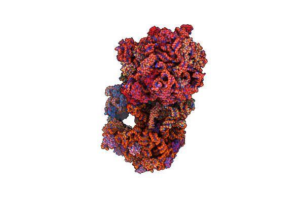
Structure Of The Wild-Type 70S E Coli Ribosome Bound To Tigecycline
Organism: Escherichia coli, Escherichia coli k-12, Escherichia coli (strain k12)
Method: X-RAY DIFFRACTION Resolution:2.96 Å Release Date: 2016-07-06 Classification: ribosome/antibiotic Ligands: MG, PG4, MPD, PUT, T1C, ZN, PEG, EDO, PGE, SPD, 1PE, ACY, GUN, TRS |
Organism: Escherichia coli, Escherichia coli k-12, Escherichia coli (strain k12)
Method: X-RAY DIFFRACTION
Release Date: 2016-07-06
Ligands: MG, PG4, MPD, PUT, T1C, ZN, PEG, EDO, PGE, SPD, 1PE, ACY, GUN, TRS
 |
Structure Of The Escherichia Coli Ribosome With The U1052G Mutation In The 16S Rrna
|
 |
Crystal Structure Of P. Aeruginosa Ampc
Organism: Pseudomonas aeruginosa (strain atcc 15692 / pao1 / 1c / prs 101 / lmg 12228)
Method: X-RAY DIFFRACTION Resolution:1.28 Å Release Date: 2015-08-05 Classification: hydrolase/hydrolase inhibitor Ligands: GOL, DMS, 3VT |
Organism: Pseudomonas aeruginosa (strain atcc 15692 / pao1 / 1c / prs 101 / lmg 12228)
Method: X-RAY DIFFRACTION
Release Date: 2015-08-05
Ligands: GOL, DMS, 3VT
 |
Crystal Structure Of P. Aeruginosa Ampc
Organism: Pseudomonas aeruginosa pao1
Method: X-RAY DIFFRACTION Resolution:1.05 Å Release Date: 2015-08-05 Classification: hydrolase/hydrolase inhibitor Ligands: GOL, 3VU |
Organism: Pseudomonas aeruginosa pao1
Method: X-RAY DIFFRACTION
Release Date: 2015-08-05
Ligands: GOL, 3VU
 |
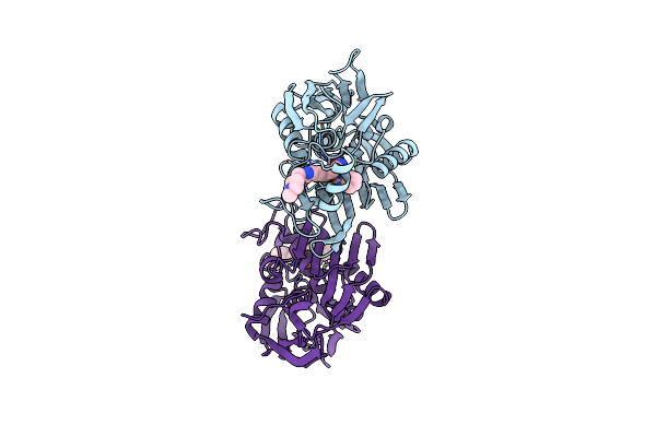
Crystal Structure Of P. Aeruginosa Oxa10
Organism: Pseudomonas aeruginosa
Method: X-RAY DIFFRACTION Resolution:1.60 Å Release Date: 2015-08-05 Classification: hydrolase/hydrolase inhibitor Ligands: SO4, 3VU, CO2 |
Organism: Pseudomonas aeruginosa
Method: X-RAY DIFFRACTION
Release Date: 2015-08-05
Ligands: SO4, 3VU, CO2
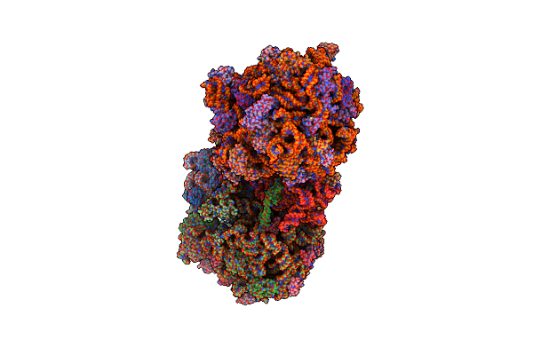 |
Crystal Structure Of The E. Coli Ribosome Bound To Negamycin.
Organism: Escherichia coli str. k-12 substr. mg1655, Escherichia coli, Thermus thermophilus
Method: X-RAY DIFFRACTION Resolution:3.09 Å Release Date: 2014-11-05 Classification: Ribosome/Antibiotic Ligands: MG, ZN, NEG |
Organism: Escherichia coli str. k-12 substr. mg1655, Escherichia coli, Thermus thermophilus
Method: X-RAY DIFFRACTION
Release Date: 2014-11-05
Ligands: MG, ZN, NEG
 |
Lpxc From P.Aeruginosa With The Inhibitor 6-(Benzimidazol-1-Yl)-5-[4-[2-[6-[(4-Methylpiperazin-1-Yl)Methyl]-3-Pyridyl]Ethynyl]Phenyl]Pyridine-3-Carbohydroxamic Acid
Organism: Pseudomonas aeruginosa
Method: X-RAY DIFFRACTION Resolution:2.06 Å Release Date: 2014-09-24 Classification: HYDROLASE/HYDROLASE INHIBITOR Ligands: ZN, 2SZ |
Organism: Pseudomonas aeruginosa
Method: X-RAY DIFFRACTION
Release Date: 2014-09-24
Ligands: ZN, 2SZ
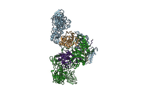 |
Apo Phers From P. Aeuriginosa
Organism: Pseudomonas aeruginosa
Method: X-RAY DIFFRACTION Resolution:2.79 Å Release Date: 2014-06-25 Classification: LIGASE |
Organism: Pseudomonas aeruginosa
Method: X-RAY DIFFRACTION
Release Date: 2014-06-25
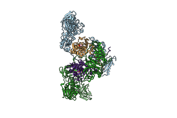 |
Phers In Complex With Compound 2A
Organism: Pseudomonas aeruginosa
Method: X-RAY DIFFRACTION Resolution:2.62 Å Release Date: 2014-06-25 Classification: LIGASE/LIGASE INHIBITOR Ligands: 2NL |
Organism: Pseudomonas aeruginosa
Method: X-RAY DIFFRACTION
Release Date: 2014-06-25
Ligands: 2NL