

Search Count: 214
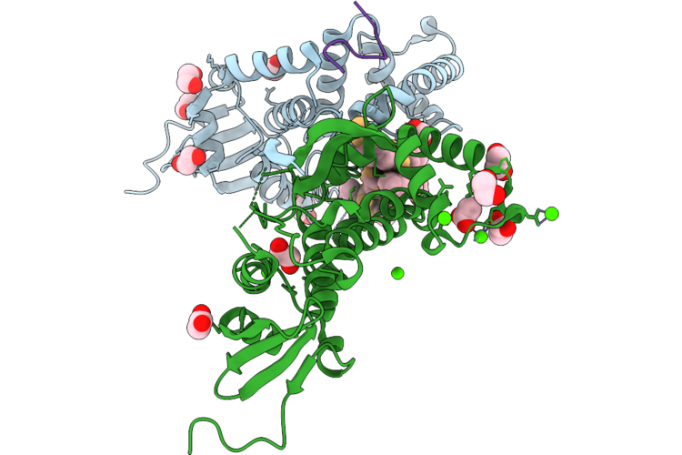 |
Crystal Structure Of Human Mlh1 C-Terminal Domain With Cyclic Peptide (2S)-N-[(6S,9S,12S,15S,18S,26Ar)-12-(4-Aminobutyl)-18-Benzyl-21-(2-Carbamoylethyl)-9-(Hydroxymethyl)- 15-[(4-Hydroxyphenyl)Methyl]-1,7,10,13,16,19,22-Heptaoxo-Hexacosahydropyrrolo[2,1-C]1,4,7,10,13,16,19-Heptaazacyclotetracosan-6-Yl]-2-[(2S)-2-Acetamidopropanamido]-3-Methylbutanamide
Organism: Homo sapiens, Synthetic construct
Method: X-RAY DIFFRACTION Resolution:2.28 Å Release Date: 2026-07-08 Classification: DNA BINDING PROTEIN Ligands: EDO, CA, ACT |
Organism: Homo sapiens, Synthetic construct
Method: X-RAY DIFFRACTION
Release Date: 2026-07-08
Ligands: EDO, CA, ACT
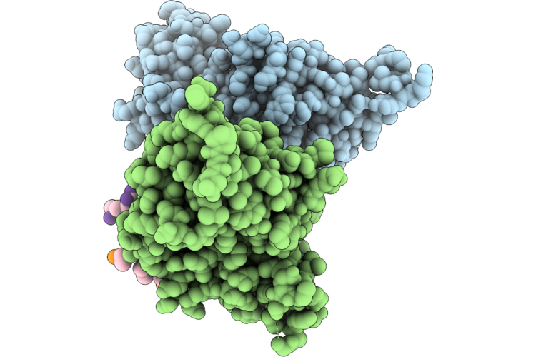 |
Crystal Structure Of Mlh1-Ctd With Peptide Qavlsrffq
Organism: Homo sapiens
Method: X-RAY DIFFRACTION Resolution:2.44 Å Release Date: 2026-06-24 Classification: DNA BINDING PROTEIN |
Organism: Homo sapiens
Method: X-RAY DIFFRACTION
Release Date: 2026-06-24
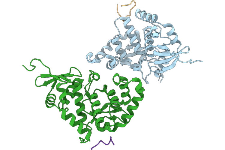 |
Crystal Structure Of Mlh1-Ctd With Chdi-00915848
Organism: Homo sapiens
Method: X-RAY DIFFRACTION Resolution:2.25 Å Release Date: 2026-06-24 Classification: DNA BINDING PROTEIN |
Organism: Homo sapiens
Method: X-RAY DIFFRACTION
Release Date: 2026-06-24
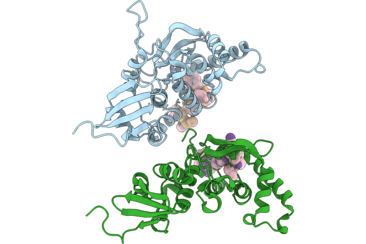 |
Crystal Structure Of Mlh1-Ctd With Chdi-00916630
Organism: Homo sapiens, Synthetic construct
Method: X-RAY DIFFRACTION Resolution:1.93 Å Release Date: 2026-06-24 Classification: DNA BINDING PROTEIN |
Organism: Homo sapiens, Synthetic construct
Method: X-RAY DIFFRACTION
Release Date: 2026-06-24
 |
Tulane Virus Protease Without Added Ligands
Organism: Tulane virus
Method: X-RAY DIFFRACTION Resolution:2.10 Å Release Date: 2026-02-25 Classification: VIRAL PROTEIN |
Organism: Tulane virus
Method: X-RAY DIFFRACTION
Release Date: 2026-02-25
 |
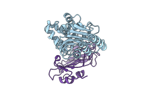
Tulane Virus Protease Complexed With Rupintrivir
Organism: Tulane virus
Method: X-RAY DIFFRACTION Resolution:1.90 Å Release Date: 2026-02-25 Classification: VIRAL PROTEIN Ligands: AG7 |
Organism: Tulane virus
Method: X-RAY DIFFRACTION
Release Date: 2026-02-25
Ligands: AG7
 |
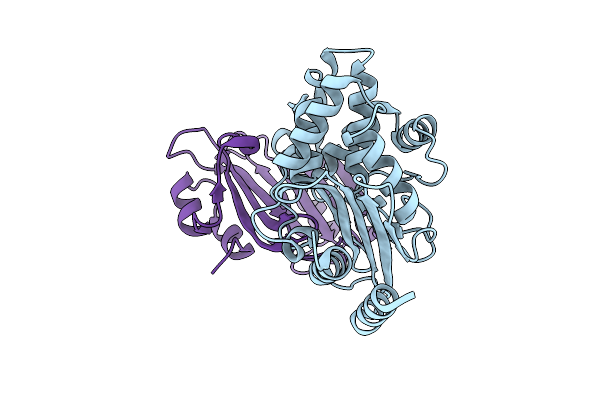
Ctx-M-14 Wt In Complex With Blip E73W
Organism: Escherichia coli, Streptomyces clavuligerus
Method: X-RAY DIFFRACTION Resolution:1.65 Å Release Date: 2025-11-05 Classification: HYDROLASE/PROTEIN BINDING |
Organism: Escherichia coli, Streptomyces clavuligerus
Method: X-RAY DIFFRACTION
Release Date: 2025-11-05
 |
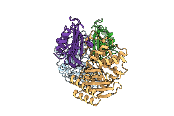
Ctx-M-15 Wt In Complex With Blip E73W
Organism: Escherichia coli, Streptomyces clavuligerus
Method: X-RAY DIFFRACTION Resolution:1.58 Å Release Date: 2025-11-05 Classification: HYDROLASE/PROTEIN BINDING |
Organism: Escherichia coli, Streptomyces clavuligerus
Method: X-RAY DIFFRACTION
Release Date: 2025-11-05
 |
Tem-1 Wt In Complex With Blip E73W
Organism: Escherichia coli, Streptomyces clavuligerus
Method: X-RAY DIFFRACTION Resolution:1.78 Å Release Date: 2025-11-05 Classification: HYDROLASE/PROTEIN BINDING |
Organism: Escherichia coli, Streptomyces clavuligerus
Method: X-RAY DIFFRACTION
Release Date: 2025-11-05
 |
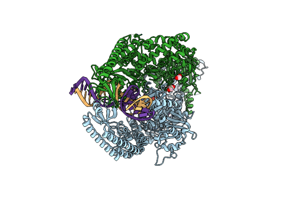
The Crystal Structure Of Dna-Bound Human Mutsbeta (Msh2/Msh3) In The Canonical Mismatch Bound Conformation With Adp Bound In Msh2 And Msh3
Organism: Homo sapiens, Synthetic construct
Method: X-RAY DIFFRACTION Resolution:2.33 Å Release Date: 2025-06-11 Classification: DNA BINDING PROTEIN Ligands: ADP, MG, EDO |
Organism: Homo sapiens, Synthetic construct
Method: X-RAY DIFFRACTION
Release Date: 2025-06-11
Ligands: ADP, MG, EDO
 |
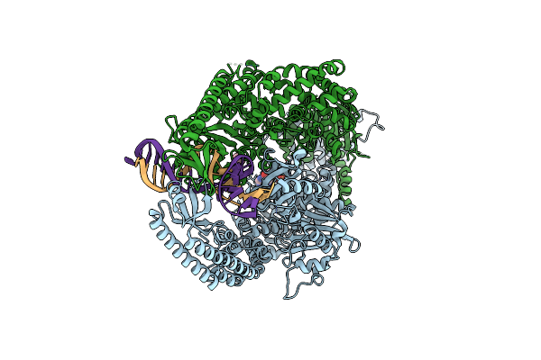
The Crystal Structure Of Dna-Bound Human Mutsbeta (Msh2/Msh3) In The Canonical Mismatch Bound Conformation With Adp Bound In Msh2
Organism: Homo sapiens, Synthetic construct
Method: X-RAY DIFFRACTION Resolution:2.63 Å Release Date: 2025-06-11 Classification: DNA BINDING PROTEIN Ligands: ADP, MG, NA, EDO |
Organism: Homo sapiens, Synthetic construct
Method: X-RAY DIFFRACTION
Release Date: 2025-06-11
Ligands: ADP, MG, NA, EDO
 |
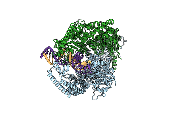
The Crystal Structure Of Dna-Bound Human Mutsbeta (Msh2/Msh3_G901D) In The Canonical Mismatch Bound Conformation With Adp Bound In Msh2
Organism: Homo sapiens, Synthetic construct
Method: X-RAY DIFFRACTION Resolution:2.69 Å Release Date: 2025-06-11 Classification: DNA BINDING PROTEIN Ligands: ADP, EDO |
Organism: Homo sapiens, Synthetic construct
Method: X-RAY DIFFRACTION
Release Date: 2025-06-11
Ligands: ADP, EDO
 |
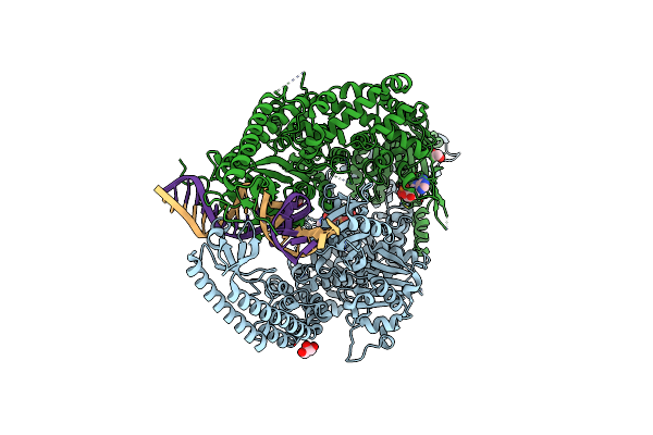
The Crystal Structure Of Dna-Bound Human Mutsbeta (Msh2_G674D/Msh3) In The Canonical Mismatch Bound Conformation, Nucleotide Free
Organism: Homo sapiens, Synthetic construct
Method: X-RAY DIFFRACTION Resolution:3.16 Å Release Date: 2025-06-11 Classification: DNA BINDING PROTEIN |
Organism: Homo sapiens, Synthetic construct
Method: X-RAY DIFFRACTION
Release Date: 2025-06-11
 |
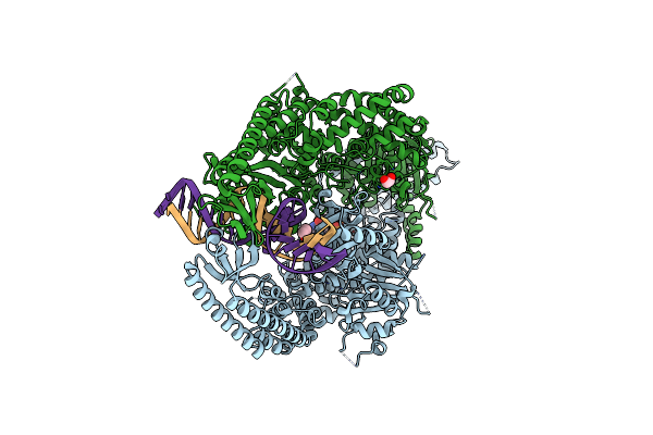
The Crystal Structure Of Dna-Bound Human Mutsbeta (Msh2_E749A/Msh3_E976A) In The Canonical Mismatch Bound Conformation With Adp Bound In Msh2 And Msh3
Organism: Homo sapiens, Synthetic construct
Method: X-RAY DIFFRACTION Resolution:2.84 Å Release Date: 2025-06-11 Classification: DNA BINDING PROTEIN Ligands: ADP, GOL, CL, EDO |
Organism: Homo sapiens, Synthetic construct
Method: X-RAY DIFFRACTION
Release Date: 2025-06-11
Ligands: ADP, GOL, CL, EDO
 |
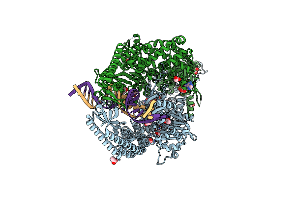
The Crystal Structure Of Dna-Bound Human Mutsbeta (Msh2_E749A/Msh3) In The Canonical Mismatch Bound Conformation With Adp Bound In Msh2
Organism: Homo sapiens, Synthetic construct
Method: X-RAY DIFFRACTION Resolution:2.67 Å Release Date: 2025-06-11 Classification: DNA BINDING PROTEIN Ligands: ADP, MG, CL, EDO |
Organism: Homo sapiens, Synthetic construct
Method: X-RAY DIFFRACTION
Release Date: 2025-06-11
Ligands: ADP, MG, CL, EDO
 |
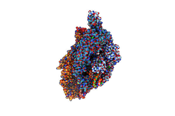
The Crystal Structure Of Dna-Bound Human Mutsbeta (Msh2_K675R/Msh3_K902R) In The Canonical Mismatch Bound Conformation With Adp Bound In Msh2 And Msh3
Organism: Homo sapiens, Synthetic construct
Method: X-RAY DIFFRACTION Resolution:2.09 Å Release Date: 2025-06-11 Classification: DNA BINDING PROTEIN Ligands: ADP, MG, EDO, ACT, CL |
Organism: Homo sapiens, Synthetic construct
Method: X-RAY DIFFRACTION
Release Date: 2025-06-11
Ligands: ADP, MG, EDO, ACT, CL
 |
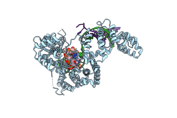
The Crystal Structure Of Dna-Bound Human Mutsbeta (Msh2_E749A/Msh3_E976A) In The Canonical Mismatch Bound Conformation With Adp Bound In Msh2
Organism: Homo sapiens, Synthetic construct
Method: X-RAY DIFFRACTION Resolution:2.53 Å Release Date: 2025-06-11 Classification: DNA BINDING PROTEIN Ligands: ADP, CL, NA, PEG, EDO, GOL |
Organism: Homo sapiens, Synthetic construct
Method: X-RAY DIFFRACTION
Release Date: 2025-06-11
Ligands: ADP, CL, NA, PEG, EDO, GOL
 |
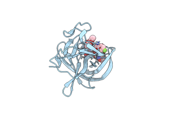
The Crystal Structure Of Fan1 Nuclease Bound To 5' Phosphorylated P(Dg)/3'(Dt-Dt-Dt-Dt) Double Flap Dna
Organism: Homo sapiens, Synthetic construct
Method: X-RAY DIFFRACTION Resolution:2.65 Å Release Date: 2025-03-05 Classification: DNA Ligands: CA, CL |
Organism: Homo sapiens, Synthetic construct
Method: X-RAY DIFFRACTION
Release Date: 2025-03-05
Ligands: CA, CL
 |
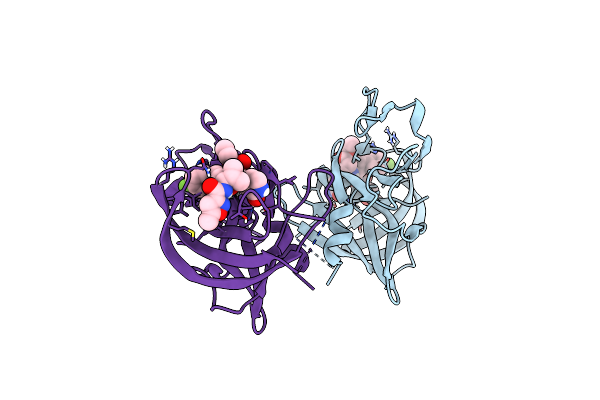
Human Norovirus Gii.4 Sydney Protease R112A Mutant In Complex With Rupintrivir
Organism: Norovirus
Method: X-RAY DIFFRACTION Resolution:2.00 Å Release Date: 2025-02-19 Classification: VIRAL PROTEIN Ligands: AG7, IOD |
Organism: Norovirus
Method: X-RAY DIFFRACTION
Release Date: 2025-02-19
Ligands: AG7, IOD
 |
Human Norovirus Gi.1 Norwalk Protease In Complex With Rupintrivir
Organism: Norovirus
Method: X-RAY DIFFRACTION Resolution:1.80 Å Release Date: 2025-02-19 Classification: VIRAL PROTEIN Ligands: AG7 |
Organism: Norovirus
Method: X-RAY DIFFRACTION
Release Date: 2025-02-19
Ligands: AG7