

Search Count: 28
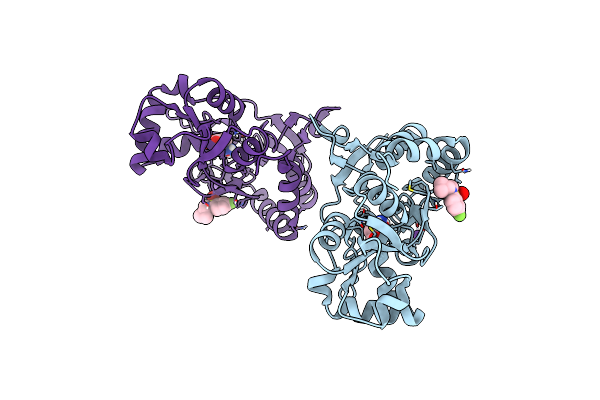 |
Crystal Structure Of The Gluk2 Ligand-Binding Domain In Complex With L-Glutamate And Bpam344 At 1.60 A Resolution
Organism: Rattus norvegicus
Method: X-RAY DIFFRACTION Resolution:1.60 Å Release Date: 2024-09-18 Classification: MEMBRANE PROTEIN Ligands: GLU, 2J9, CL, NA |
Organism: Rattus norvegicus
Method: X-RAY DIFFRACTION
Release Date: 2024-09-18
Ligands: GLU, 2J9, CL, NA
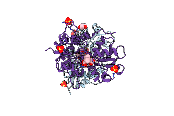 |
Crystal Structure Of The Gluk1 Ligand-Binding Domain In Complex With Kainate And Bpam538 At 1.90 A Resolution
Organism: Rattus norvegicus
Method: X-RAY DIFFRACTION Resolution:1.90 Å Release Date: 2024-08-14 Classification: MEMBRANE PROTEIN Ligands: KAI, SO4, CL, GOL, 9TE |
Organism: Rattus norvegicus
Method: X-RAY DIFFRACTION
Release Date: 2024-08-14
Ligands: KAI, SO4, CL, GOL, 9TE
 |
Crystal Structure Of The Ampa Receptor Glua2-L504Y-N775S Ligand Binding Domain In Complex With L-Glutamate And Positive Allosteric Modulator Bpam395 At 1.55A Resolution
Organism: Rattus norvegicus
Method: X-RAY DIFFRACTION Resolution:1.55 Å Release Date: 2023-12-27 Classification: MEMBRANE PROTEIN Ligands: GLU, ZN, ACT, UF5, CL, GOL, CAC |
Organism: Rattus norvegicus
Method: X-RAY DIFFRACTION
Release Date: 2023-12-27
Ligands: GLU, ZN, ACT, UF5, CL, GOL, CAC
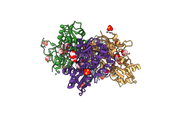 |
Crystal Structure Of The Kainate Receptor Gluk3-H523A Ligand Binding Domain In Complex With Kainate At 2.7A Resolution
Organism: Rattus norvegicus
Method: X-RAY DIFFRACTION Resolution:2.70 Å Release Date: 2023-12-13 Classification: MEMBRANE PROTEIN Ligands: ACT, ZN, CL, SO4, PEG, KAI, GOL |
Organism: Rattus norvegicus
Method: X-RAY DIFFRACTION
Release Date: 2023-12-13
Ligands: ACT, ZN, CL, SO4, PEG, KAI, GOL
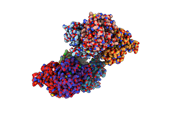 |
Crystal Structure Of The Kainate Receptor Gluk3-H523A Ligand Binding Domain In Complex With Kainate And The Positive Allosteric Modulator Bpam344 At 2.9A Resolution
Organism: Rattus norvegicus
Method: X-RAY DIFFRACTION Resolution:2.90 Å Release Date: 2023-12-13 Classification: MEMBRANE PROTEIN Ligands: 2J9, ZN, GOL, CL, SO4, KAI, ACT |
Organism: Rattus norvegicus
Method: X-RAY DIFFRACTION
Release Date: 2023-12-13
Ligands: 2J9, ZN, GOL, CL, SO4, KAI, ACT
 |
Structure Of The Glua2 Ligand-Binding Domain (L483Y-N754S) In Complex With Glutamate And Bpam549
Organism: Rattus norvegicus
Method: X-RAY DIFFRACTION Resolution:1.90 Å Release Date: 2021-08-11 Classification: SIGNALING PROTEIN Ligands: ZN, CAC, ACT, GLU, QSZ, QSW, GOL, CL |
Organism: Rattus norvegicus
Method: X-RAY DIFFRACTION
Release Date: 2021-08-11
Ligands: ZN, CAC, ACT, GLU, QSZ, QSW, GOL, CL
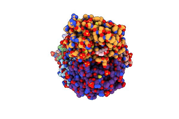 |
Human Kallikrein 7 With Aromatic Coumarinic Ester Compound 1 Covalently Bound To H57
Organism: Homo sapiens
Method: X-RAY DIFFRACTION Resolution:2.00 Å Release Date: 2020-05-20 Classification: HYDROLASE Ligands: PGE, SO4, SH7, EPE |
Organism: Homo sapiens
Method: X-RAY DIFFRACTION
Release Date: 2020-05-20
Ligands: PGE, SO4, SH7, EPE
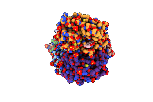 |
Human Kallikrein 7 With Aromatic Coumarinic Ester Compound 2 Covalently Bound To H57
Organism: Homo sapiens
Method: X-RAY DIFFRACTION Resolution:1.85 Å Release Date: 2020-05-20 Classification: HYDROLASE Ligands: PGE, SH8, SO4, EPE |
Organism: Homo sapiens
Method: X-RAY DIFFRACTION
Release Date: 2020-05-20
Ligands: PGE, SH8, SO4, EPE
 |
Human Kallikrein 7 With Aromatic Coumarinic Ester Compound 3 Covalently Bound To H57
Organism: Homo sapiens
Method: X-RAY DIFFRACTION Resolution:1.97 Å Release Date: 2020-05-20 Classification: HYDROLASE Ligands: PGE, SO4, LFW, EPE |
Organism: Homo sapiens
Method: X-RAY DIFFRACTION
Release Date: 2020-05-20
Ligands: PGE, SO4, LFW, EPE
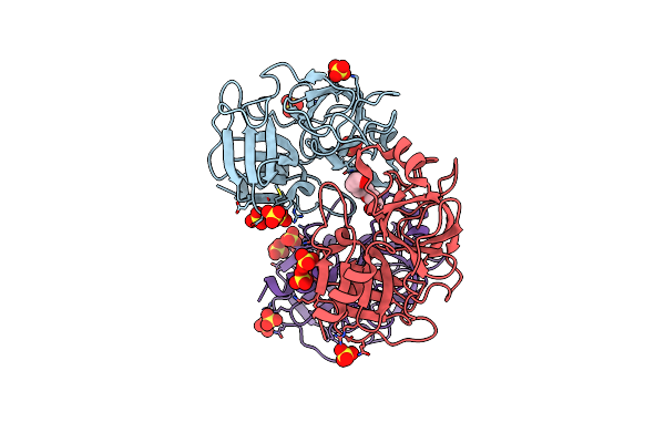 |
Human Kallikrein-Related Peptidase 7 (Klk7) In The Unliganded State
Organism: Homo sapiens
Method: X-RAY DIFFRACTION Resolution:2.23 Å Release Date: 2020-05-20 Classification: HYDROLASE Ligands: PGE, SO4 |
Organism: Homo sapiens
Method: X-RAY DIFFRACTION
Release Date: 2020-05-20
Ligands: PGE, SO4
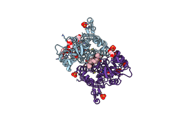 |
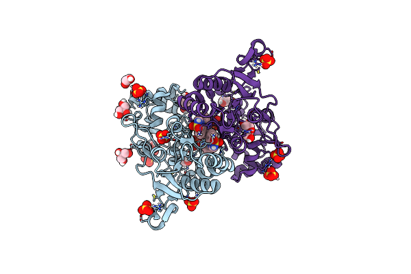
Structure Of Glua2 Ligand-Binding Domain (S1S2J-L504Y-N775S) In Complex With Glutamate And Tdpam02 At 2.4 A Resolution.
Organism: Rattus norvegicus
Method: X-RAY DIFFRACTION Resolution:2.40 Å Release Date: 2019-04-03 Classification: MEMBRANE PROTEIN Ligands: GOL, CL, GLU, SO4, PEG, PGE, FXW |
Organism: Rattus norvegicus
Method: X-RAY DIFFRACTION
Release Date: 2019-04-03
Ligands: GOL, CL, GLU, SO4, PEG, PGE, FXW
 |
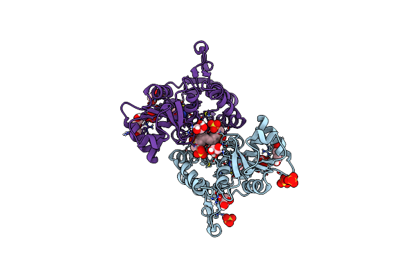
Structure Of Glua2 Ligand-Binding Domain (S1S1J) In Complex With Positive Allosteric Modulator Tdpam02 At 1.8 A Resolution
Organism: Rattus norvegicus
Method: X-RAY DIFFRACTION Resolution:1.88 Å Release Date: 2019-04-03 Classification: MEMBRANE PROTEIN Ligands: SO4, GOL, CL, FXW, PEG |
Organism: Rattus norvegicus
Method: X-RAY DIFFRACTION
Release Date: 2019-04-03
Ligands: SO4, GOL, CL, FXW, PEG
 |
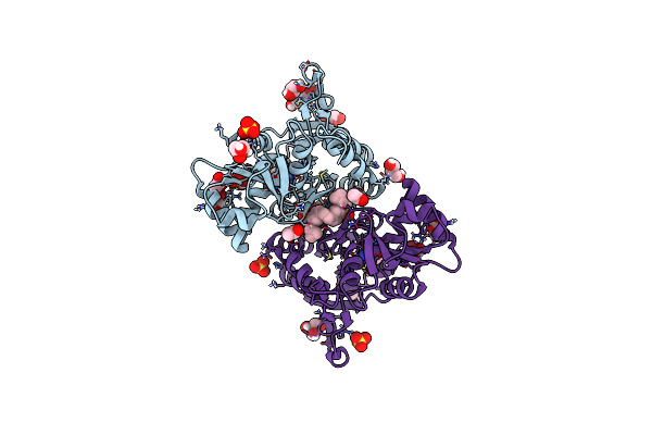
Structure Of Glua2 Ligand-Binding Domain (S1S2J-N775S) In Complex With Glutamate And Tdpam01 At 1.9 A Resolution.
Organism: Rattus norvegicus
Method: X-RAY DIFFRACTION Resolution:1.90 Å Release Date: 2019-04-03 Classification: MEMBRANE PROTEIN Ligands: SO4, GLU, GOL, CL, PEG, D45 |
Organism: Rattus norvegicus
Method: X-RAY DIFFRACTION
Release Date: 2019-04-03
Ligands: SO4, GLU, GOL, CL, PEG, D45
 |
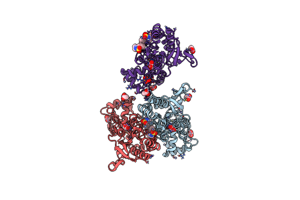
Structure Of Glua2 Ligand-Binding Domain (S1S2J-N775S) In Complex With Glutamate And Tdpam02 At 1.6 A Resolution.
Organism: Rattus norvegicus
Method: X-RAY DIFFRACTION Resolution:1.62 Å Release Date: 2019-04-03 Classification: MEMBRANE PROTEIN Ligands: FXW, GLU, SO4, GOL, PEG, CL, ACT |
Organism: Rattus norvegicus
Method: X-RAY DIFFRACTION
Release Date: 2019-04-03
Ligands: FXW, GLU, SO4, GOL, PEG, CL, ACT
 |
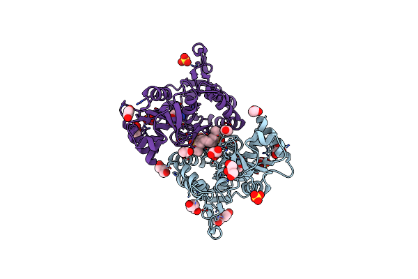
Structure Of Glua2 Ligand-Binding Domain (S1S2J-L504Y-N775S) In Complex With Glutamate And Tdpam01 At 1.6 A Resolution.
Organism: Rattus norvegicus
Method: X-RAY DIFFRACTION Resolution:1.60 Å Release Date: 2019-04-03 Classification: MEMBRANE PROTEIN Ligands: GLU, ZN, D45, ACT, GOL, CL, SO4 |
Organism: Rattus norvegicus
Method: X-RAY DIFFRACTION
Release Date: 2019-04-03
Ligands: GLU, ZN, D45, ACT, GOL, CL, SO4
 |
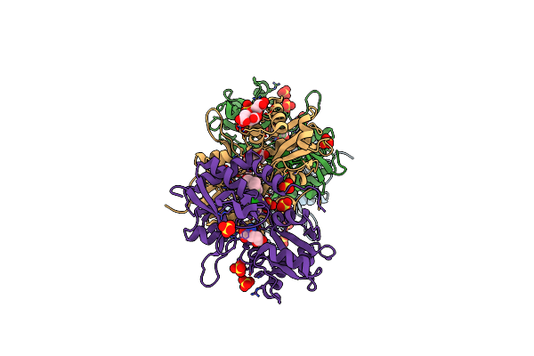
Crystal Structure Of The Glua2 Ligand-Binding Domain (S1S2J) In Complex With The Positive Allosteric Modulator Tdpam01 At 1.4 A Resolution.
Organism: Rattus norvegicus
Method: X-RAY DIFFRACTION Resolution:1.40 Å Release Date: 2018-11-21 Classification: MEMBRANE PROTEIN Ligands: SO4, PEG, GOL, CL, ACT, GLU, EDO, D45 |
Organism: Rattus norvegicus
Method: X-RAY DIFFRACTION
Release Date: 2018-11-21
Ligands: SO4, PEG, GOL, CL, ACT, GLU, EDO, D45
 |
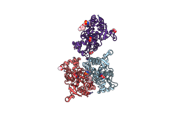
Crystal Structure Of The Glua2 Ligand-Binding Domain (S1S2J-L504Y-N775S) In Complex With Glutamate And Bpam121 At 1.78 A Resolution
Organism: Rattus norvegicus
Method: X-RAY DIFFRACTION Resolution:1.78 Å Release Date: 2018-01-03 Classification: MEMBRANE PROTEIN Ligands: SO4, 7M6, CL, GOL, GLU, EDO, CIT, PEG |
Organism: Rattus norvegicus
Method: X-RAY DIFFRACTION
Release Date: 2018-01-03
Ligands: SO4, 7M6, CL, GOL, GLU, EDO, CIT, PEG
 |
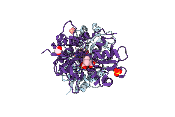
Crystal Structure Of The Glua2 Ligand-Binding Domain (S1S2J) In Complex With Glutamate And Positive Allosteric Modulator Bpam538
Organism: Rattus norvegicus
Method: X-RAY DIFFRACTION Resolution:2.00 Å Release Date: 2018-01-03 Classification: MEMBRANE PROTEIN Ligands: GLU, ZN, ACT, 9TE |
Organism: Rattus norvegicus
Method: X-RAY DIFFRACTION
Release Date: 2018-01-03
Ligands: GLU, ZN, ACT, 9TE
 |
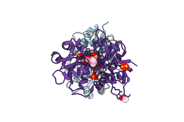
Crystal Structure Of The Gluk1 Ligand-Binding Domain In Complex With Kainate And Bpam-344 At 1.90 A Resolution
Organism: Rattus norvegicus
Method: X-RAY DIFFRACTION Resolution:1.90 Å Release Date: 2017-04-12 Classification: MEMBRANE PROTEIN Ligands: 2J9, KAI, CL, GOL, SO4 |
Organism: Rattus norvegicus
Method: X-RAY DIFFRACTION
Release Date: 2017-04-12
Ligands: 2J9, KAI, CL, GOL, SO4
 |
Crystal Structure Of The Gluk1 Ligand-Binding Domain In Complex With Kainate And Bpam-521 At 2.18 A Resolution
Organism: Rattus norvegicus
Method: X-RAY DIFFRACTION Resolution:2.18 Å Release Date: 2017-04-12 Classification: MEMBRANE PROTEIN Ligands: 5PX, KAI, CL, SO4, GOL, ACT |
Organism: Rattus norvegicus
Method: X-RAY DIFFRACTION
Release Date: 2017-04-12
Ligands: 5PX, KAI, CL, SO4, GOL, ACT