

Search Count: 77
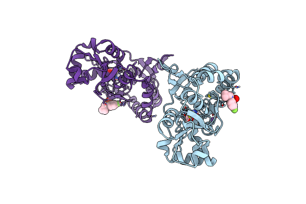 |
Crystal Structure Of The Gluk2 Ligand-Binding Domain In Complex With L-Glutamate And Bpam344 At 1.60 A Resolution
Organism: Rattus norvegicus
Method: X-RAY DIFFRACTION Resolution:1.60 Å Release Date: 2024-09-18 Classification: MEMBRANE PROTEIN Ligands: GLU, 2J9, CL, NA |
Organism: Rattus norvegicus
Method: X-RAY DIFFRACTION
Release Date: 2024-09-18
Ligands: GLU, 2J9, CL, NA
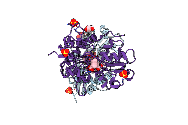 |
Crystal Structure Of The Gluk1 Ligand-Binding Domain In Complex With Kainate And Bpam538 At 1.90 A Resolution
Organism: Rattus norvegicus
Method: X-RAY DIFFRACTION Resolution:1.90 Å Release Date: 2024-08-14 Classification: MEMBRANE PROTEIN Ligands: KAI, SO4, CL, GOL, 9TE |
Organism: Rattus norvegicus
Method: X-RAY DIFFRACTION
Release Date: 2024-08-14
Ligands: KAI, SO4, CL, GOL, 9TE
 |
Crystal Structure Of The Ampa Receptor Glua2-L504Y-N775S Ligand Binding Domain In Complex With L-Glutamate And Positive Allosteric Modulator Bpam395 At 1.55A Resolution
Organism: Rattus norvegicus
Method: X-RAY DIFFRACTION Resolution:1.55 Å Release Date: 2023-12-27 Classification: MEMBRANE PROTEIN Ligands: GLU, ZN, ACT, UF5, CL, GOL, CAC |
Organism: Rattus norvegicus
Method: X-RAY DIFFRACTION
Release Date: 2023-12-27
Ligands: GLU, ZN, ACT, UF5, CL, GOL, CAC
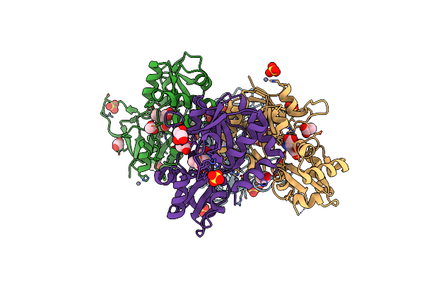 |
Crystal Structure Of The Kainate Receptor Gluk3-H523A Ligand Binding Domain In Complex With Kainate At 2.7A Resolution
Organism: Rattus norvegicus
Method: X-RAY DIFFRACTION Resolution:2.70 Å Release Date: 2023-12-13 Classification: MEMBRANE PROTEIN Ligands: ACT, ZN, CL, SO4, PEG, KAI, GOL |
Organism: Rattus norvegicus
Method: X-RAY DIFFRACTION
Release Date: 2023-12-13
Ligands: ACT, ZN, CL, SO4, PEG, KAI, GOL
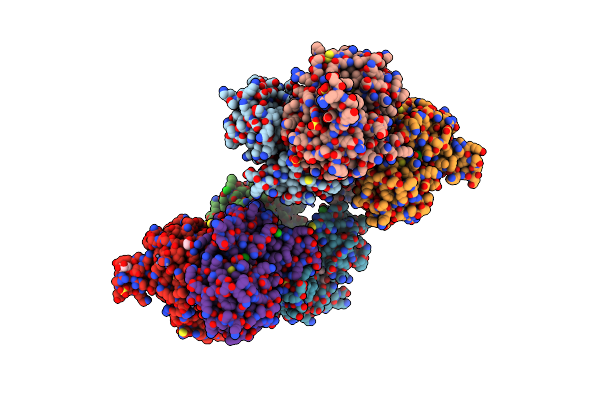 |
Crystal Structure Of The Kainate Receptor Gluk3-H523A Ligand Binding Domain In Complex With Kainate And The Positive Allosteric Modulator Bpam344 At 2.9A Resolution
Organism: Rattus norvegicus
Method: X-RAY DIFFRACTION Resolution:2.90 Å Release Date: 2023-12-13 Classification: MEMBRANE PROTEIN Ligands: 2J9, ZN, GOL, CL, SO4, KAI, ACT |
Organism: Rattus norvegicus
Method: X-RAY DIFFRACTION
Release Date: 2023-12-13
Ligands: 2J9, ZN, GOL, CL, SO4, KAI, ACT
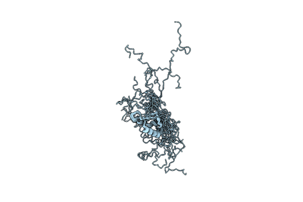 |
Structure Of N-Terminal Of Schistosoma Japonicum Asparaginyl-Trna Synthetase
Organism: Schistosoma japonicum
Method: SOLUTION NMR Release Date: 2023-09-06 Classification: LIGASE |
Organism: Schistosoma japonicum
Method: SOLUTION NMR
Release Date: 2023-09-06
 |
Truncated Ac-Aip-2
Organism: Ancylostoma caninum
Method: SOLUTION NMR Release Date: 2022-10-19 Classification: UNKNOWN FUNCTION |
Organism: Ancylostoma caninum
Method: SOLUTION NMR
Release Date: 2022-10-19
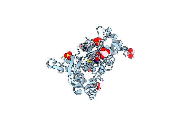 |
Structure Of The Ampa Receptor Glua2O Ligand-Binding Domain (S1S2J) In Complex With The Compound (S)-1-[2'-Amino-2'-Carboxyethyl]-5,7-Dihydrothieno[3,4-D]Pyrimidin- 2,4(1H,3H)-Dione At Resolution 1.60A
Organism: Rattus norvegicus
Method: X-RAY DIFFRACTION Resolution:1.61 Å Release Date: 2020-06-03 Classification: MEMBRANE PROTEIN Ligands: SO4, GOL, CGW, CL |
Organism: Rattus norvegicus
Method: X-RAY DIFFRACTION
Release Date: 2020-06-03
Ligands: SO4, GOL, CGW, CL
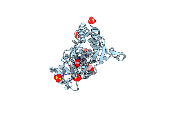 |
Structure Of The Ampa Receptor Glua2O Ligand-Binding Domain (S1S2J) In Complex With The Compound ( S) - 1- [2'-Amino-2'-Carboxyethyl]-5 ,7- Dihydropyrrolo[3,4-D]Pyrimidin-2,4(1H,3H)-Dione At Resolution 1.20A
Organism: Rattus norvegicus
Method: X-RAY DIFFRACTION Resolution:1.20 Å Release Date: 2020-06-03 Classification: MEMBRANE PROTEIN Ligands: GOL, SO4, PVQ, NH4, CL |
Organism: Rattus norvegicus
Method: X-RAY DIFFRACTION
Release Date: 2020-06-03
Ligands: GOL, SO4, PVQ, NH4, CL
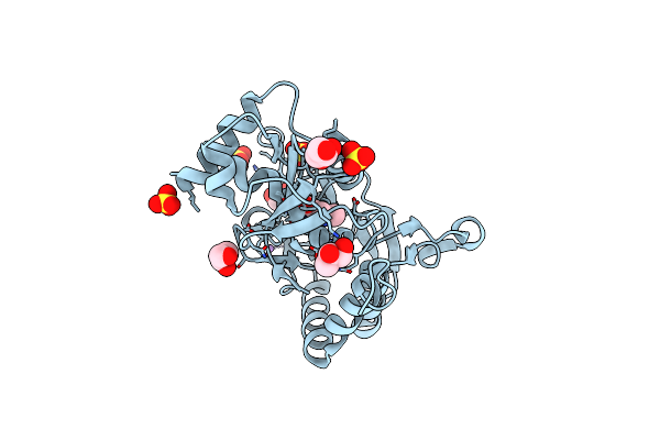 |
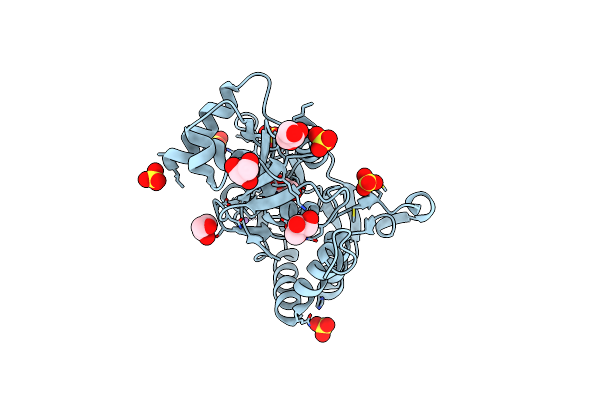
Structure Of The Ampa Receptor Glua2O Ligand-Binding Domain (S1S2J) In Complex With The Compound ( S) - 1- [2'-Amino-2'-Carboxyethyl]-6-Methyl-5 ,7- Dihydropyrrolo[3,4-D]Pyrimidin-2,4(1H,3H)-Dione At Resolution 1.00A
Organism: Rattus norvegicus
Method: X-RAY DIFFRACTION Resolution:1.00 Å Release Date: 2020-06-03 Classification: MEMBRANE PROTEIN Ligands: SO4, GOL, LI, CG8, CL |
Organism: Rattus norvegicus
Method: X-RAY DIFFRACTION
Release Date: 2020-06-03
Ligands: SO4, GOL, LI, CG8, CL
 |
Structure Of The Ampa Receptor Glua2O Ligand-Binding Domain (S1S2J) In Complex With The Compound (S)-1-(2'-Amino-2'-Carboxyethyl)-5,7-Dihydrofuro[3,4-D]- Pyrimidine-2,4(1H,3H)-Dione At Resolution 1.15A
Organism: Rattus norvegicus
Method: X-RAY DIFFRACTION Resolution:1.15 Å Release Date: 2020-06-03 Classification: MEMBRANE PROTEIN Ligands: GOL, SO4, LI, PVK, CL |
Organism: Rattus norvegicus
Method: X-RAY DIFFRACTION
Release Date: 2020-06-03
Ligands: GOL, SO4, LI, PVK, CL
 |
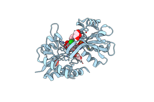
Structure Of The Ampa Receptor Glua2O Ligand-Binding Domain (S1S2J) In Complex With The Compound (S)-1-(2'-Amino-2'-Carboxyethyl)Furo[3,4-D]Pyrimidin-2,4-Dione At Resolution 1.47A
Organism: Rattus norvegicus
Method: X-RAY DIFFRACTION Resolution:1.47 Å Release Date: 2020-06-03 Classification: MEMBRANE PROTEIN Ligands: SO4, GOL, LI, CL, OUB |
Organism: Rattus norvegicus
Method: X-RAY DIFFRACTION
Release Date: 2020-06-03
Ligands: SO4, GOL, LI, CL, OUB
 |
Structure Of Gluk1 Ligand-Binding Domain (S1S2) In Complex With N-(7-(1H-Imidazol-1-Yl)-2,3-Dioxo-6-(Trifluoromethyl)-3,4-Dihydroquinoxalin-1(2H)-Yl Benzamide At 2.3 A Resolution
Organism: Rattus norvegicus
Method: X-RAY DIFFRACTION Resolution:2.30 Å Release Date: 2019-10-30 Classification: MEMBRANE PROTEIN Ligands: SO4, L5H, CL, GOL |
Organism: Rattus norvegicus
Method: X-RAY DIFFRACTION
Release Date: 2019-10-30
Ligands: SO4, L5H, CL, GOL
 |
Structure Of Glua2 Ligand-Binding Domain (S1S2J) In Complex With The Agonist (S)-2-Amino-3-(1-Ethyl-4-Hydroxy-1H-1,2,3-Triazol-5-Yl)Propanoic Acid At 1.4 A Resolution
Organism: Rattus norvegicus
Method: X-RAY DIFFRACTION Resolution:1.40 Å Release Date: 2019-04-17 Classification: MEMBRANE PROTEIN Ligands: SO4, HJ8, GOL, PGE, CL, CIT, LI, PEG |
Organism: Rattus norvegicus
Method: X-RAY DIFFRACTION
Release Date: 2019-04-17
Ligands: SO4, HJ8, GOL, PGE, CL, CIT, LI, PEG
 |
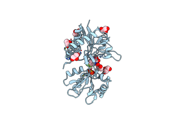
Structure Of Glua2 Ligand-Binding Domain (S1S2J) In Complex With The Agonist (S)-2-Amino-3-(2-Methyl-5-Hydroxy-2H-1,2,3-Triazol-4-Yl)Propanoic Acid At 1.55 A Resolution
Organism: Rattus norvegicus
Method: X-RAY DIFFRACTION Resolution:1.55 Å Release Date: 2019-04-17 Classification: MEMBRANE PROTEIN Ligands: HJH, SO4, GOL, CL, LI |
Organism: Rattus norvegicus
Method: X-RAY DIFFRACTION
Release Date: 2019-04-17
Ligands: HJH, SO4, GOL, CL, LI
 |
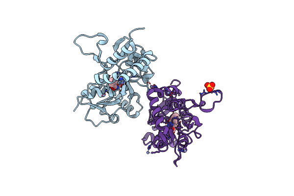
Structure Of Gluk1 Ligand-Binding Domain In Complex With N-(7-Fluoro-2,3-Dioxo-6-(Trifluoromethyl)-3,4-Dihydroquinoxalin-1(2H)-Yl)-2-Hydroxybenzamide At 1.85 A Resolution
Organism: Rattus norvegicus
Method: X-RAY DIFFRACTION Resolution:1.85 Å Release Date: 2019-01-23 Classification: MEMBRANE PROTEIN Ligands: GOL, SO4, EC8, CL |
Organism: Rattus norvegicus
Method: X-RAY DIFFRACTION
Release Date: 2019-01-23
Ligands: GOL, SO4, EC8, CL
 |
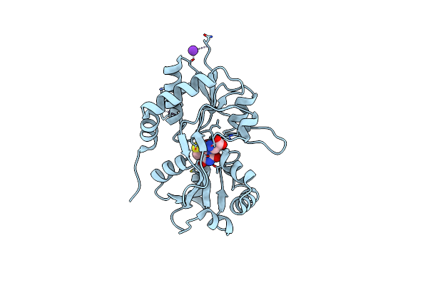
Crystal Structure Of The Kainate Receptor Gluk3 Ligand Binding Domain In Complex With (S)-1-[2'-Amino-2'-Carboxyethyl]-6-Methyl-5,7-Dihydropyrrolo[3,4-D]Pyrimidin-2,4(1H,3H)-Dione At Resolution 2.4A
Organism: Rattus norvegicus
Method: X-RAY DIFFRACTION Resolution:2.40 Å Release Date: 2018-02-28 Classification: MEMBRANE PROTEIN Ligands: ZN, CG8, CL, SO4 |
Organism: Rattus norvegicus
Method: X-RAY DIFFRACTION
Release Date: 2018-02-28
Ligands: ZN, CG8, CL, SO4
 |
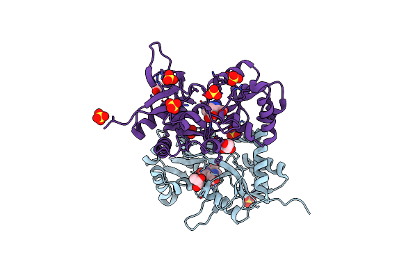
Crystal Structure Of The Kainate Receptor Gluk3 Ligand Binding Domain In Complex With (S)-1-[2-Amino-2-Carboxyethyl]-5,7-Dihydrothieno[3,4-D]Pyrimidin-2,4(1H,3H)-Dione At Resolution 2.6A
Organism: Rattus norvegicus
Method: X-RAY DIFFRACTION Resolution:2.60 Å Release Date: 2018-02-28 Classification: MEMBRANE PROTEIN Ligands: CGW, K, CL |
Organism: Rattus norvegicus
Method: X-RAY DIFFRACTION
Release Date: 2018-02-28
Ligands: CGW, K, CL
 |
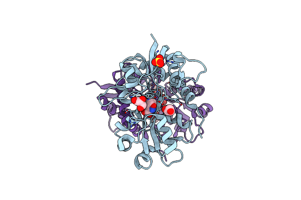
Structure Of Gluk1 Ligand-Binding Domain (S1S2) In Complex With Lm-12B At 2.05 A Resolution
Organism: Rattus norvegicus
Method: X-RAY DIFFRACTION Resolution:2.05 Å Release Date: 2017-07-26 Classification: MEMBRANE PROTEIN Ligands: SO4, CL, GOL, 8VE, ACT |
Organism: Rattus norvegicus
Method: X-RAY DIFFRACTION
Release Date: 2017-07-26
Ligands: SO4, CL, GOL, 8VE, ACT
 |
Structure Of Gluk1 Ligand-Binding Domain (S1S2) In Complex With Cip-As At 2.85 A Resolution
Organism: Rattus norvegicus
Method: X-RAY DIFFRACTION Resolution:2.85 Å Release Date: 2017-07-26 Classification: MEMBRANE PROTEIN Ligands: CL, 8VN, GOL, SO4 |
Organism: Rattus norvegicus
Method: X-RAY DIFFRACTION
Release Date: 2017-07-26
Ligands: CL, 8VN, GOL, SO4