
Search Count: 41
 |
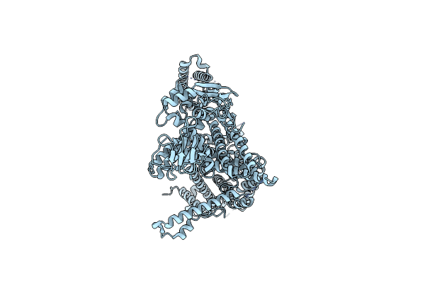
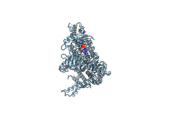
Organism: Vibrio cholerae, Vicugna pacos
Method: X-RAY DIFFRACTION Resolution:2.05 Å Release Date: 2024-11-06 Classification: TRANSPORT PROTEIN Ligands: IMD, GOL, PGE |
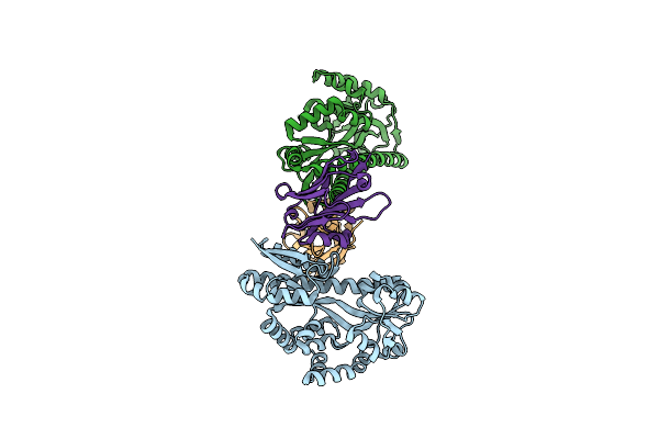 |
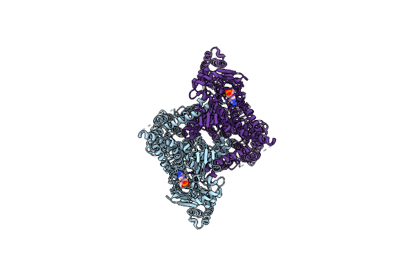
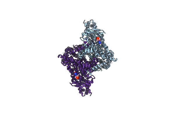
Organism: Vibrio cholerae, Vicugna pacos
Method: X-RAY DIFFRACTION Resolution:2.64 Å Release Date: 2024-11-06 Classification: TRANSPORT PROTEIN |
 |
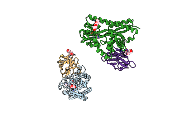
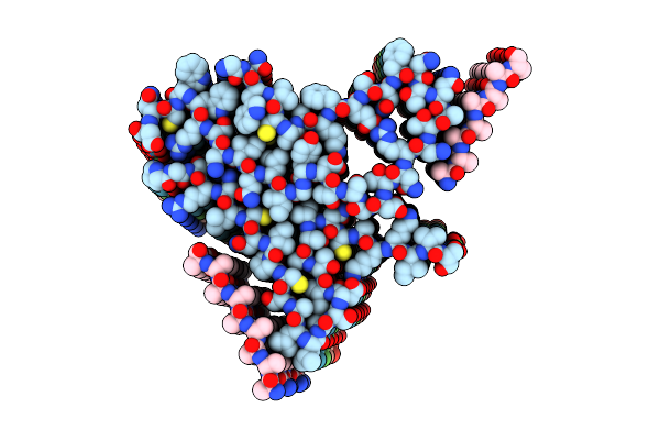
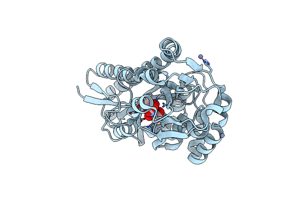
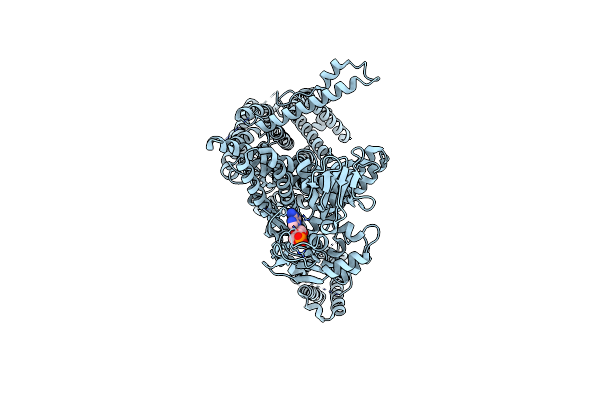
Crystal Structure Of Vcsiap W73A Mutant In Complex With Sialic Acid And A Vhh Antibody (Vhh_Vcp#2)
Organism: Vicugna pacos
Method: X-RAY DIFFRACTION Resolution:2.81 Å Release Date: 2024-11-06 Classification: TRANSPORT PROTEIN Ligands: SLB, GOL |
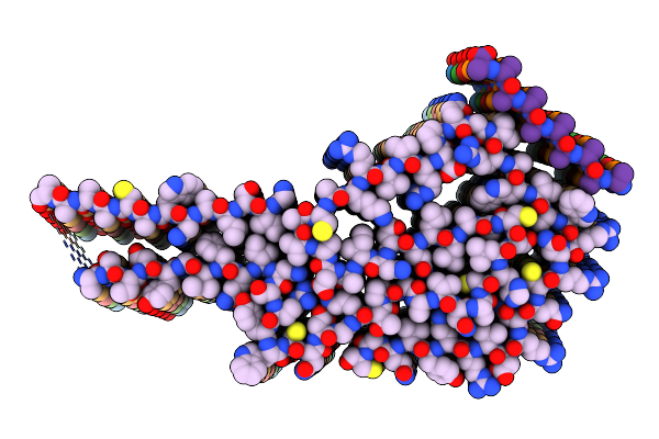 |
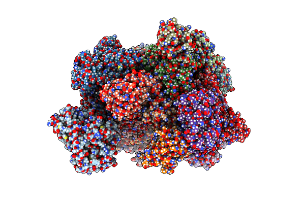
Cryo-Em Structure Of The Heat-Irreversible Amyloid Fibrils Of Human Lysozyme
Organism: Homo sapiens
Method: ELECTRON MICROSCOPY Release Date: 2024-09-18 Classification: PROTEIN FIBRIL |
 |
Cryo-Em Structure Of The Heat-Irreversible Amyloid Fibrils Of Hen Egg-White Lysozyme
Organism: Gallus gallus
Method: ELECTRON MICROSCOPY Release Date: 2024-09-18 Classification: PROTEIN FIBRIL |
 |
Organism: Homo sapiens, Synthetic construct
Method: ELECTRON MICROSCOPY Release Date: 2024-06-19 Classification: MEMBRANE PROTEIN Ligands: PLC |
 |
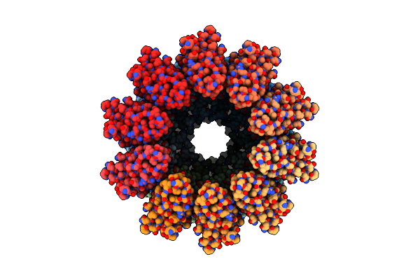
Structure Of Heteromeric Calhm2/4 Channel In Complex With Synthetic Nanobody Sbc4
Organism: Homo sapiens, Synthetic construct
Method: ELECTRON MICROSCOPY Release Date: 2024-06-19 Classification: MEMBRANE PROTEIN |
 |
Structure Of Heteromeric Calhm2/4 Channel In Complex With Synthetic Nanobodies Sbc2 And Sbc4
Organism: Homo sapiens, Synthetic construct
Method: ELECTRON MICROSCOPY Release Date: 2024-06-19 Classification: MEMBRANE PROTEIN Ligands: PLC |
 |
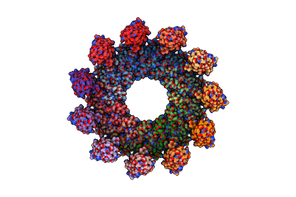
Cryo-Em Structure Of A Dimer Of Decameric Human Calhm4 In Complex With Synthetic Nanobody Sbc4
Organism: Homo sapiens, Synthetic construct
Method: ELECTRON MICROSCOPY Release Date: 2024-06-19 Classification: MEMBRANE PROTEIN |
 |
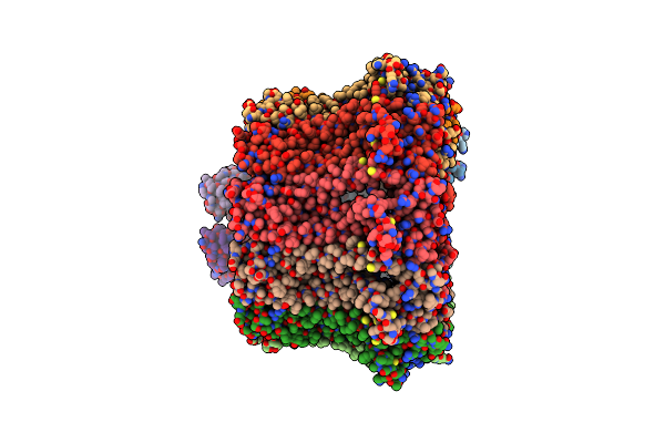
Organism: Haemophilus influenzae
Method: X-RAY DIFFRACTION Resolution:1.90 Å Release Date: 2023-12-27 Classification: SUGAR BINDING PROTEIN Ligands: SLB, ZN |
 |
Organism: Strongylocentrotus purpuratus
Method: ELECTRON MICROSCOPY Release Date: 2023-11-08 Classification: MEMBRANE PROTEIN |
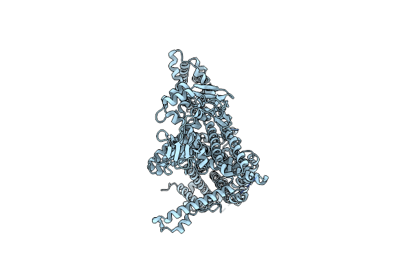 |
Organism: Strongylocentrotus purpuratus
Method: ELECTRON MICROSCOPY Release Date: 2023-11-08 Classification: MEMBRANE PROTEIN |
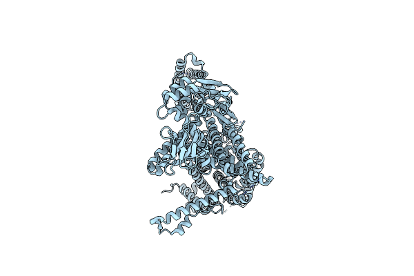 |
Organism: Strongylocentrotus purpuratus
Method: ELECTRON MICROSCOPY Release Date: 2023-11-08 Classification: MEMBRANE PROTEIN |
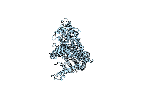 |
Organism: Strongylocentrotus purpuratus
Method: ELECTRON MICROSCOPY Release Date: 2023-11-08 Classification: MEMBRANE PROTEIN |
 |
Organism: Strongylocentrotus purpuratus
Method: ELECTRON MICROSCOPY Release Date: 2023-11-08 Classification: MEMBRANE PROTEIN |
 |
Organism: Strongylocentrotus purpuratus
Method: ELECTRON MICROSCOPY Release Date: 2023-11-08 Classification: MEMBRANE PROTEIN Ligands: CMP |
 |
Organism: Strongylocentrotus purpuratus
Method: ELECTRON MICROSCOPY Release Date: 2023-11-08 Classification: MEMBRANE PROTEIN Ligands: CMP |
 |
Organism: Strongylocentrotus purpuratus
Method: ELECTRON MICROSCOPY Release Date: 2023-11-08 Classification: MEMBRANE PROTEIN Ligands: PCG |
 |
Organism: Strongylocentrotus purpuratus
Method: ELECTRON MICROSCOPY Release Date: 2023-11-08 Classification: MEMBRANE PROTEIN Ligands: PCG |
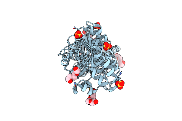 |
Organism: Sulfurihydrogenibium sp. yo3aop1
Method: X-RAY DIFFRACTION Resolution:2.10 Å Release Date: 2022-11-16 Classification: PEPTIDE BINDING PROTEIN Ligands: SO4, PGE, PG4 |