

Search Count: 16
 |
Human Cardiac Interacting Heads Motif, E525K Mutant
Organism: Homo sapiens
Method: ELECTRON MICROSCOPY Release Date: 2026-06-17 Classification: MOTOR PROTEIN Ligands: ADP, PO4, MG Modified Residue(s): M3L |
Organism: Homo sapiens
Method: ELECTRON MICROSCOPY
Release Date: 2026-06-17
Ligands: ADP, PO4, MG
 |
Human Cardiac Interacting Heads Motif, Conformation B
Organism: Homo sapiens
Method: ELECTRON MICROSCOPY Release Date: 2026-06-17 Classification: MOTOR PROTEIN Ligands: ADP, PO4, MG Modified Residue(s): M3L |
Organism: Homo sapiens
Method: ELECTRON MICROSCOPY
Release Date: 2026-06-17
Ligands: ADP, PO4, MG
 |
Human Cardiac Interacting Heads Motif, Conformation A
Organism: Homo sapiens
Method: ELECTRON MICROSCOPY Resolution:3.60 Å Release Date: 2026-06-17 Classification: MOTOR PROTEIN Ligands: ADP, PO4, MG Modified Residue(s): M3L |
Organism: Homo sapiens
Method: ELECTRON MICROSCOPY
Release Date: 2026-06-17
Ligands: ADP, PO4, MG
 |
Crystal Structure Of Bacillus Subtilis Glyceraldehyde-3-Phosphate Dehydrogenase Gapb
Organism: Bacillus subtilis subsp. subtilis str. 168
Method: X-RAY DIFFRACTION Resolution:2.30 Å Release Date: 2024-10-02 Classification: OXIDOREDUCTASE |
Organism: Bacillus subtilis subsp. subtilis str. 168
Method: X-RAY DIFFRACTION
Release Date: 2024-10-02
 |
Structure Of The Human Beta-Cardiac Myosin Folded-Back Off State
Organism: Homo sapiens
Method: ELECTRON MICROSCOPY Release Date: 2023-06-07 Classification: CONTRACTILE PROTEIN Ligands: ADP, PO4, MG Modified Residue(s): M3L |
Organism: Homo sapiens
Method: ELECTRON MICROSCOPY
Release Date: 2023-06-07
Ligands: ADP, PO4, MG
 |
Crystal Structure Of Gigantea In Complex With Lkp2
Organism: Arabidopsis thaliana
Method: X-RAY DIFFRACTION Resolution:3.50 Å Release Date: 2022-04-27 Classification: CIRCADIAN CLOCK PROTEIN Ligands: FMN Modified Residue(s): MSE |
Organism: Arabidopsis thaliana
Method: X-RAY DIFFRACTION
Release Date: 2022-04-27
Ligands: FMN
 |
Crystal Structure Of The Tandem B-Box Domain Of Arabidopsis Thaliana Constans
Organism: Arabidopsis thaliana
Method: X-RAY DIFFRACTION Resolution:2.40 Å Release Date: 2022-03-02 Classification: PLANT PROTEIN Ligands: ZN |
Organism: Arabidopsis thaliana
Method: X-RAY DIFFRACTION
Release Date: 2022-03-02
Ligands: ZN
 |
The Crystal Structure Of B. Subtilis Rsbv/Rsbw Complex In The Monoclinic Crystal Form
Organism: Bacillus subtilis (strain 168)
Method: X-RAY DIFFRACTION Resolution:3.40 Å Release Date: 2020-07-15 Classification: SIGNALING PROTEIN |
Organism: Bacillus subtilis (strain 168)
Method: X-RAY DIFFRACTION
Release Date: 2020-07-15
 |
The Crystal Structure Of B. Subtilis Rsbv/Rsbw Complex In The Hexagonal Crystal Form
Organism: Bacillus subtilis (strain 168)
Method: X-RAY DIFFRACTION Resolution:3.10 Å Release Date: 2020-07-15 Classification: SIGNALING PROTEIN |
Organism: Bacillus subtilis (strain 168)
Method: X-RAY DIFFRACTION
Release Date: 2020-07-15
 |
Crystal Structure Of Mimivirus Ung Y322L In Complex With Ugi
Organism: Acanthamoeba polyphaga mimivirus, Bacillus phage pbs2
Method: X-RAY DIFFRACTION Resolution:2.60 Å Release Date: 2020-07-08 Classification: DNA BINDING PROTEIN/INHIBITOR |
Organism: Acanthamoeba polyphaga mimivirus, Bacillus phage pbs2
Method: X-RAY DIFFRACTION
Release Date: 2020-07-08
 |
Crystal Structure Of Mimivirus Ung Y322F In Complex With Ugi
Organism: Acanthamoeba polyphaga mimivirus, Bacillus phage pbs2
Method: X-RAY DIFFRACTION Resolution:3.10 Å Release Date: 2020-07-08 Classification: DNA BINDING PROTEIN/INHIBITOR |
Organism: Acanthamoeba polyphaga mimivirus, Bacillus phage pbs2
Method: X-RAY DIFFRACTION
Release Date: 2020-07-08
 |
Crystal Structure Of Bacillus Subtilis Sigw Domain 4 In Complexed With -35 Element Dna
Organism: Bacillus subtilis (strain 168), Synthetic construct
Method: X-RAY DIFFRACTION Resolution:3.10 Å Release Date: 2020-01-01 Classification: TRANSCRIPTION/DNA |
Organism: Bacillus subtilis (strain 168), Synthetic construct
Method: X-RAY DIFFRACTION
Release Date: 2020-01-01
 |
Crystal Structure Of Bacillus Subtilis Rsbs
Organism: Bacillus subtilis
Method: X-RAY DIFFRACTION Resolution:3.10 Å Release Date: 2019-09-11 Classification: PROTEIN BINDING Modified Residue(s): SEP |
Organism: Bacillus subtilis
Method: X-RAY DIFFRACTION
Release Date: 2019-09-11
 |
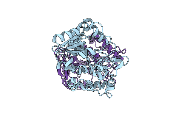
Crystal Structure Of Usp35 C450S In Complex With Ubiquitin
Organism: Homo sapiens
Method: X-RAY DIFFRACTION Resolution:1.84 Å Release Date: 2018-05-09 Classification: HYDROLASE Ligands: SO4, EDO, ZN |
Organism: Homo sapiens
Method: X-RAY DIFFRACTION
Release Date: 2018-05-09
Ligands: SO4, EDO, ZN
 |
Crystal Structure Of Mimivirus Uracil-Dna Glycosylase
Organism: Acanthamoeba polyphaga mimivirus
Method: X-RAY DIFFRACTION Resolution:2.30 Å Release Date: 2017-08-09 Classification: HYDROLASE |
Organism: Acanthamoeba polyphaga mimivirus
Method: X-RAY DIFFRACTION
Release Date: 2017-08-09
 |
Dienelactone Hydrolase At 2.8 Angstroms
Organism: Pseudomonas knackmussii
Method: X-RAY DIFFRACTION Resolution:1.80 Å Release Date: 1996-08-17 Classification: HYDROLYTIC ENZYME Modified Residue(s): CSD |
Organism: Pseudomonas knackmussii
Method: X-RAY DIFFRACTION
Release Date: 1996-08-17