

Search Count: 24
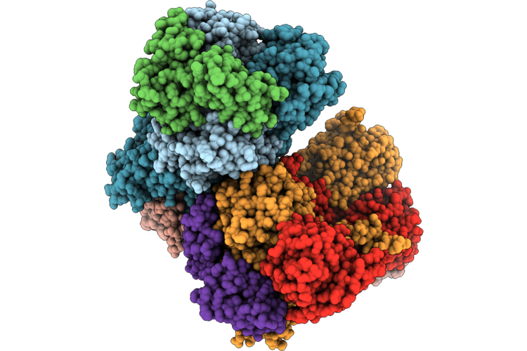 |
Crystal Structure Of Fused Glycerol Dehydratase
Organism: Klebsiella pneumoniae
Method: X-RAY DIFFRACTION Resolution:2.91 Å Release Date: 2026-06-24 Classification: LYASE Ligands: B12 |
Organism: Klebsiella pneumoniae
Method: X-RAY DIFFRACTION
Release Date: 2026-06-24
Ligands: B12
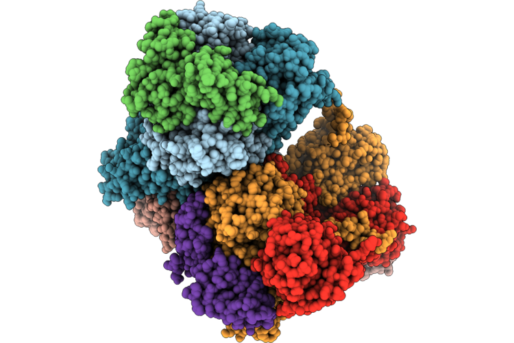 |
Crystal Structure Of Fused Glycerol Dehydratase M158W Variant
Organism: Klebsiella pneumoniae
Method: X-RAY DIFFRACTION Resolution:3.68 Å Release Date: 2026-06-24 Classification: LYASE Ligands: B12 |
Organism: Klebsiella pneumoniae
Method: X-RAY DIFFRACTION
Release Date: 2026-06-24
Ligands: B12
 |
Crystal Structure Of Fused Glycerol Dehydratase L113W Variant
Organism: Klebsiella pneumoniae
Method: X-RAY DIFFRACTION Resolution:3.02 Å Release Date: 2026-06-24 Classification: LYASE Ligands: B12 |
Organism: Klebsiella pneumoniae
Method: X-RAY DIFFRACTION
Release Date: 2026-06-24
Ligands: B12
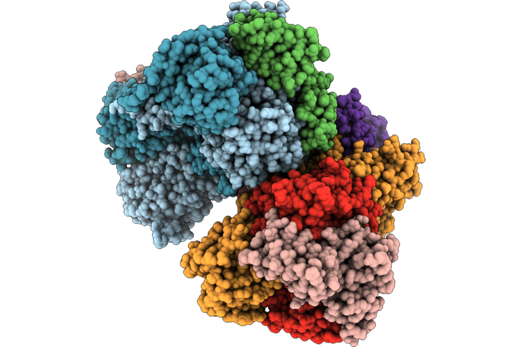 |
Crystal Structure Of Fused Glycerol Dehydratase A177M Variant
Organism: Klebsiella pneumoniae
Method: X-RAY DIFFRACTION Resolution:3.56 Å Release Date: 2026-06-24 Classification: LYASE Ligands: B12 |
Organism: Klebsiella pneumoniae
Method: X-RAY DIFFRACTION
Release Date: 2026-06-24
Ligands: B12
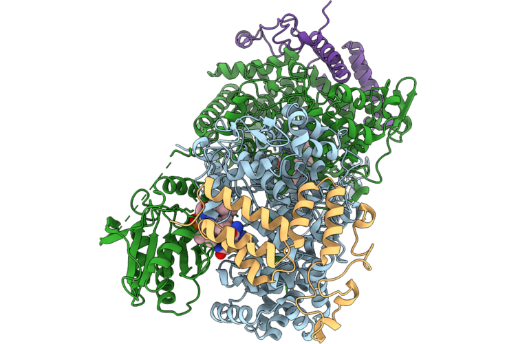 |
Crystal Structure Of Fused Glycerol Dehydratase A177M/M158W Variant
Organism: Klebsiella pneumoniae
Method: X-RAY DIFFRACTION Resolution:3.58 Å Release Date: 2026-06-24 Classification: LYASE Ligands: B12 |
Organism: Klebsiella pneumoniae
Method: X-RAY DIFFRACTION
Release Date: 2026-06-24
Ligands: B12
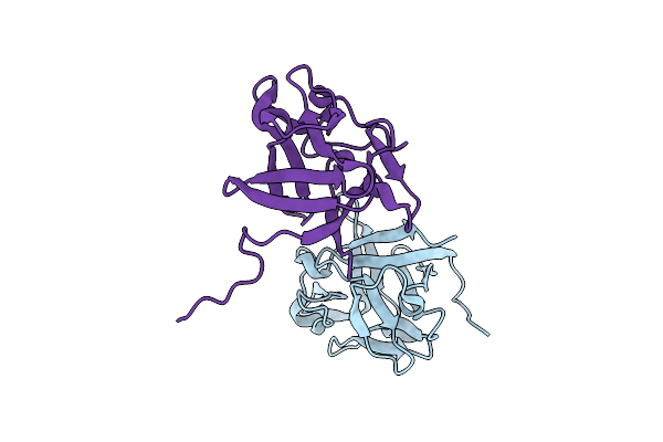 |
Linear Form Of Fgf10 From Homo Sapiens
Organism: Homo sapiens
Method: X-RAY DIFFRACTION Resolution:2.56 Å Release Date: 2025-03-26 Classification: HORMONE |
Organism: Homo sapiens
Method: X-RAY DIFFRACTION
Release Date: 2025-03-26
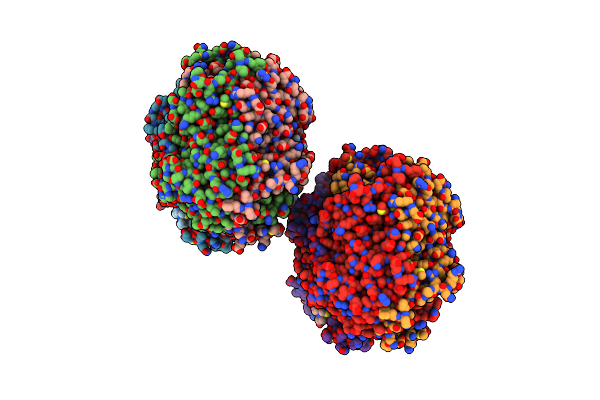 |
The Mutant Variant Of Pngm-1, H93 Was Substituuted For Alanine To Study Metal Coordination
Organism: Uncultured bacterium
Method: X-RAY DIFFRACTION Resolution:1.61 Å Release Date: 2023-01-18 Classification: HYDROLASE Ligands: ZN |
Organism: Uncultured bacterium
Method: X-RAY DIFFRACTION
Release Date: 2023-01-18
Ligands: ZN
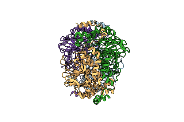 |
The Mutant Variant Of Pngm-1. H279A Was Substituted For Alanine To Study Metal Coordination.
Organism: Uncultured bacterium
Method: X-RAY DIFFRACTION Resolution:1.96 Å Release Date: 2021-04-28 Classification: ANTIBIOTIC Ligands: ZN |
Organism: Uncultured bacterium
Method: X-RAY DIFFRACTION
Release Date: 2021-04-28
Ligands: ZN
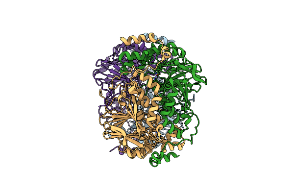 |
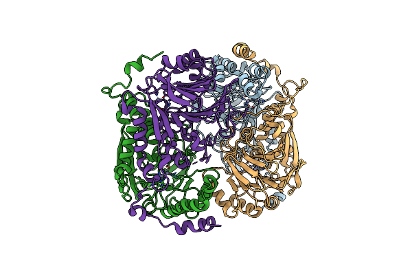
The Mutant Variant Of Pngm-1. H96 Was Substituted For Alanine To Study Metal Coordination.
Organism: Uncultured bacterium
Method: X-RAY DIFFRACTION Resolution:2.45 Å Release Date: 2021-04-28 Classification: ANTIBIOTIC Ligands: ZN |
Organism: Uncultured bacterium
Method: X-RAY DIFFRACTION
Release Date: 2021-04-28
Ligands: ZN
 |
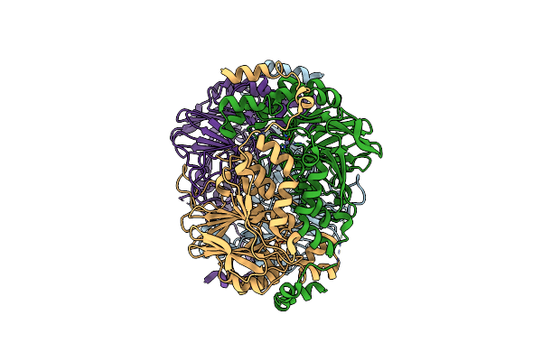
The Mutant Variant Of Pngm-1. H257 Was Substituted For Alanine To Study Substrate Binding.
Organism: Uncultured bacterium
Method: X-RAY DIFFRACTION Resolution:2.00 Å Release Date: 2021-04-28 Classification: ANTIBIOTIC Ligands: ZN |
Organism: Uncultured bacterium
Method: X-RAY DIFFRACTION
Release Date: 2021-04-28
Ligands: ZN
 |
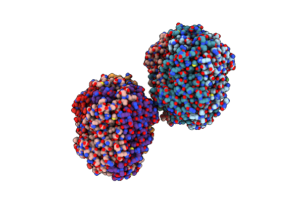
The Mutant Variant Of Pngm-1. H279 Was Substituted For Alanine To Study Metal Coordination.
Organism: Uncultured bacterium
Method: X-RAY DIFFRACTION Resolution:2.16 Å Release Date: 2021-04-28 Classification: ANTIBIOTIC Ligands: ZN |
Organism: Uncultured bacterium
Method: X-RAY DIFFRACTION
Release Date: 2021-04-28
Ligands: ZN
 |
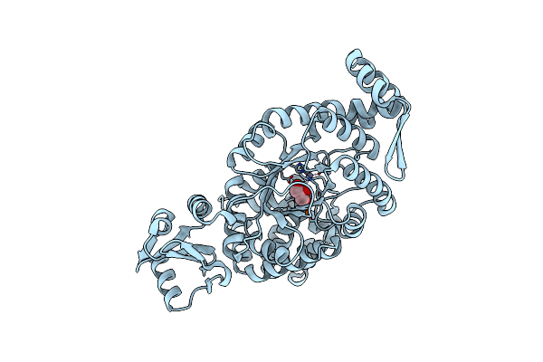
The Mutant Variant Of Pngm-1. H91 Was Substituted For Alanine To Study Metal Coordination.
Organism: Uncultured bacterium
Method: X-RAY DIFFRACTION Resolution:1.94 Å Release Date: 2021-04-28 Classification: ANTIBIOTIC Ligands: ZN |
Organism: Uncultured bacterium
Method: X-RAY DIFFRACTION
Release Date: 2021-04-28
Ligands: ZN
 |
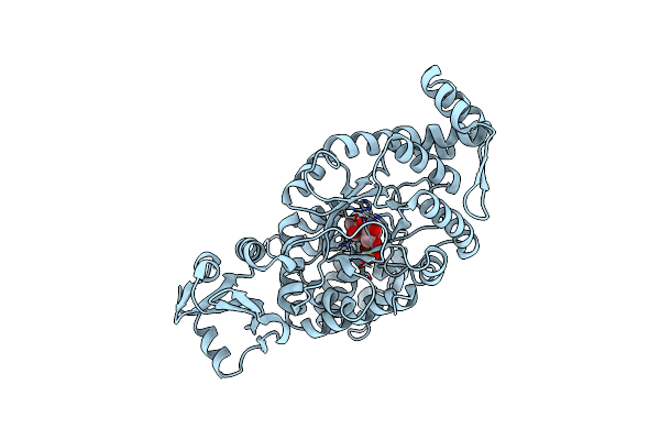
One Glycerol Complexed Crystal Structure Of Fructuronate-Tagaturonate Epimerase Uxae From Cohnella Laeviribosi
Organism: Cohnella laeviribosi
Method: X-RAY DIFFRACTION Resolution:1.72 Å Release Date: 2019-10-23 Classification: ISOMERASE Ligands: GOL, ZN |
Organism: Cohnella laeviribosi
Method: X-RAY DIFFRACTION
Release Date: 2019-10-23
Ligands: GOL, ZN
 |
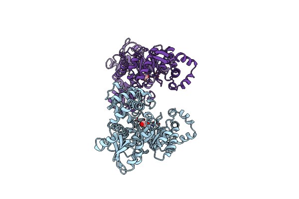
Two Glycerol Complexed Crystal Structure Of Fructuronate-Tagaturonate Epimerase Uxae From Cohnella Laeviribosi
Organism: Cohnella laeviribosi
Method: X-RAY DIFFRACTION Resolution:2.69 Å Release Date: 2019-10-23 Classification: ISOMERASE Ligands: GOL, ZN, PO4 |
Organism: Cohnella laeviribosi
Method: X-RAY DIFFRACTION
Release Date: 2019-10-23
Ligands: GOL, ZN, PO4
 |
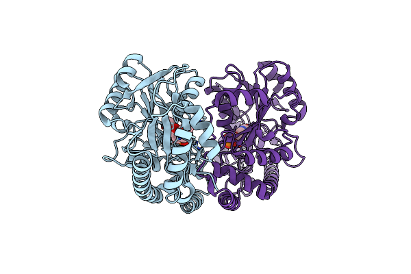
Native Crystal Structure Of Fructuronate-Tagaturonate Epimerase Uxae From Cohnella Laeviribosi
Organism: Cohnella laeviribosi
Method: X-RAY DIFFRACTION Resolution:2.51 Å Release Date: 2019-10-23 Classification: ISOMERASE Ligands: EDO, ZN |
Organism: Cohnella laeviribosi
Method: X-RAY DIFFRACTION
Release Date: 2019-10-23
Ligands: EDO, ZN
 |
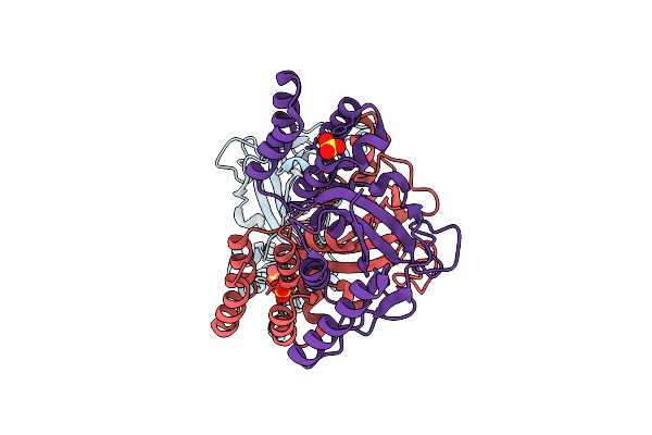
Crystal Structure Of The C-Terminal Domain Of Bacillus Subtilis Gabr Reveals A Closed Conformation By The Binding Of Gamma-Aminobutyric Acid, Inducing The Transcriptional Activation
Organism: Bacillus subtilis (strain 168)
Method: X-RAY DIFFRACTION Resolution:2.00 Å Release Date: 2017-05-24 Classification: TRANSCRIPTION Ligands: PLP, ABU |
Organism: Bacillus subtilis (strain 168)
Method: X-RAY DIFFRACTION
Release Date: 2017-05-24
Ligands: PLP, ABU
 |
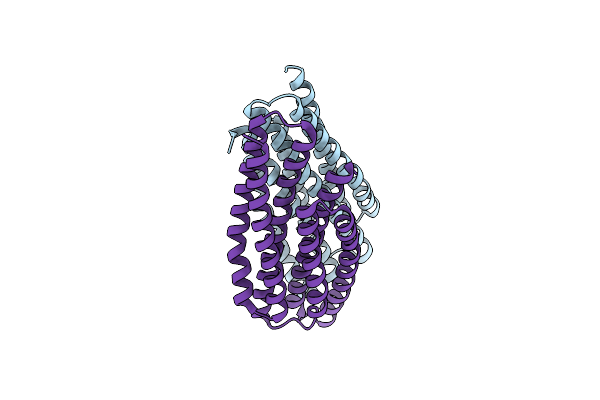
Induced Dimer Structure Of Methionine Sulfoxide Reductase U16C From Clostridium Oremlandii
Organism: Alkaliphilus oremlandii ohilas
Method: X-RAY DIFFRACTION Resolution:2.90 Å Release Date: 2015-07-15 Classification: OXIDOREDUCTASE Ligands: SO4 |
Organism: Alkaliphilus oremlandii ohilas
Method: X-RAY DIFFRACTION
Release Date: 2015-07-15
Ligands: SO4
 |
Crystal Structure Of Phou From Pseudomonas Aeruginosa
Organism: Pseudomonas aeruginosa
Method: X-RAY DIFFRACTION Resolution:2.28 Å Release Date: 2015-02-18 Classification: UNKNOWN FUNCTION |
Organism: Pseudomonas aeruginosa
Method: X-RAY DIFFRACTION
Release Date: 2015-02-18
 |
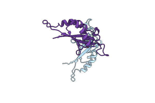
Structure Of 6-Pyruvoyltetrahydropterin Synthase
Organism: Escherichia coli
Method: X-RAY DIFFRACTION Resolution:2.34 Å Release Date: 2011-12-07 Classification: LYASE Ligands: ZN |
Organism: Escherichia coli
Method: X-RAY DIFFRACTION
Release Date: 2011-12-07
Ligands: ZN
 |
Crystal Structure Of A 6-Pyruvoyltetrahydropterin Synthase Homologue From Esherichia Coli
Organism: Escherichia coli
Method: X-RAY DIFFRACTION Resolution:2.93 Å Release Date: 2011-12-07 Classification: LYASE Ligands: ZN |
Organism: Escherichia coli
Method: X-RAY DIFFRACTION
Release Date: 2011-12-07
Ligands: ZN