

Search Count: 17
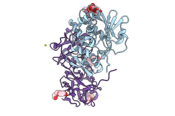 |
The Structure Of The Monoclinic Crystal Form Of The Type Ii Ribosome Inactivating Protein From Winter Aconite Eranthis Hyemalis.
Organism: Eranthis hyemalis
Method: X-RAY DIFFRACTION Resolution:1.50 Å Release Date: 2026-01-21 Classification: TOXIN Ligands: NAG, FUC, PG4, MG |
Organism: Eranthis hyemalis
Method: X-RAY DIFFRACTION
Release Date: 2026-01-21
Ligands: NAG, FUC, PG4, MG
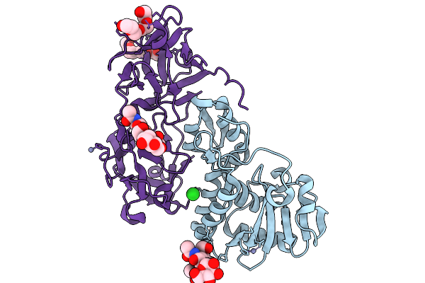 |
The Structure Of The Orthorhombic Crystal Form Of The Type Ii Ribosome Inactivating Protein From Winter Aconite Eranthis Hyemalis.
Organism: Eranthis hyemalis
Method: X-RAY DIFFRACTION Resolution:1.80 Å Release Date: 2026-01-21 Classification: TOXIN Ligands: NAG, MAN, FUC, ZN, CL, PG4, AH2 |
Organism: Eranthis hyemalis
Method: X-RAY DIFFRACTION
Release Date: 2026-01-21
Ligands: NAG, MAN, FUC, ZN, CL, PG4, AH2
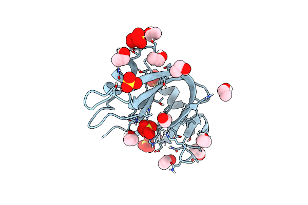 |
Ultra High Resolution X-Ray Structure Of Orthorhombic Bovine Pancreatic Ribonuclease At 100K
Organism: Bos taurus
Method: X-RAY DIFFRACTION Resolution:0.85 Å Release Date: 2022-07-27 Classification: HYDROLASE Ligands: EOH, SO4 |
Organism: Bos taurus
Method: X-RAY DIFFRACTION
Release Date: 2022-07-27
Ligands: EOH, SO4
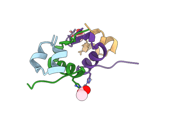 |
X-Ray Structure Of Human Recombinant 2Zn Insulin At 0.92 Angstrom
Organism: Homo sapiens
Method: X-RAY DIFFRACTION Resolution:0.95 Å Release Date: 2016-10-12 Classification: IMMUNE SYSTEM Ligands: ZN, ACT, POL |
Organism: Homo sapiens
Method: X-RAY DIFFRACTION
Release Date: 2016-10-12
Ligands: ZN, ACT, POL
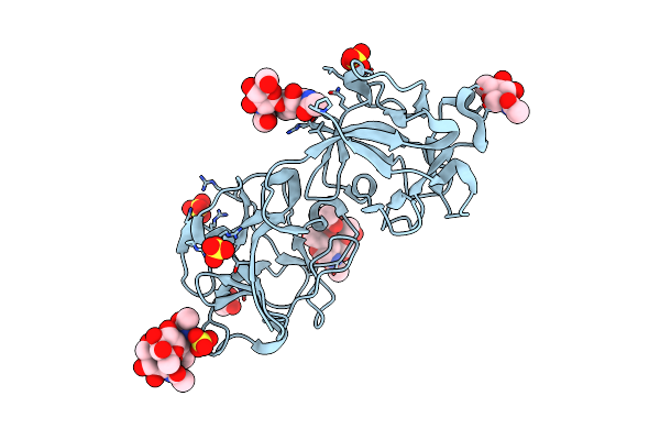 |
Sambucus Nigra Agglutinin Ii (Sna-Ii), Tetragonal Crystal Form
Organism: Sambucus nigra
Method: X-RAY DIFFRACTION Resolution:1.35 Å Release Date: 2008-11-25 Classification: Sugar binding protein, Plant protein Ligands: NAG, SO4, ACT |
Organism: Sambucus nigra
Method: X-RAY DIFFRACTION
Release Date: 2008-11-25
Ligands: NAG, SO4, ACT
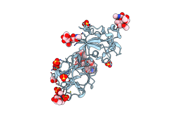 |
Sambucus Nigra Agglutinin Ii (Sna-Ii), Hexagonal Crystal Form
Organism: Sambucus nigra
Method: X-RAY DIFFRACTION Resolution:1.95 Å Release Date: 2008-11-25 Classification: SUGAR BINDING PROTEIN, Plant protein Ligands: SO4, ACT |
Organism: Sambucus nigra
Method: X-RAY DIFFRACTION
Release Date: 2008-11-25
Ligands: SO4, ACT
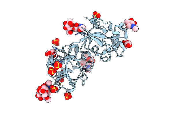 |
Sambucus Nigra Agglutinin Ii (Sna-Ii)- Tetragonal Crystal Form- Complexed To Galactose
Organism: Sambucus nigra
Method: X-RAY DIFFRACTION Resolution:1.55 Å Release Date: 2008-11-25 Classification: Sugar binding protein, Plant protein Ligands: NAG, GLA, SO4, ACT |
Organism: Sambucus nigra
Method: X-RAY DIFFRACTION
Release Date: 2008-11-25
Ligands: NAG, GLA, SO4, ACT
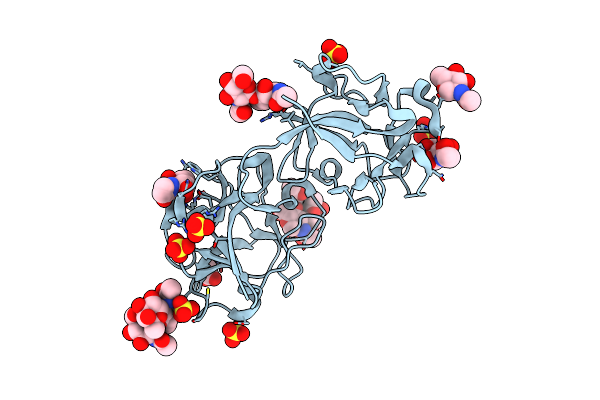 |
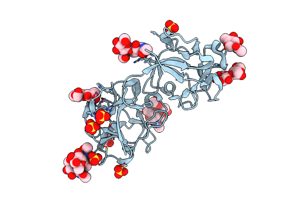
Crystal Structure Of Sambucus Nigra Agglutinin Ii (Sna-Ii)-Tetragonal Crystal Form- Complexed To N-Acetylgalactosamine
Organism: Sambucus nigra
Method: X-RAY DIFFRACTION Resolution:1.55 Å Release Date: 2008-11-25 Classification: SUGAR BINDING PROTEIN, Plant protein Ligands: NAG, A2G, NGA, SO4, ACT |
Organism: Sambucus nigra
Method: X-RAY DIFFRACTION
Release Date: 2008-11-25
Ligands: NAG, A2G, NGA, SO4, ACT
 |
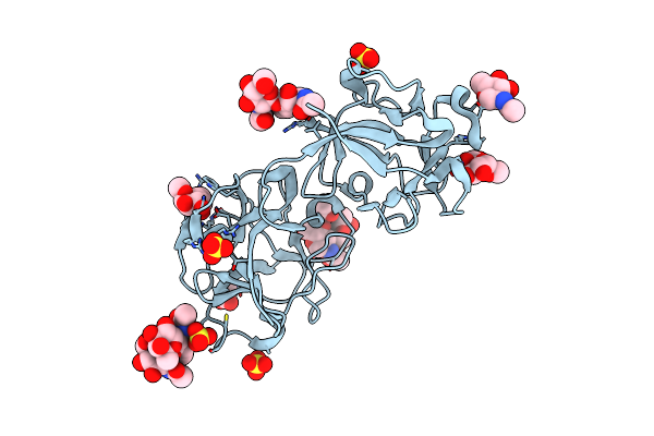
Sambucus Nigra Agglutinin Ii, Tetragonal Crystal Form- Complexed To Lactose
Organism: Sambucus nigra
Method: X-RAY DIFFRACTION Resolution:1.55 Å Release Date: 2008-11-25 Classification: SUGAR BINDING PROTEIN, Plant Protein Ligands: NAG, SO4, ACT |
Organism: Sambucus nigra
Method: X-RAY DIFFRACTION
Release Date: 2008-11-25
Ligands: NAG, SO4, ACT
 |
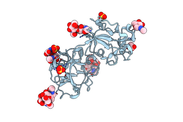
Crystal Structure Of Sambucus Nigra Agglutinin Ii (Sna-Ii)-Tetragonal Crystal Form- Complexed To Alpha1 Methylgalactose
Organism: Sambucus nigra
Method: X-RAY DIFFRACTION Resolution:1.55 Å Release Date: 2008-11-25 Classification: SUGAR BINDING PROTEIN, Plant protein Ligands: NAG, AMG, SO4, ACT |
Organism: Sambucus nigra
Method: X-RAY DIFFRACTION
Release Date: 2008-11-25
Ligands: NAG, AMG, SO4, ACT
 |
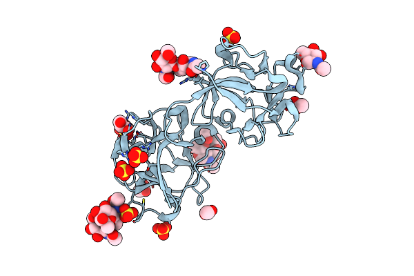
Sambucus Nigra Agglutinin Ii (Sna-Ii)- Tetragonal Crystal Form- Complexed To Tn Antigen
Organism: Sambucus nigra
Method: X-RAY DIFFRACTION Resolution:1.40 Å Release Date: 2008-11-25 Classification: SUGAR BINDING PROTEIN, Plant Protein Ligands: NAG, A2G, SO4, ACT, SER |
Organism: Sambucus nigra
Method: X-RAY DIFFRACTION
Release Date: 2008-11-25
Ligands: NAG, A2G, SO4, ACT, SER
 |
Sambucus Nigra Aggutinin Ii. Tetragonal Crystal Form- Complexed To Fucose
Organism: Sambucus nigra
Method: X-RAY DIFFRACTION Resolution:1.55 Å Release Date: 2008-11-25 Classification: SUGAR BINDING PROTEIN, Plant Protein Ligands: NAG, FCA, SO4, ACT |
Organism: Sambucus nigra
Method: X-RAY DIFFRACTION
Release Date: 2008-11-25
Ligands: NAG, FCA, SO4, ACT
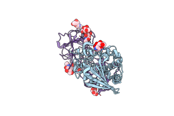 |
Mistletoe Lectin I From Viscum Album Complexed With Galactose
Organism: Viscum album
Method: X-RAY DIFFRACTION Resolution:3.00 Å Release Date: 2003-07-01 Classification: HYDROLASE/SUGAR BINDING PROTEIN Ligands: NAG, GAL |
Organism: Viscum album
Method: X-RAY DIFFRACTION
Release Date: 2003-07-01
Ligands: NAG, GAL
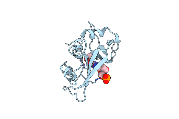 |
Structure Of The Crystalline Complex Of Deoxycytidylyl-3',5'-Guanosine (3',5'-Dcpdg) Co-Crystalised With Ribonuclease At 1.9 Angstroms Resolution. Retrobinding In Pancreatic Rnasea Is Independent Of Mode Of Inhibitor Intromission
Organism: Bos taurus
Method: X-RAY DIFFRACTION Resolution:1.90 Å Release Date: 1995-09-15 Classification: HYDROLASE (ENDORIBONUCLEASE) Ligands: PO4, CGP |
Organism: Bos taurus
Method: X-RAY DIFFRACTION
Release Date: 1995-09-15
Ligands: PO4, CGP
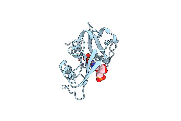 |
Newly Observed Binding Mode In Pancreatic Ribonuclease
Organism: Bos taurus
Method: X-RAY DIFFRACTION Resolution:1.50 Å Release Date: 1994-01-31 Classification: HYDROLASE(ENDORIBONUCLEASE) Ligands: SO4, 5GP |
Organism: Bos taurus
Method: X-RAY DIFFRACTION
Release Date: 1994-01-31
Ligands: SO4, 5GP
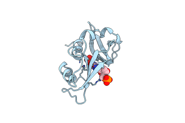 |
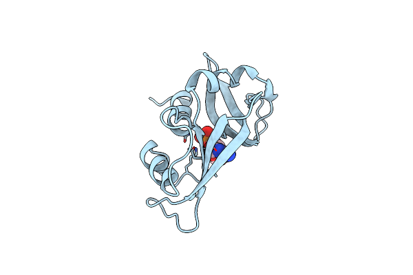
Newly Observed Binding Mode In Pancreatic Ribonuclease
Organism: Bos taurus
Method: X-RAY DIFFRACTION Resolution:1.50 Å Release Date: 1994-01-31 Classification: HYDROLASE(ENDORIBONUCLEASE) Ligands: SO4, DGP |
Organism: Bos taurus
Method: X-RAY DIFFRACTION
Release Date: 1994-01-31
Ligands: SO4, DGP
 |
Structure Of The Crystalline Complex Of Cytidylic Acid (2'-Cmp) With Ribonuclease At 1.6 Angstroms Resolution
Organism: Bos taurus
Method: X-RAY DIFFRACTION Resolution:1.60 Å Release Date: 1994-01-31 Classification: HYDROLASE(ENDORIBONUCLEASE) Ligands: C2P |
Organism: Bos taurus
Method: X-RAY DIFFRACTION
Release Date: 1994-01-31
Ligands: C2P