

Search Count: 235
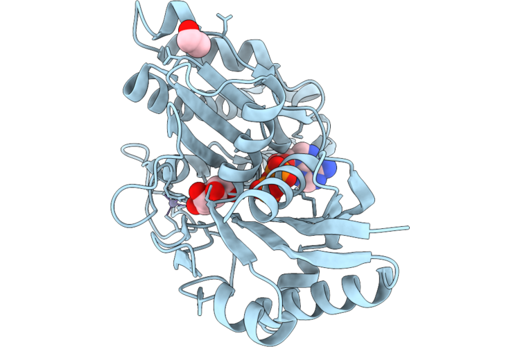 |
X-Ray Structure Of Paenibacillus Kribbensis D-Allose Kinase
Organism: Paenibacillus kribbensis
Method: X-RAY DIFFRACTION Resolution:1.58 Å Release Date: 2026-09-23 Classification: TRANSFERASE Ligands: ZN, ALL, ADP, EDO |
Organism: Paenibacillus kribbensis
Method: X-RAY DIFFRACTION
Release Date: 2026-09-23
Ligands: ZN, ALL, ADP, EDO
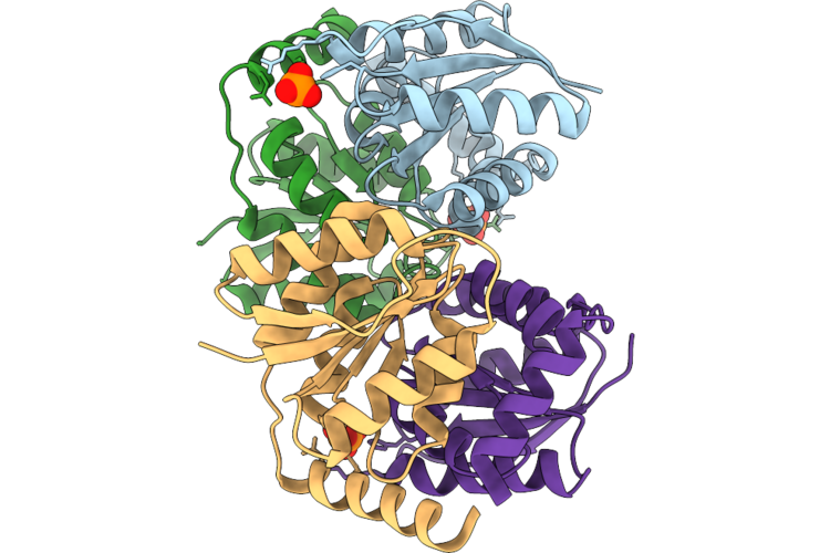 |
X-Ray Structure Of Paenibacillus Kribbensis D-Ribose-5-Phosphate Isomerase B
Organism: Paenibacillus kribbensis
Method: X-RAY DIFFRACTION Resolution:1.94 Å Release Date: 2026-09-23 Classification: ISOMERASE Ligands: PO4 |
Organism: Paenibacillus kribbensis
Method: X-RAY DIFFRACTION
Release Date: 2026-09-23
Ligands: PO4
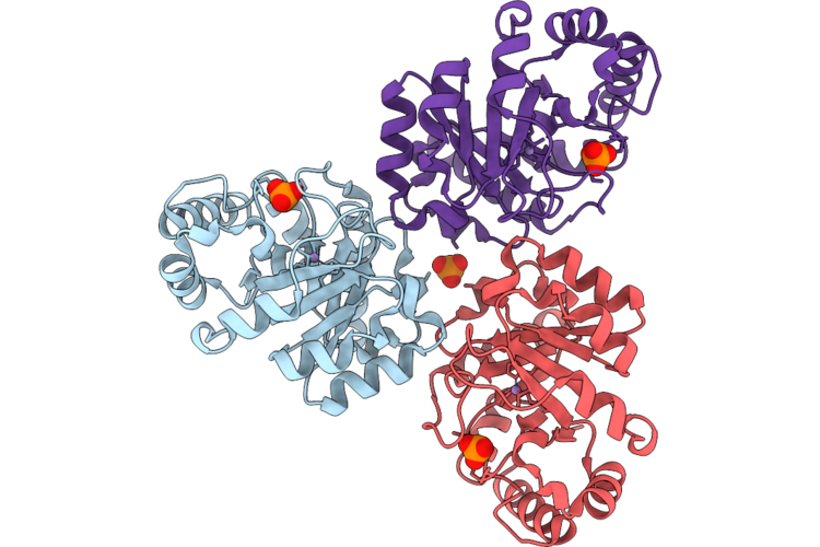 |
X-Ray Structure Of Paenibacillus Kribbensis D-Allulose-6-Phosphate 3-Epimerase
Organism: Paenibacillus kribbensis
Method: X-RAY DIFFRACTION Resolution:1.46 Å Release Date: 2026-09-23 Classification: ISOMERASE Ligands: MN, PO4 |
Organism: Paenibacillus kribbensis
Method: X-RAY DIFFRACTION
Release Date: 2026-09-23
Ligands: MN, PO4
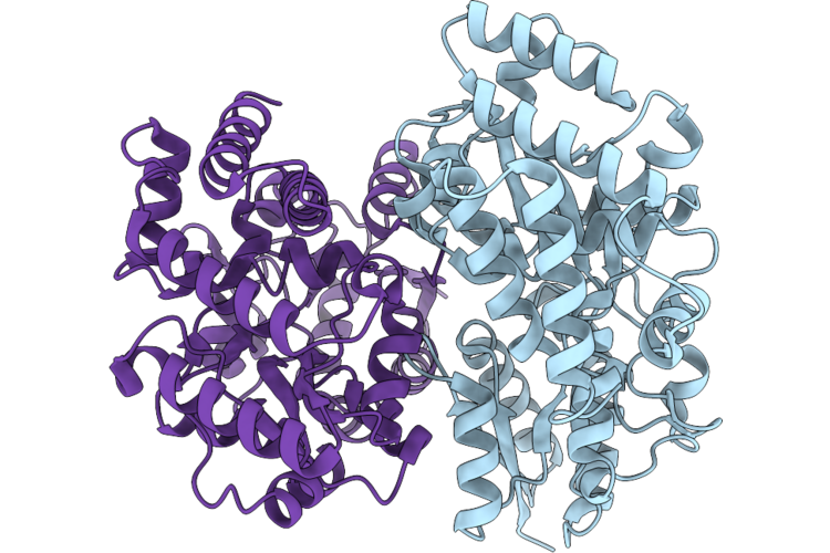 |
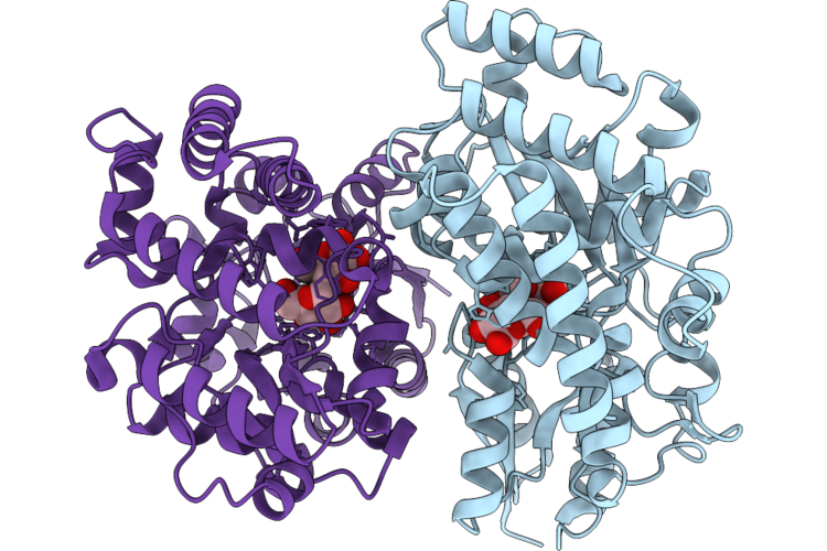
Paenibacillus Maltose-Binding Protein
Organism: Paenibacillus sp. fpu-7
Method: X-RAY DIFFRACTION Resolution:1.44 Å Release Date: 2026-07-15 Classification: SUGAR BINDING PROTEIN |
Organism: Paenibacillus sp. fpu-7
Method: X-RAY DIFFRACTION
Release Date: 2026-07-15
 |
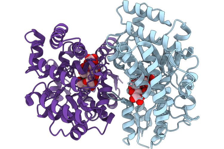
Maltose-Binding Protein
Organism: Paenibacillus sp. fpu-7
Method: X-RAY DIFFRACTION Resolution:1.70 Å Release Date: 2026-07-15 Classification: SUGAR BINDING PROTEIN |
Organism: Paenibacillus sp. fpu-7
Method: X-RAY DIFFRACTION
Release Date: 2026-07-15
 |
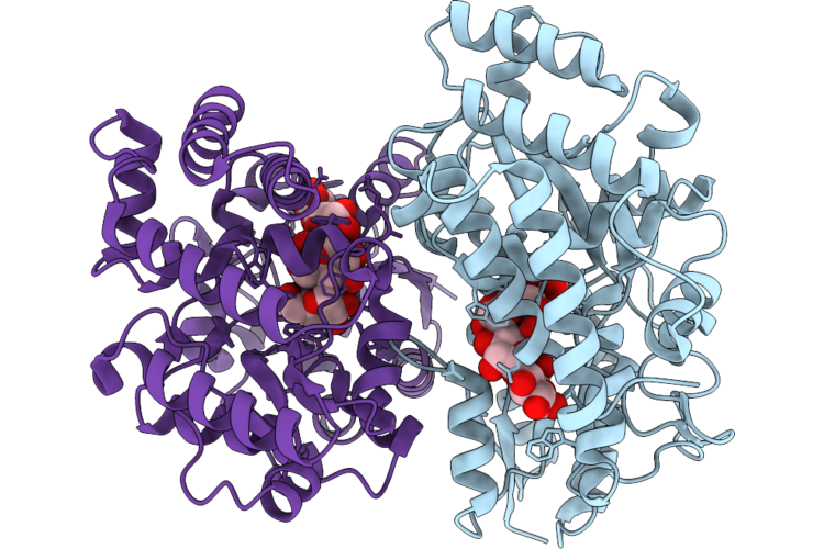
Maltose-Binding Protein
Organism: Paenibacillus sp. fpu-7
Method: X-RAY DIFFRACTION Resolution:1.60 Å Release Date: 2026-07-15 Classification: SUGAR BINDING PROTEIN |
Organism: Paenibacillus sp. fpu-7
Method: X-RAY DIFFRACTION
Release Date: 2026-07-15
 |
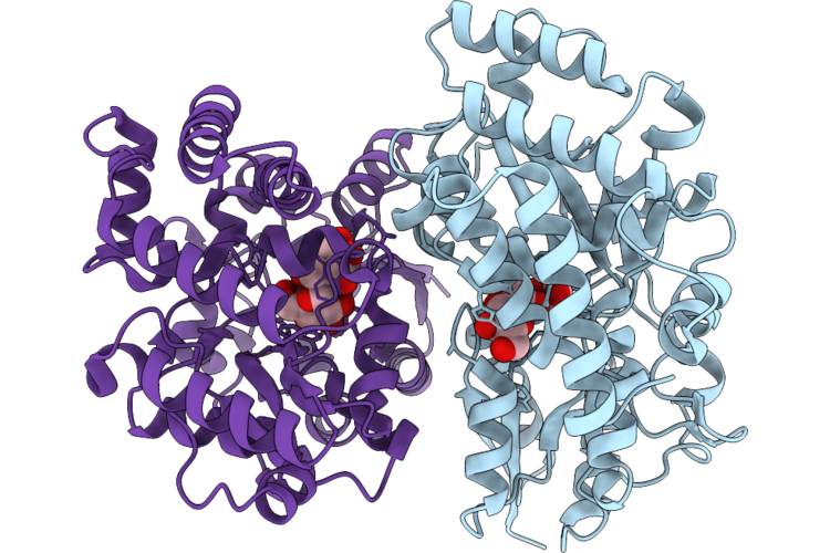
Maltose-Binding Protein
Organism: Paenibacillus sp. fpu-7
Method: X-RAY DIFFRACTION Resolution:1.50 Å Release Date: 2026-07-15 Classification: SUGAR BINDING PROTEIN |
Organism: Paenibacillus sp. fpu-7
Method: X-RAY DIFFRACTION
Release Date: 2026-07-15
 |
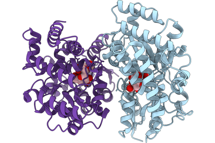
Maltose-Binding Protein
Organism: Paenibacillus sp. fpu-7
Method: X-RAY DIFFRACTION Resolution:1.92 Å Release Date: 2026-07-15 Classification: SUGAR BINDING PROTEIN |
Organism: Paenibacillus sp. fpu-7
Method: X-RAY DIFFRACTION
Release Date: 2026-07-15
 |
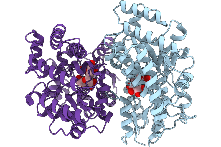
Maltose-Binding Protein
Organism: Paenibacillus sp. fpu-7
Method: X-RAY DIFFRACTION Resolution:1.93 Å Release Date: 2026-07-15 Classification: SUGAR BINDING PROTEIN |
Organism: Paenibacillus sp. fpu-7
Method: X-RAY DIFFRACTION
Release Date: 2026-07-15
 |
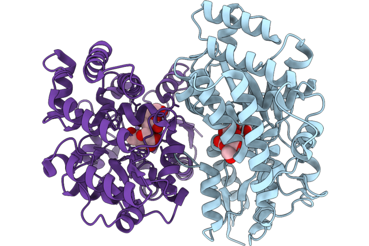
Maltose-Binding Protein
Organism: Paenibacillus sp. fpu-7
Method: X-RAY DIFFRACTION Resolution:1.35 Å Release Date: 2026-07-15 Classification: SUGAR BINDING PROTEIN |
Organism: Paenibacillus sp. fpu-7
Method: X-RAY DIFFRACTION
Release Date: 2026-07-15
 |
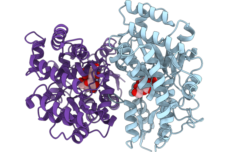
Maltose-Binding Protein
Organism: Paenibacillus sp. fpu-7
Method: X-RAY DIFFRACTION Resolution:2.10 Å Release Date: 2026-07-15 Classification: SUGAR BINDING PROTEIN |
Organism: Paenibacillus sp. fpu-7
Method: X-RAY DIFFRACTION
Release Date: 2026-07-15
 |
Maltose-Binding Protein
Organism: Paenibacillus sp. fpu-7
Method: X-RAY DIFFRACTION Resolution:2.10 Å Release Date: 2026-07-15 Classification: SUGAR BINDING PROTEIN |
Organism: Paenibacillus sp. fpu-7
Method: X-RAY DIFFRACTION
Release Date: 2026-07-15
 |
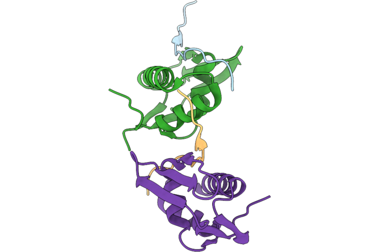
Lppb In Complex With Lppa Leader Peptide
Organism: Paenibacillus alginolyticus
Method: X-RAY DIFFRACTION Resolution:2.10 Å Release Date: 2026-05-27 Classification: PEPTIDE BINDING PROTEIN |
Organism: Paenibacillus alginolyticus
Method: X-RAY DIFFRACTION
Release Date: 2026-05-27
 |
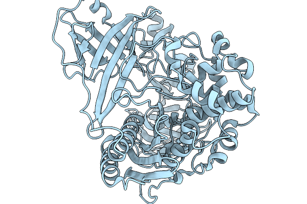
Pbab1 In Complex With Pbaa Leader Peptide
Organism: Paenibacillus alginolyticus
Method: X-RAY DIFFRACTION Resolution:2.00 Å Release Date: 2026-05-27 Classification: PEPTIDE BINDING PROTEIN |
Organism: Paenibacillus alginolyticus
Method: X-RAY DIFFRACTION
Release Date: 2026-05-27
 |
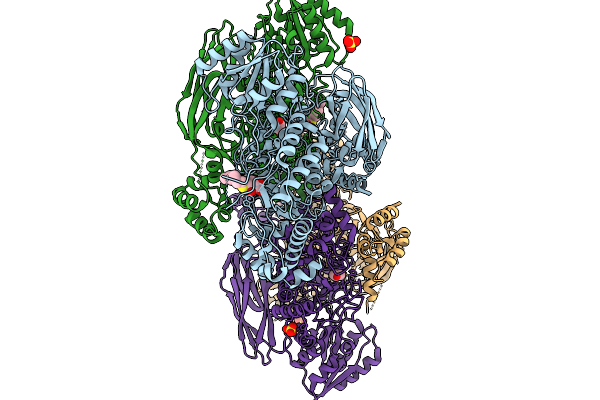
Lip3 Dl-Peptidase In The Apo-State
Organism: Paenibacillus
Method: X-RAY DIFFRACTION Resolution:2.04 Å Release Date: 2026-02-18 Classification: HYDROLASE |
Organism: Paenibacillus
Method: X-RAY DIFFRACTION
Release Date: 2026-02-18
 |
Structure Of Stg-Hydrolyzing Beta-Glucosidase 1 (Pstg1) Complexed With Heptyl 1-Thio-Beta-D-Glucopyranoside
Organism: Paenibacillus relictisesami
Method: X-RAY DIFFRACTION Resolution:3.35 Å Release Date: 2026-02-04 Classification: HYDROLASE Ligands: HTG, SO4, EDO, GOL |
Organism: Paenibacillus relictisesami
Method: X-RAY DIFFRACTION
Release Date: 2026-02-04
Ligands: HTG, SO4, EDO, GOL
 |
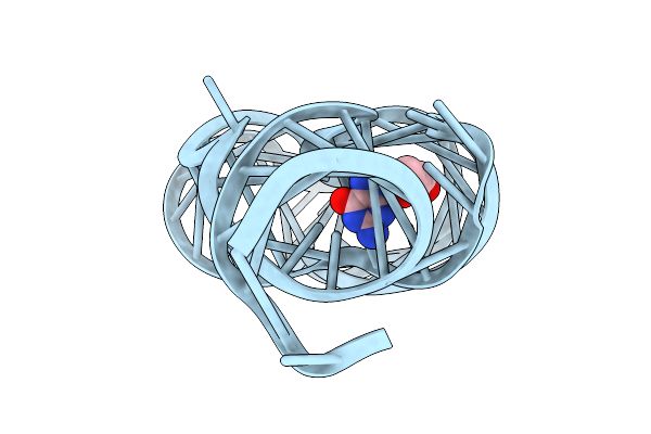
Crystal Structure Of Guanine-Ii Riboswitch In Complex With Guanine
Organism: Paenibacillus sp. fsl e2-0178
Method: X-RAY DIFFRACTION Resolution:2.75 Å Release Date: 2025-11-26 Classification: RNA Ligands: GUN, MG |
Organism: Paenibacillus sp. fsl e2-0178
Method: X-RAY DIFFRACTION
Release Date: 2025-11-26
Ligands: GUN, MG
 |
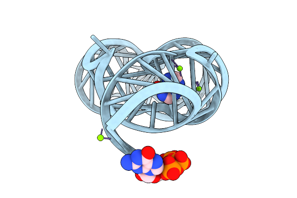
Crystal Structure Of Guanine-Ii Riboswitch In Complex With 2'-Deoxyguanosine
Organism: Paenibacillus sp. fsl e2-0178
Method: X-RAY DIFFRACTION Resolution:2.61 Å Release Date: 2025-11-26 Classification: RNA Ligands: MG, GNG |
Organism: Paenibacillus sp. fsl e2-0178
Method: X-RAY DIFFRACTION
Release Date: 2025-11-26
Ligands: MG, GNG
 |
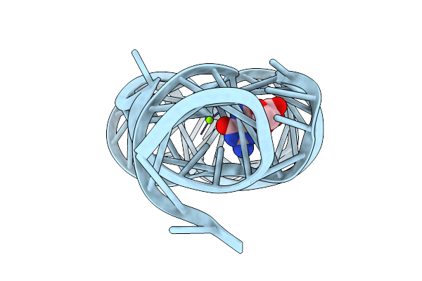
Crystal Structure Of Guanine-Ii Riboswitch In Complex With Hypoxanthine
Organism: Paenibacillus sp. fsl e2-0178
Method: X-RAY DIFFRACTION Resolution:2.26 Å Release Date: 2025-11-26 Classification: RNA Ligands: HPA, GTP, MG |
Organism: Paenibacillus sp. fsl e2-0178
Method: X-RAY DIFFRACTION
Release Date: 2025-11-26
Ligands: HPA, GTP, MG
 |
Crystal Structure Of Guanine-Ii Riboswitch In Complex With Guanosine
Organism: Paenibacillus sp. fsl e2-0178
Method: X-RAY DIFFRACTION Resolution:2.83 Å Release Date: 2025-11-26 Classification: RNA Ligands: GMP, MG |
Organism: Paenibacillus sp. fsl e2-0178
Method: X-RAY DIFFRACTION
Release Date: 2025-11-26
Ligands: GMP, MG