

Search Count: 618
 |
Parm From R1 Plasmid In Complex With Peptide From C-Terminus Of Parr
Organism: Escherichia coli
Method: X-RAY DIFFRACTION Resolution:2.20 Å Release Date: 2012-11-07 Classification: TRANSPORT PROTEIN Ligands: ANP, MG |
Organism: Escherichia coli
Method: X-RAY DIFFRACTION
Release Date: 2012-11-07
Ligands: ANP, MG
 |
Parm From Plasmid R1 In Complex With Amppnp
Organism: Escherichia coli
Method: X-RAY DIFFRACTION Resolution:2.00 Å Release Date: 2012-11-07 Classification: TRANSPORT PROTEIN Ligands: ANP, MG |
Organism: Escherichia coli
Method: X-RAY DIFFRACTION
Release Date: 2012-11-07
Ligands: ANP, MG
 |
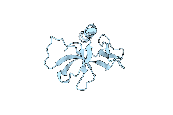
Ccdb, A Topoisomerase Poison From E. Coli
Organism: Escherichia coli
Method: X-RAY DIFFRACTION Resolution:2.60 Å Release Date: 1998-07-15 Classification: PLASMID Ligands: CL |
Organism: Escherichia coli
Method: X-RAY DIFFRACTION
Release Date: 1998-07-15
Ligands: CL
 |
Ccdb, A Topoisomerase Poison From Escherichia Coli
Organism: Escherichia coli
Method: X-RAY DIFFRACTION Resolution:1.45 Å Release Date: 1998-10-14 Classification: PLASMID Ligands: CL |
Organism: Escherichia coli
Method: X-RAY DIFFRACTION
Release Date: 1998-10-14
Ligands: CL
 |
Structure Of Sopb(155-323)-18Mer Dna Complex, I23 Form
Organism: Escherichia coli
Method: X-RAY DIFFRACTION Resolution:2.86 Å Release Date: 2010-05-05 Classification: DNA BINDING PROTEIN/DNA Ligands: SO4 |
Organism: Escherichia coli
Method: X-RAY DIFFRACTION
Release Date: 2010-05-05
Ligands: SO4
 |
Mobm Relaxase Domain (Mobv; Mob_Pre) Bound To 26Nt Pmv158 Orit Dna
Organism: Streptococcus agalactiae, Plasmid pmv158
Method: X-RAY DIFFRACTION Resolution:2.00 Å Release Date: 2017-04-12 Classification: DNA BINDING PROTEIN Ligands: CL, MG, GOL, NA Modified Residue(s): MSE |
Organism: Streptococcus agalactiae, Plasmid pmv158
Method: X-RAY DIFFRACTION
Release Date: 2017-04-12
Ligands: CL, MG, GOL, NA
 |
Structure Of Sopb(155-272)-18Mer Complex, P21 Form
Organism: Escherichia coli
Method: X-RAY DIFFRACTION Resolution:2.98 Å Release Date: 2010-05-05 Classification: DNA-BINDING PROTEIN/DNA Ligands: CA |
Organism: Escherichia coli
Method: X-RAY DIFFRACTION
Release Date: 2010-05-05
Ligands: CA
 |
Structure Of Sopb(155-272)-18Mer Complex, I23 Form
Organism: Escherichia coli
Method: X-RAY DIFFRACTION Resolution:2.99 Å Release Date: 2010-05-05 Classification: DNA BINDING PROTEIN/DNA Ligands: SO4 |
Organism: Escherichia coli
Method: X-RAY DIFFRACTION
Release Date: 2010-05-05
Ligands: SO4
 |
Crystal Structure Of A Plasmid Partition Protein From Borrelia Burgdorferi At 2.25A Resolution, Iodide Soak
Organism: Borrelia burgdorferi
Method: X-RAY DIFFRACTION Resolution:2.25 Å Release Date: 2009-11-03 Classification: BIOSYNTHETIC PROTEIN |
Organism: Borrelia burgdorferi
Method: X-RAY DIFFRACTION
Release Date: 2009-11-03
 |
X-Ray Crystal Structure Of The Passenger Domain Of Plasmid Encoded Toxin, An Autrotansporter Enterotoxin From Enteroaggregative Escherichia Coli (Eaec)
Organism: Escherichia coli
Method: X-RAY DIFFRACTION Resolution:2.30 Å Release Date: 2014-03-12 Classification: HYDROLASE |
Organism: Escherichia coli
Method: X-RAY DIFFRACTION
Release Date: 2014-03-12
 |
Chromosomal Parde Ta System From P. Aeruginosa
Organism: Pseudomonas aeruginosa
Method: X-RAY DIFFRACTION Resolution:1.77 Å Release Date: 2021-07-28 Classification: TOXIN/ANTITOXIN Ligands: GOL, 2PE, BEN |
Organism: Pseudomonas aeruginosa
Method: X-RAY DIFFRACTION
Release Date: 2021-07-28
Ligands: GOL, 2PE, BEN
 |
Parr From Plasmid Pb171
Organism: Escherichia coli
Method: X-RAY DIFFRACTION Resolution:2.80 Å Release Date: 2007-10-09 Classification: DNA BINDING PROTEIN |
Organism: Escherichia coli
Method: X-RAY DIFFRACTION
Release Date: 2007-10-09
 |
Plasmid Partitioning Protein Tubr From Bacillus Cereus
Organism: Bacillus cereus
Method: X-RAY DIFFRACTION Resolution:2.00 Å Release Date: 2019-06-26 Classification: TRANSCRIPTION Modified Residue(s): MSE |
Organism: Bacillus cereus
Method: X-RAY DIFFRACTION
Release Date: 2019-06-26
 |
Crystal Structure Of The Paaa2-Pare2 Antitoxin-Toxin Complex
Organism: Escherichia coli o157
Method: X-RAY DIFFRACTION Resolution:2.67 Å Release Date: 2016-06-01 Classification: TOXIN Ligands: SO4 |
Organism: Escherichia coli o157
Method: X-RAY DIFFRACTION
Release Date: 2016-06-01
Ligands: SO4
 |
Crystal Structure Of The Paaa2-Pare2 Antitoxin-Toxin Complex
Organism: Escherichia coli o157
Method: X-RAY DIFFRACTION Resolution:2.83 Å Release Date: 2016-06-01 Classification: TOXIN Ligands: GOL Modified Residue(s): MSE |
Organism: Escherichia coli o157
Method: X-RAY DIFFRACTION
Release Date: 2016-06-01
Ligands: GOL
 |
Crystal Structure Of The Paaa2-Pare2 Antitoxin-Toxin Complex
Organism: Escherichia coli o157:h7 str. ss52
Method: X-RAY DIFFRACTION Resolution:3.82 Å Release Date: 2016-08-24 Classification: TOXIN Modified Residue(s): MSE |
Organism: Escherichia coli o157:h7 str. ss52
Method: X-RAY DIFFRACTION
Release Date: 2016-08-24
 |
Tran, A Repressor Of An Enterococcus Conjugative Type Iv Secretion System
Organism: Enterococcus faecalis, Plasmid pip501
Method: X-RAY DIFFRACTION Resolution:1.93 Å Release Date: 2018-07-25 Classification: DNA BINDING PROTEIN Ligands: EPE |
Organism: Enterococcus faecalis, Plasmid pip501
Method: X-RAY DIFFRACTION
Release Date: 2018-07-25
Ligands: EPE
 |
Structure Of Cdomain
Organism: Escherichia coli
Method: X-RAY DIFFRACTION Resolution:1.58 Å Release Date: 2010-03-31 Classification: DNA BINDING PROTEIN Ligands: ACT |
Organism: Escherichia coli
Method: X-RAY DIFFRACTION
Release Date: 2010-03-31
Ligands: ACT
 |
Crystal Structure Of Catalase-Peroxidase Katg Of Burkholderia Pseudomallei Treated With Inh
Organism: Burkholderia pseudomallei (strain 1710b)
Method: X-RAY DIFFRACTION Resolution:1.70 Å Release Date: 2016-08-24 Classification: OXIDOREDUCTASE Ligands: HP5, NA, OXY, MPD |
Organism: Burkholderia pseudomallei (strain 1710b)
Method: X-RAY DIFFRACTION
Release Date: 2016-08-24
Ligands: HP5, NA, OXY, MPD
 |
A 3.3 Angstrom Cryo-Em Structure Of An Engineered High-Affinity Human Prothrombinase Complex
Organism: Homo sapiens
Method: ELECTRON MICROSCOPY Release Date: 2025-11-26 Classification: BLOOD CLOTTING Ligands: CA, NA, 0GJ, CU Modified Residue(s): TYS |
Organism: Homo sapiens
Method: ELECTRON MICROSCOPY
Release Date: 2025-11-26
Ligands: CA, NA, 0GJ, CU