

Search Count: 15
 |
The Crystal Structure Of The Phthalate Hydrolase Estj6 In Complex With Phthalic Acid
Organism: Unidentified
Method: X-RAY DIFFRACTION Resolution:1.90 Å Release Date: 2026-07-08 Classification: HYDROLASE Ligands: PHT |
Organism: Unidentified
Method: X-RAY DIFFRACTION
Release Date: 2026-07-08
Ligands: PHT
 |
Crystal Structure Of Cysteine-Dependent Hydrolase (Csdh) From Rhodococcus Opacus In Complex With Phthalate
Organism: Rhodococcus opacus
Method: X-RAY DIFFRACTION Resolution:2.90 Å Release Date: 2026-06-17 Classification: HYDROLASE Ligands: PHT |
Organism: Rhodococcus opacus
Method: X-RAY DIFFRACTION
Release Date: 2026-06-17
Ligands: PHT
 |
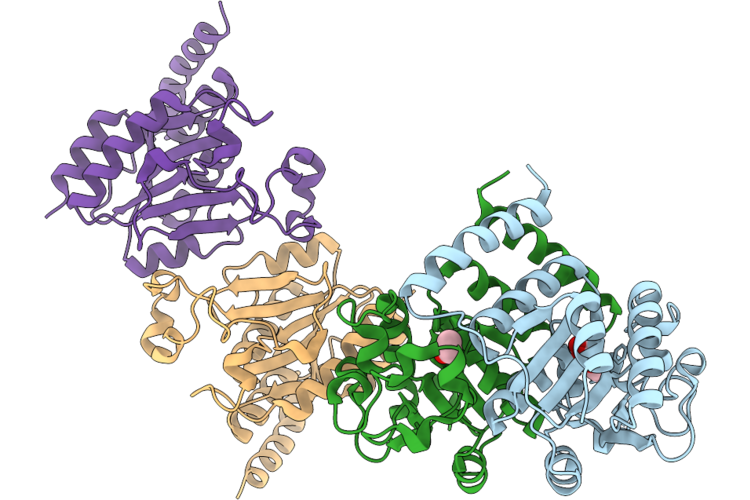
Crystal Structure Of Cysteine-Dependent Hydrolase (Csdh) From Rhodococcus Opacus In Complex With Monobutylphthalate (Mbp)
Organism: Rhodococcus opacus
Method: X-RAY DIFFRACTION Resolution:2.90 Å Release Date: 2026-06-10 Classification: HYDROLASE Ligands: PHT |
Organism: Rhodococcus opacus
Method: X-RAY DIFFRACTION
Release Date: 2026-06-10
Ligands: PHT
 |
Crystal Structure Of Cysteine-Dependent Hydrolase (Csdh) From Rhodococcus Opacus In Complex With Degradation Products Of Diethyl Phthalate
Organism: Rhodococcus opacus
Method: X-RAY DIFFRACTION Resolution:2.90 Å Release Date: 2026-06-10 Classification: HYDROLASE Ligands: PHT, EOH |
Organism: Rhodococcus opacus
Method: X-RAY DIFFRACTION
Release Date: 2026-06-10
Ligands: PHT, EOH
 |
Crystal Structure Of Monoalkyl Phthalate Hydrolase In Complex With Mbp From Rhodococcus Sp. Eg-5
Organism: Rhodococcus sp. eg-5
Method: X-RAY DIFFRACTION Resolution:2.80 Å Release Date: 2026-05-27 Classification: HYDROLASE Ligands: PEG, EDO, PHT, 1BO, MLI |
Organism: Rhodococcus sp. eg-5
Method: X-RAY DIFFRACTION
Release Date: 2026-05-27
Ligands: PEG, EDO, PHT, 1BO, MLI
 |
Ests1 Phthalate Ester Degrading Esterase From Sulfobacillus Acidophilus In Complex With Phthalate
Organism: Sulfobacillus acidophilus dsm 10332
Method: X-RAY DIFFRACTION Resolution:1.50 Å Release Date: 2024-12-25 Classification: HYDROLASE Ligands: EDO, PHT |
Organism: Sulfobacillus acidophilus dsm 10332
Method: X-RAY DIFFRACTION
Release Date: 2024-12-25
Ligands: EDO, PHT
 |
Crystal Structure Of Mehph In Complex With Mbp
Organism: Gordonia sp. p8219
Method: X-RAY DIFFRACTION Resolution:2.80 Å Release Date: 2023-03-15 Classification: HYDROLASE Ligands: 1BO, PHT |
Organism: Gordonia sp. p8219
Method: X-RAY DIFFRACTION
Release Date: 2023-03-15
Ligands: 1BO, PHT
 |
Crystal Structure Of Phthalate Dioxygenase In Complex With Phthalate
Organism: Comamonas testosteroni (strain dsm 14576 / kf-1)
Method: X-RAY DIFFRACTION Resolution:2.74 Å Release Date: 2021-12-15 Classification: OXIDOREDUCTASE Ligands: FE2, FES, PHT |
Organism: Comamonas testosteroni (strain dsm 14576 / kf-1)
Method: X-RAY DIFFRACTION
Release Date: 2021-12-15
Ligands: FE2, FES, PHT
 |
Structure Of The Y21F Variant Of Quinolinate Synthase In Complex With Phthalate
Organism: Thermotoga maritima (strain atcc 43589 / msb8 / dsm 3109 / jcm 10099)
Method: X-RAY DIFFRACTION Resolution:2.00 Å Release Date: 2018-04-25 Classification: TRANSFERASE Ligands: SF4, PHT |
Organism: Thermotoga maritima (strain atcc 43589 / msb8 / dsm 3109 / jcm 10099)
Method: X-RAY DIFFRACTION
Release Date: 2018-04-25
Ligands: SF4, PHT
 |
Pandda Analysis Group Deposition -- Crystal Structure Of Jmjd2D In Complex With N09552A
Organism: Homo sapiens
Method: X-RAY DIFFRACTION Resolution:1.40 Å Release Date: 2017-03-15 Classification: OXIDOREDUCTASE Ligands: ZN, NI, OGA, EDO, SO4, PHT, 1XN |
Organism: Homo sapiens
Method: X-RAY DIFFRACTION
Release Date: 2017-03-15
Ligands: ZN, NI, OGA, EDO, SO4, PHT, 1XN
 |
The Crystal Structure Of Human Quinolinic Acid Phosphoribosyltransferase In Complex With Its Inhibitor Phthalic Acid
Organism: Homo sapiens
Method: X-RAY DIFFRACTION Resolution:2.55 Å Release Date: 2013-10-02 Classification: TRANSFERASE/TRANSFERASE INHIBITOR Ligands: PHT |
Organism: Homo sapiens
Method: X-RAY DIFFRACTION
Release Date: 2013-10-02
Ligands: PHT
 |
Crystal Structure Of The Quinolinate Phosphoribosyl Transferase (Bna6) From Sachharomyces Cerevisiae Complexed With The Inhibitor Phthalate
Organism: Saccharomyces cerevisiae
Method: X-RAY DIFFRACTION Resolution:2.40 Å Release Date: 2008-04-01 Classification: TRANSFERASE Ligands: PHT |
Organism: Saccharomyces cerevisiae
Method: X-RAY DIFFRACTION
Release Date: 2008-04-01
Ligands: PHT
 |
Crystal Structure Of The Quinolinate Phosphoribosyl Transferase (Bna6) From Saccharomyces Cerevisiae Complexed With Prpp And The Inhibitor Phthalate
Organism: Saccharomyces cerevisiae
Method: X-RAY DIFFRACTION Resolution:2.29 Å Release Date: 2008-04-01 Classification: TRANSFERASE Ligands: PRP, PHT |
Organism: Saccharomyces cerevisiae
Method: X-RAY DIFFRACTION
Release Date: 2008-04-01
Ligands: PRP, PHT
 |
Crystal Structure Of Quinolinic Acid Phosphoribosyltransferase From Helicobacter Pylori With Phthalic Acid
Organism: Helicobacter pylori
Method: X-RAY DIFFRACTION Resolution:2.51 Å Release Date: 2006-02-14 Classification: TRANSFERASE Ligands: SO4, PHT |
Organism: Helicobacter pylori
Method: X-RAY DIFFRACTION
Release Date: 2006-02-14
Ligands: SO4, PHT
 |
Quinolinate Phosphoribosyltransferase (Qaprtase) From Mycobacterium Tuberculosis In Complex With Phthalate And Prpcp
Organism: Mycobacterium tuberculosis h37rv
Method: X-RAY DIFFRACTION Resolution:2.45 Å Release Date: 1998-10-21 Classification: TRANSFERASE Ligands: MN, PHT, PPC |
Organism: Mycobacterium tuberculosis h37rv
Method: X-RAY DIFFRACTION
Release Date: 1998-10-21
Ligands: MN, PHT, PPC