
Search Count: 2,354
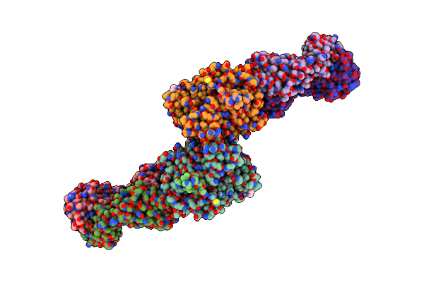 |
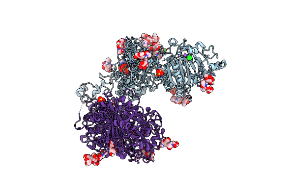
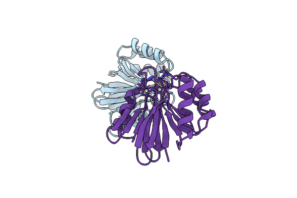
Domain Organization And Conformational Plasticity Of The G Protein Effector, Pde6
Organism: Bos taurus, Mus musculus
Method: ELECTRON MICROSCOPY Release Date: 2015-06-10 Classification: HYDROLASE/IMMUNE SYSTEM Ligands: IBM |
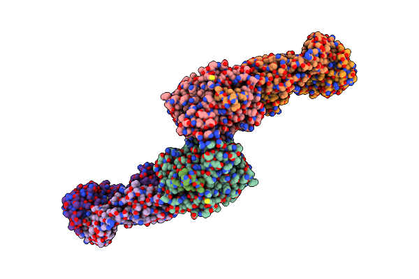 |
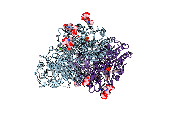
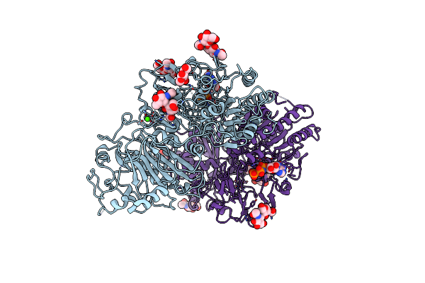
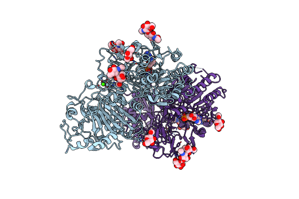
Domain Organization And Conformational Plasticity Of The G Protein Effector, Pde6
Organism: Mus musculus, Bos taurus
Method: ELECTRON MICROSCOPY Release Date: 2015-09-30 Classification: HYDROLASE/IMMUNE SYSTEM |
 |
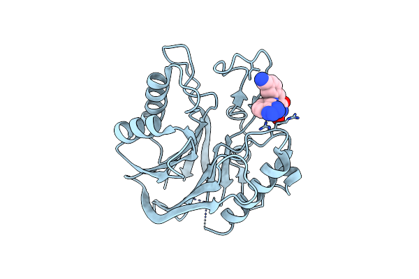
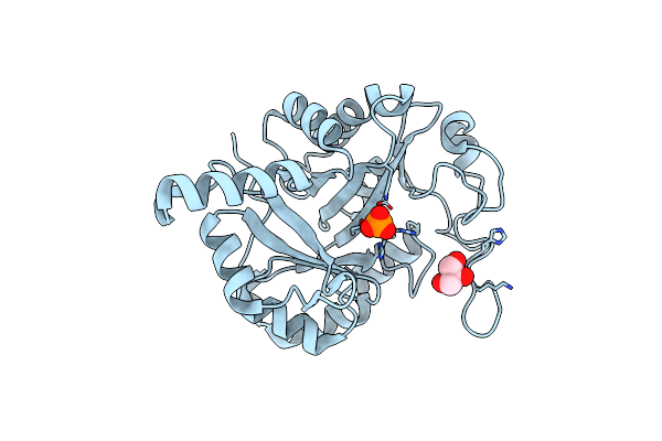
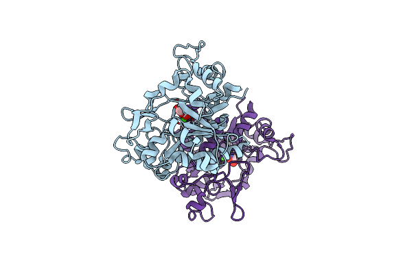
Crystal Structure Of The Catalytic Domain Of Human Tyrosyl Dna Phosphodiesterase 2
Organism: Homo sapiens
Method: X-RAY DIFFRACTION Resolution:3.10 Å Release Date: 2016-05-04 Classification: HYDROLASE Ligands: MG, GOL |
 |
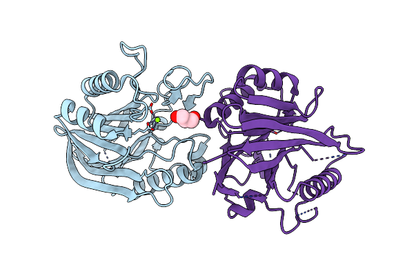
Crystal Structure Of The Catalytic Domain Of Human Tyrosyl Dna Phosphodiesterase 2 In Complex With A Small Molecule Inhibitor
Organism: Homo sapiens
Method: X-RAY DIFFRACTION Resolution:3.40 Å Release Date: 2016-05-04 Classification: HYDROLASE Ligands: 6FQ |
 |
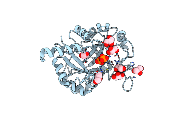
Crystal Structure Of Staphylococcus Aureus Lysophosphatidylglycerol Phospholipase D
Organism: Staphylococcus aureus
Method: X-RAY DIFFRACTION Resolution:2.40 Å Release Date: 2023-11-01 Classification: HYDROLASE Ligands: 2HP, GOL |
 |
Crystal Structure Of Staphylococcus Aureus Lysophosphatidylglycerol Phospholipase D
Organism: Staphylococcus aureus
Method: X-RAY DIFFRACTION Resolution:2.10 Å Release Date: 2023-11-01 Classification: HYDROLASE Ligands: 2HP, GOL, CL |
 |
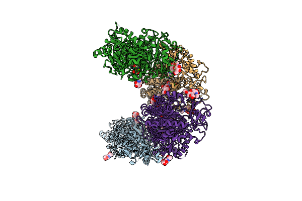
Organism: Bos taurus
Method: ELECTRON MICROSCOPY Resolution:3.06 Å Release Date: 2026-05-06 Classification: HYDROLASE Ligands: VIA, PCG, MG, ZN |
 |
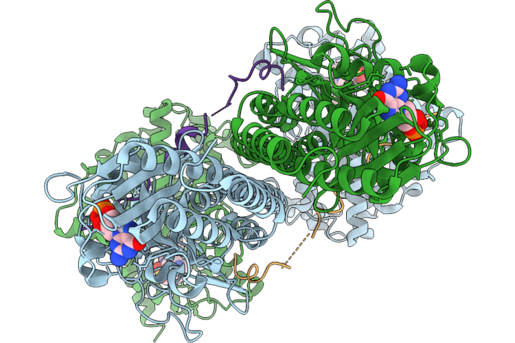
Human Ectonucleotide Pyrophosphatase / Phosphodiesterase 3 (Enpp3, Npp3, Cd203C)
Organism: Homo sapiens
Method: X-RAY DIFFRACTION Resolution:2.30 Å Release Date: 2018-05-02 Classification: HYDROLASE Ligands: ZN, CA, NAG, NA |
 |
Human Ectonucleotide Pyrophosphatase / Phosphodiesterase 3 (Enpp3, Npp3, Cd203C), Inactive (T205A), N594S, With Alpha,Beta-Methylene-Atp (Ampcpp)
Organism: Homo sapiens
Method: X-RAY DIFFRACTION Resolution:1.94 Å Release Date: 2018-05-02 Classification: HYDROLASE Ligands: ZN, CA, NA, CL, SO4, APC |
 |
Crystal Structure Of Ectonucleotide Phosphodiesterase/Pyrophosphatase-3 (Npp3)
Organism: Rattus norvegicus
Method: X-RAY DIFFRACTION Resolution:2.40 Å Release Date: 2018-08-01 Classification: HYDROLASE Ligands: PO4, ZN, CA, NAG |
 |
Crystal Structure Of Ectonucleotide Phosphodiesterase/Pyrophosphatase-3 (Npp3) In Complex With Ap4A
Organism: Rattus norvegicus
Method: X-RAY DIFFRACTION Resolution:2.40 Å Release Date: 2018-08-01 Classification: HYDROLASE Ligands: B4P, ZN, CA, NAG |
 |
Crystal Structure Of Ectonucleotide Phosphodiesterase/Pyrophosphatase-3 (Npp3) In Complex With Udpglcnac
Organism: Rattus norvegicus
Method: X-RAY DIFFRACTION Resolution:2.30 Å Release Date: 2018-08-01 Classification: HYDROLASE Ligands: UD1, ZN, CA, NAG |
 |
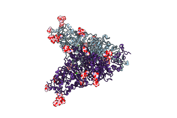
Structural And Functional Characterization Of A Novel Archaeal Phosphodiesterase
Organism: Methanocaldococcus jannaschii
Method: X-RAY DIFFRACTION Resolution:2.50 Å Release Date: 2004-08-10 Classification: PHOSPHODIESTERASE Ligands: MN |
 |
Structural And Functional Characterization Of A Novel Archaeal Phosphodiesterase
Organism: Methanocaldococcus jannaschii
Method: X-RAY DIFFRACTION Resolution:2.50 Å Release Date: 2004-08-10 Classification: PHOSPHODIESTERASE Ligands: NI |
 |
Crystal Structure Of Ectonucleotide Phosphodiesterase/Pyrophosphatase-3 (Npp3) In Complex With Ampnpp
Organism: Rattus norvegicus
Method: X-RAY DIFFRACTION Resolution:3.00 Å Release Date: 2018-08-01 Classification: HYDROLASE Ligands: ZAN, ZN, CA, NAG |
 |
Crystal Structure Of Ectonucleotide Phosphodiesterase/Pyrophosphatase-3 (Npp3) In Complex With Amp
Organism: Rattus norvegicus
Method: X-RAY DIFFRACTION Resolution:2.50 Å Release Date: 2018-08-01 Classification: HYDROLASE Ligands: AMP, ZN, CA, NAG |
 |
Full Length Ectodomain Of Ectonucleotide Phosphodiesterase/Pyrophosphatase-3 (Npp3) Including The Smb Domains But With A Partially Disordered Active Site Structure
Organism: Rattus norvegicus
Method: X-RAY DIFFRACTION Resolution:2.80 Å Release Date: 2018-11-07 Classification: HYDROLASE Ligands: CA, NAG |
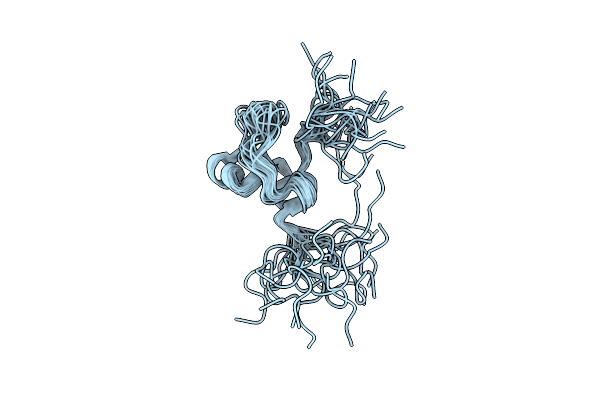 |
Solution Structure Of The Somatomedin B Domain Of Human Ectonucleotide Pyrophosphatase/Phosphodiesterase Family Member
|
 |
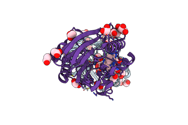
Crystal Structure Of Periplasmic Glycerophosphodiester Phosphodiesterase From Escherichia Coli
Organism: Escherichia coli
Method: X-RAY DIFFRACTION Resolution:1.70 Å Release Date: 2005-01-25 Classification: HYDROLASE Ligands: CA, GOL |
 |
Crystal Structure Of M2Htdp2-Cat In Complex With A Small Molecule Inhibitor
Organism: Mus musculus
Method: X-RAY DIFFRACTION Resolution:1.80 Å Release Date: 2016-05-04 Classification: HYDROLASE Ligands: 6FQ, MG, GOL, EDO, ACT |