

Search Count: 3,349
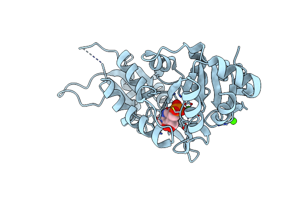 |
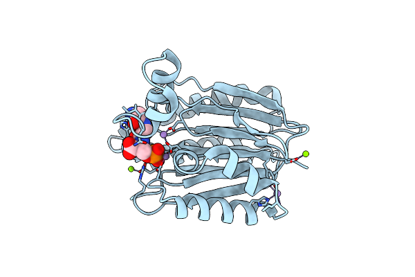
Crystal Structure Of Human Pyridoxal 5'-Phosphate Phosphatase With Its Substrate
Organism: Homo sapiens
Method: X-RAY DIFFRACTION Resolution:1.80 Å Release Date: 2007-02-27 Classification: PHOSPHATASE Ligands: PLP, CA |
Organism: Homo sapiens
Method: X-RAY DIFFRACTION
Release Date: 2007-02-27
Ligands: PLP, CA
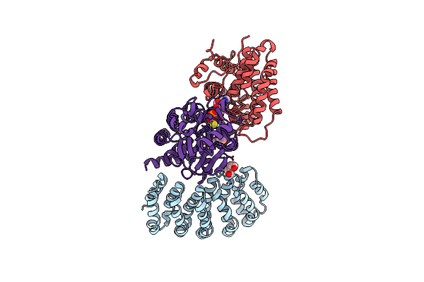 |
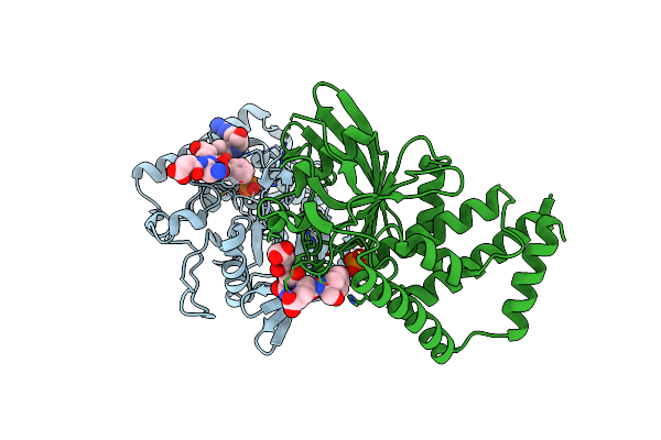
Crystal Structure Of Protein Phosphatase 2A (Pp2A) And Pp2A Phosphatase Activator (Ptpa) Complex With Atpgammas
Organism: Homo sapiens
Method: X-RAY DIFFRACTION Resolution:2.82 Å Release Date: 2013-10-09 Classification: Hydrolase/signaling protein Ligands: PEG, MN, AGS, MES |
Organism: Homo sapiens
Method: X-RAY DIFFRACTION
Release Date: 2013-10-09
Ligands: PEG, MN, AGS, MES
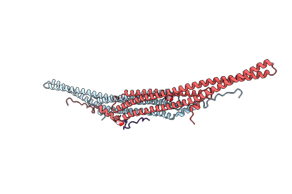 |
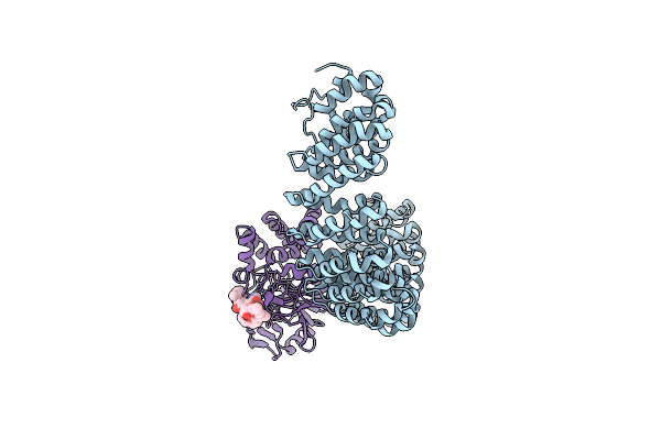
Crystal Structure Of The F-Bar Domain Of Pstipip1 Bound To The Cth Domain Of The Phosphatase Lyp
Organism: Homo sapiens
Method: X-RAY DIFFRACTION Resolution:2.15 Å Release Date: 2022-02-23 Classification: SIGNALING PROTEIN Ligands: GOL |
Organism: Homo sapiens
Method: X-RAY DIFFRACTION
Release Date: 2022-02-23
Ligands: GOL
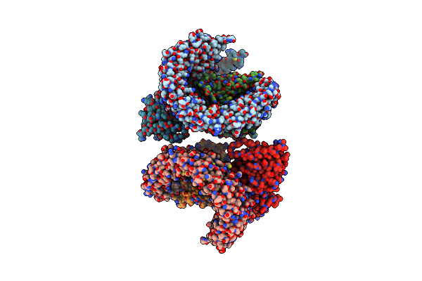 |
Structure Of The Protein Phosphatase 2A Holoenzyme
Organism: Homo sapiens, Cyanobacteria
Method: X-RAY DIFFRACTION Resolution:3.30 Å Release Date: 2006-12-12 Classification: HYDROLASE/HYDROLASE INHIBITOR Ligands: MN |
Organism: Homo sapiens, Cyanobacteria
Method: X-RAY DIFFRACTION
Release Date: 2006-12-12
Ligands: MN
 |
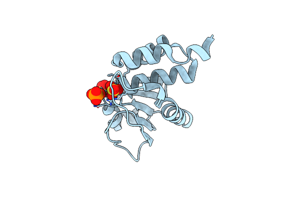
Crystal Structure Of The Co-Expressed Sds22:Pp1:I3 Complex
Organism: Homo sapiens
Method: X-RAY DIFFRACTION Resolution:3.20 Å Release Date: 2023-12-06 Classification: HYDROLASE Ligands: FE, PO4 |
Organism: Homo sapiens
Method: X-RAY DIFFRACTION
Release Date: 2023-12-06
Ligands: FE, PO4
 |
Crystal Structrue Of A Novel Mitogen-Activated Protein Kinase Phosphatase, Skrp1
Organism: Homo sapiens
Method: X-RAY DIFFRACTION Resolution:1.26 Å Release Date: 2012-04-04 Classification: HYDROLASE Ligands: PO4, SO4 |
Organism: Homo sapiens
Method: X-RAY DIFFRACTION
Release Date: 2012-04-04
Ligands: PO4, SO4
 |
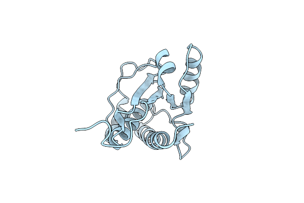
Crystal Structure Of Human Dual Specificity Phosphatase 14
Organism: Homo sapiens
Method: X-RAY DIFFRACTION Resolution:1.88 Å Release Date: 2009-10-06 Classification: HYDROLASE Ligands: PO4 |
Organism: Homo sapiens
Method: X-RAY DIFFRACTION
Release Date: 2009-10-06
Ligands: PO4
 |
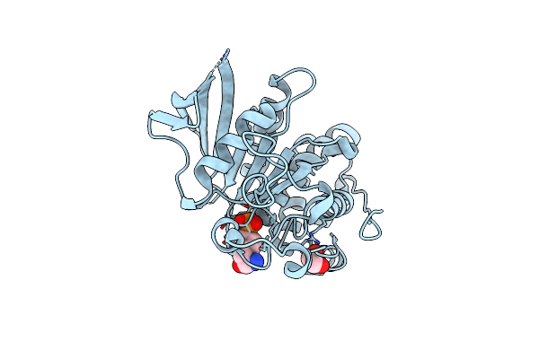
Crystal Structure Of The Apo Low Molecular Weight Protein Tyrosine Phosphatase Isoform A
Organism: Homo sapiens
Method: X-RAY DIFFRACTION Resolution:2.30 Å Release Date: 2015-07-15 Classification: HYDROLASE |
Organism: Homo sapiens
Method: X-RAY DIFFRACTION
Release Date: 2015-07-15
 |
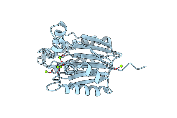
Crystal Structure Of The C472S Mutant Of Human Protein Tyrosine Phosphatase Ptpn5 (Step, Striatum Enriched Phosphatase) In Complex With Phosphotyrosine
Organism: Homo sapiens
Method: X-RAY DIFFRACTION Resolution:1.70 Å Release Date: 2006-05-16 Classification: HYDROLASE Ligands: PTR, EDO |
Organism: Homo sapiens
Method: X-RAY DIFFRACTION
Release Date: 2006-05-16
Ligands: PTR, EDO
 |
Crystal Structure Of Bacterial Serine Phosphatase
Organism: Staphylococcus aureus
Method: X-RAY DIFFRACTION Resolution:1.57 Å Release Date: 2019-08-07 Classification: HYDROLASE Ligands: MG |
Organism: Staphylococcus aureus
Method: X-RAY DIFFRACTION
Release Date: 2019-08-07
Ligands: MG
 |
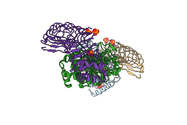
Crystal Structure Of Bacterial Serine Phosphatase With His Tag
Organism: Staphylococcus aureus
Method: X-RAY DIFFRACTION Resolution:1.35 Å Release Date: 2019-08-07 Classification: HYDROLASE Ligands: MG, PO4 |
Organism: Staphylococcus aureus
Method: X-RAY DIFFRACTION
Release Date: 2019-08-07
Ligands: MG, PO4
 |
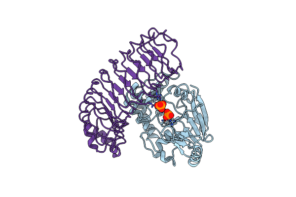
The Crystal Structure Of Coexpressed Sds22:Pp1 Complex
Organism: Homo sapiens
Method: X-RAY DIFFRACTION Resolution:2.70 Å Release Date: 2019-09-18 Classification: HYDROLASE Ligands: PO4, FE, CL, MPD |
Organism: Homo sapiens
Method: X-RAY DIFFRACTION
Release Date: 2019-09-18
Ligands: PO4, FE, CL, MPD
 |
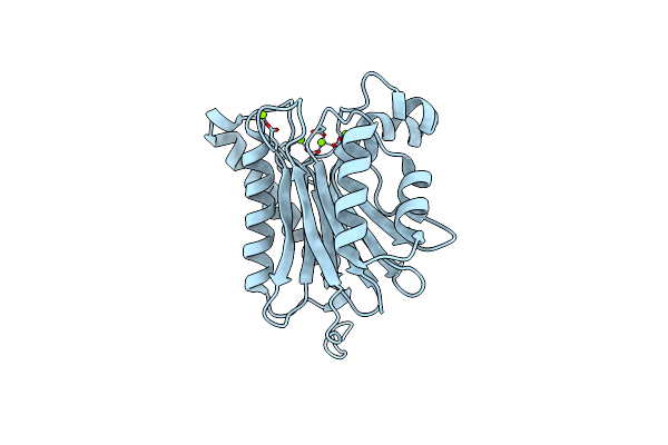
Reconstituted Pp1 Holoenzyme
Organism: Homo sapiens
Method: X-RAY DIFFRACTION Resolution:2.70 Å Release Date: 2019-09-18 Classification: HYDROLASE Ligands: PO4, MN, CL |
Organism: Homo sapiens
Method: X-RAY DIFFRACTION
Release Date: 2019-09-18
Ligands: PO4, MN, CL
 |
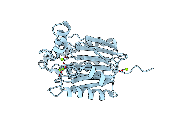
Crystal Structure Of Bacterial Serine Phosphatase With His-Tag Mutation
Organism: Staphylococcus aureus
Method: X-RAY DIFFRACTION Resolution:1.40 Å Release Date: 2019-08-07 Classification: HYDROLASE Ligands: MG |
Organism: Staphylococcus aureus
Method: X-RAY DIFFRACTION
Release Date: 2019-08-07
Ligands: MG
 |
Crystal Structure Of Bacterial Serine Phosphatase Bearing R161K Mutation
Organism: Staphylococcus aureus
Method: X-RAY DIFFRACTION Resolution:1.55 Å Release Date: 2019-08-07 Classification: HYDROLASE Ligands: MG |
Organism: Staphylococcus aureus
Method: X-RAY DIFFRACTION
Release Date: 2019-08-07
Ligands: MG
 |
Crystal Structure Of Bacterial Serine Phosphatase Bearing R161A Mutation
Organism: Staphylococcus aureus
Method: X-RAY DIFFRACTION Resolution:1.84 Å Release Date: 2019-08-07 Classification: HYDROLASE Ligands: MG |
Organism: Staphylococcus aureus
Method: X-RAY DIFFRACTION
Release Date: 2019-08-07
Ligands: MG
 |
Crystal Structure Of Bacterial Serine Phosphatase Bound With Phosphorylated Peptide
Organism: Staphylococcus aureus, Peptide display vector fth1
Method: X-RAY DIFFRACTION Resolution:1.57 Å Release Date: 2019-08-07 Classification: HYDROLASE Ligands: MG, MN |
Organism: Staphylococcus aureus, Peptide display vector fth1
Method: X-RAY DIFFRACTION
Release Date: 2019-08-07
Ligands: MG, MN
 |
Crystal Structure Of Bacterial Serine Phosphatase Bearing R161E Mutation
Organism: Staphylococcus aureus
Method: X-RAY DIFFRACTION Resolution:1.90 Å Release Date: 2019-08-07 Classification: HYDROLASE Ligands: MG |
Organism: Staphylococcus aureus
Method: X-RAY DIFFRACTION
Release Date: 2019-08-07
Ligands: MG
 |
Protein Tyrosine Phosphatase Ptpn-22 (Lyp) Bound To The Mono-Phosphorylated Lck Active Site Peptide
Organism: Homo sapiens
Method: X-RAY DIFFRACTION Resolution:2.20 Å Release Date: 2009-02-17 Classification: HYDROLASE Ligands: PO4 |
Organism: Homo sapiens
Method: X-RAY DIFFRACTION
Release Date: 2009-02-17
Ligands: PO4
 |
Structure Of The Protein Phosphatase 2A Core Enzyme Bound To Okadaic Acid
Organism: Homo sapiens
Method: X-RAY DIFFRACTION Resolution:2.60 Å Release Date: 2006-11-07 Classification: SIGNALING PROTEIN,HYDROLASE Ligands: MN, OKA |
Organism: Homo sapiens
Method: X-RAY DIFFRACTION
Release Date: 2006-11-07
Ligands: MN, OKA