

Search Count: 114
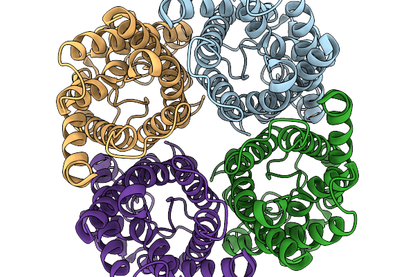 |
Cryo-Em Structure Of Aquaporin 3 At Ph 8.0 With Hydrogen Peroxide
Organism: Homo sapiens
Method: ELECTRON MICROSCOPY Resolution:3.00 Å Release Date: 2026-02-18 Classification: MEMBRANE PROTEIN Ligands: PEO |
Organism: Homo sapiens
Method: ELECTRON MICROSCOPY
Release Date: 2026-02-18
Ligands: PEO
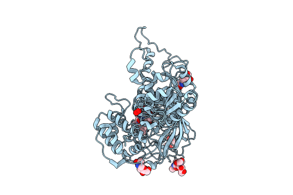 |
Heme-Containing Fungal Catalase
Organism: Aspergillus niger
Method: X-RAY DIFFRACTION Resolution:1.82 Å Release Date: 2025-12-17 Classification: OXIDOREDUCTASE Ligands: HEM, NAG, PEO, CL |
Organism: Aspergillus niger
Method: X-RAY DIFFRACTION
Release Date: 2025-12-17
Ligands: HEM, NAG, PEO, CL
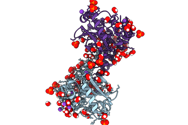 |
Finding The Exit Route Of Hydrogen Peroxide From The Manganese Superoxide Dismutase (Mnsod) Active Site
Organism: Escherichia coli
Method: X-RAY DIFFRACTION Resolution:1.33 Å Release Date: 2025-08-27 Classification: OXIDOREDUCTASE Ligands: PEO, PO4, K, MN |
Organism: Escherichia coli
Method: X-RAY DIFFRACTION
Release Date: 2025-08-27
Ligands: PEO, PO4, K, MN
 |
Crystal Structure Of N Terminal Deletion Mutant Of Staphylococcal Atypical Two-Cysteine Thiol Peroxidase Complexed With Substrate Analog
Organism: Staphylococcus aureus
Method: X-RAY DIFFRACTION Resolution:1.55 Å Release Date: 2025-06-18 Classification: OXIDOREDUCTASE Ligands: PG4, PEO, A1L2G, IMD, DTO, DTU |
Organism: Staphylococcus aureus
Method: X-RAY DIFFRACTION
Release Date: 2025-06-18
Ligands: PG4, PEO, A1L2G, IMD, DTO, DTU
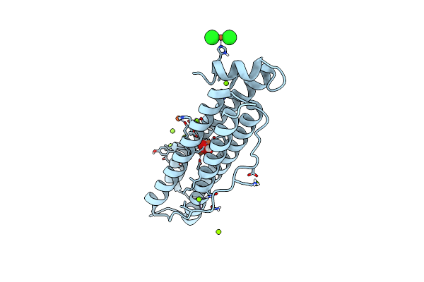 |
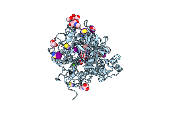
Iron Loaded Mitochondrial Ferritin Exposed To Oxygen For 2 Minutes
Organism: Homo sapiens
Method: X-RAY DIFFRACTION Resolution:1.48 Å Release Date: 2025-04-16 Classification: METAL BINDING PROTEIN Ligands: FE, MG, CL, PEO |
Organism: Homo sapiens
Method: X-RAY DIFFRACTION
Release Date: 2025-04-16
Ligands: FE, MG, CL, PEO
 |
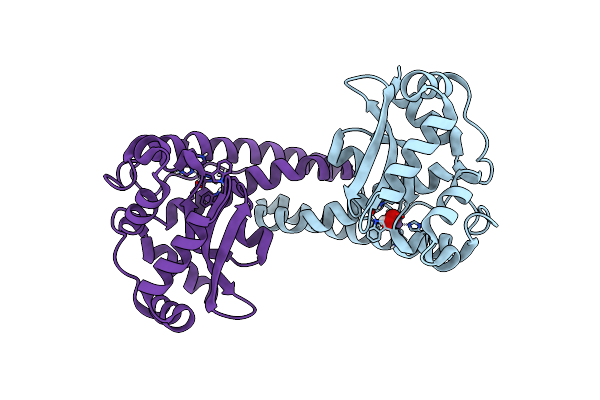
Structure Of The Intermediate Of Lactoperoxidase Formed With Thiocynate And Hydrogen Peroxidase At 1.99 A Resolution.
Organism: Bos taurus
Method: X-RAY DIFFRACTION Resolution:2.00 Å Release Date: 2025-03-26 Classification: OXIDOREDUCTASE Ligands: HEM, CA, NAG, IOD, SCN, PEO, GOL |
Organism: Bos taurus
Method: X-RAY DIFFRACTION
Release Date: 2025-03-26
Ligands: HEM, CA, NAG, IOD, SCN, PEO, GOL
 |
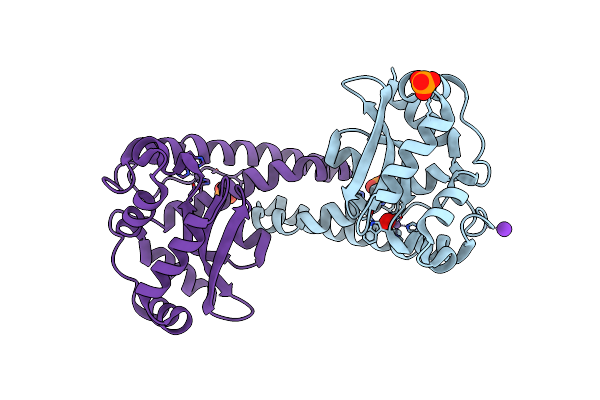
Neutron Structure Of Peroxide-Soaked Tyr34Phe Mnsod
Organism: Homo sapiens
Method: NEUTRON DIFFRACTION Resolution:2.30 Å Release Date: 2025-03-12 Classification: OXIDOREDUCTASE Ligands: MN, PEO |
Organism: Homo sapiens
Method: NEUTRON DIFFRACTION
Release Date: 2025-03-12
Ligands: MN, PEO
 |
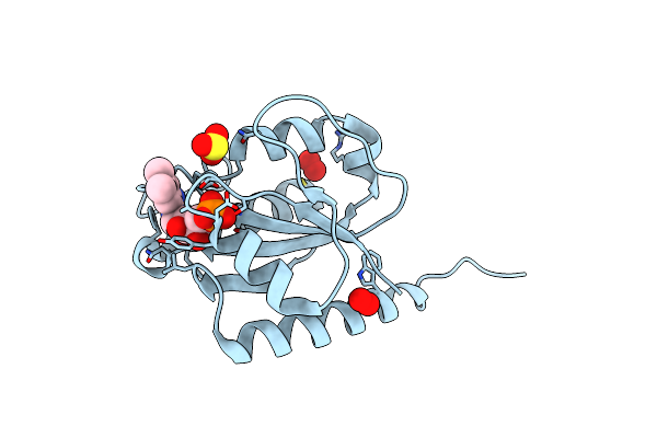
X-Ray Counterpart To The Neutron Structure Of Peroxide-Soaked Tyr34Phe Mnsod
Organism: Homo sapiens
Method: X-RAY DIFFRACTION Resolution:1.40 Å Release Date: 2025-03-12 Classification: OXIDOREDUCTASE Ligands: MN, PEO, PO4, K |
Organism: Homo sapiens
Method: X-RAY DIFFRACTION
Release Date: 2025-03-12
Ligands: MN, PEO, PO4, K
 |
Structure Of Mycobacterium Thermoresistibile Nrdi(Reduced) Determined At 1.1 Angstrom Resolution
Organism: Mycolicibacterium thermoresistibile atcc 19527
Method: X-RAY DIFFRACTION Resolution:1.21 Å Release Date: 2024-10-09 Classification: FLAVOPROTEIN Ligands: FMN, SO3, PEO, PER, CL |
Organism: Mycolicibacterium thermoresistibile atcc 19527
Method: X-RAY DIFFRACTION
Release Date: 2024-10-09
Ligands: FMN, SO3, PEO, PER, CL
 |
Crystal Structure Of Pseudomonas Aeruginosa Suhb In Complex With D-Myo-Inositol-1-Phosphate
Organism: Pseudomonas aeruginosa pao1
Method: X-RAY DIFFRACTION Resolution:2.20 Å Release Date: 2024-09-18 Classification: HYDROLASE Ligands: IPD, ACT, PEO, GOL, DTU, CL, CA |
Organism: Pseudomonas aeruginosa pao1
Method: X-RAY DIFFRACTION
Release Date: 2024-09-18
Ligands: IPD, ACT, PEO, GOL, DTU, CL, CA
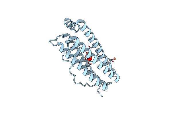 |
Azumapecten Farreri Homopolymeric Ferritin (Apf) Mutant-H2Ke Exposed To H2O2 For 2 S
Organism: Azumapecten farreri
Method: X-RAY DIFFRACTION Resolution:2.30 Å Release Date: 2024-09-11 Classification: METAL BINDING PROTEIN Ligands: FE, PEO |
Organism: Azumapecten farreri
Method: X-RAY DIFFRACTION
Release Date: 2024-09-11
Ligands: FE, PEO
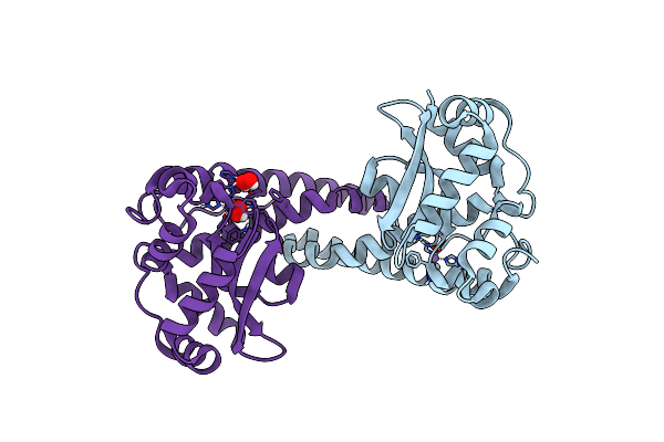 |
Neutron Structure Of Peroxide-Soaked Trp161Phe Mnsod
Organism: Homo sapiens
Method: NEUTRON DIFFRACTION Resolution:2.30 Å Release Date: 2024-07-31 Classification: OXIDOREDUCTASE Ligands: MN, PEO |
Organism: Homo sapiens
Method: NEUTRON DIFFRACTION
Release Date: 2024-07-31
Ligands: MN, PEO
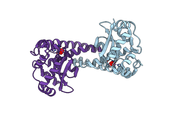 |
X-Ray Counterpart To The Neutron Structure Of Peroxide-Soaked Trp161Phe Mnsod
Organism: Homo sapiens
Method: X-RAY DIFFRACTION Resolution:1.68 Å Release Date: 2024-07-31 Classification: OXIDOREDUCTASE Ligands: MN, PEO |
Organism: Homo sapiens
Method: X-RAY DIFFRACTION
Release Date: 2024-07-31
Ligands: MN, PEO
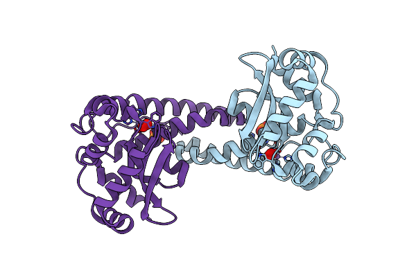 |
Peroxide-Soaked Mnsod
Organism: Homo sapiens
Method: X-RAY DIFFRACTION Resolution:1.76 Å Release Date: 2024-07-31 Classification: OXIDOREDUCTASE Ligands: MN, PEO, PO4 |
Organism: Homo sapiens
Method: X-RAY DIFFRACTION
Release Date: 2024-07-31
Ligands: MN, PEO, PO4
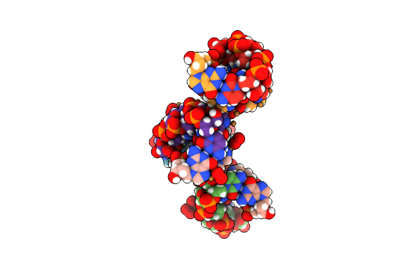 |
Crystal Structure Of The Z-Dna Duplex D(Cgcgcg) Containing Ordered Copper(Ii) And Soaked In Hydrogen Peroxide For 5 Minutes
Organism: Synthetic construct
Method: X-RAY DIFFRACTION Resolution:1.25 Å Release Date: 2024-03-27 Classification: DNA Ligands: CU, PEO |
Organism: Synthetic construct
Method: X-RAY DIFFRACTION
Release Date: 2024-03-27
Ligands: CU, PEO
 |
Crystal Structure Of The Z-Dna Duplex D(Cgcgcg) Containing Ordered Copper(Ii) And Soaked In Hydrogen Peroxide For An Hour
Organism: Synthetic construct
Method: X-RAY DIFFRACTION Resolution:1.30 Å Release Date: 2024-03-27 Classification: DNA Ligands: CU, SPM, PEO |
Organism: Synthetic construct
Method: X-RAY DIFFRACTION
Release Date: 2024-03-27
Ligands: CU, SPM, PEO
 |
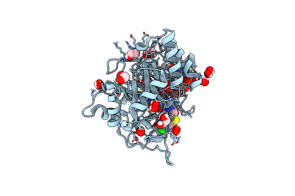
Ergothioneine Dioxygenase From Thermocatellispora Tengchongensis In Complex With Manganese
Organism: Thermocatellispora tengchongensis
Method: X-RAY DIFFRACTION Resolution:1.90 Å Release Date: 2023-12-27 Classification: OXIDOREDUCTASE Ligands: EDO, PEO, MN, MG, PG4, PEG |
Organism: Thermocatellispora tengchongensis
Method: X-RAY DIFFRACTION
Release Date: 2023-12-27
Ligands: EDO, PEO, MN, MG, PG4, PEG
 |
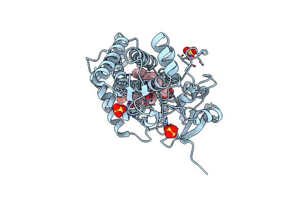
Crystal Structure Of Aldose Reductase (Akr1B1) Complexed With Nadp+ And At-003 Soaked In Hydrogen Peroxide
Organism: Homo sapiens
Method: X-RAY DIFFRACTION Resolution:1.60 Å Release Date: 2023-12-20 Classification: OXIDOREDUCTASE Ligands: NAP, XY0, EDO, PEO |
Organism: Homo sapiens
Method: X-RAY DIFFRACTION
Release Date: 2023-12-20
Ligands: NAP, XY0, EDO, PEO
 |
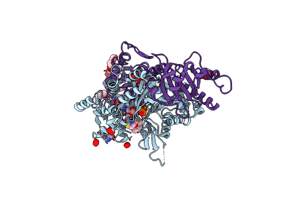
Time-Resolved Sfx-Xfel Crystal Structure Of Cyp121 Bound With Cyy Reacted With Peracetic Acid For 200 Milliseconds
Organism: Mycobacterium tuberculosis h37rv
Method: X-RAY DIFFRACTION Resolution:1.85 Å Release Date: 2023-11-22 Classification: OXIDOREDUCTASE Ligands: SO4, HEM, YTT, PEO |
Organism: Mycobacterium tuberculosis h37rv
Method: X-RAY DIFFRACTION
Release Date: 2023-11-22
Ligands: SO4, HEM, YTT, PEO
 |
Chimera Of The Inactive Exoy Nucleotidyl Cyclase Domain From Vibrio Nigripulchritudo Martx Toxin, With The Double Mutation K3528M And K3535I, Fused To A Proline-Rich-Domain (Prd) And Profilin, Bound To Latrunculin B-Adp-Mg-Actin
Organism: Vibrio nigripulchritudo, Homo sapiens, Oryctolagus cuniculus
Method: X-RAY DIFFRACTION Resolution:1.69 Å Release Date: 2023-09-20 Classification: TOXIN Ligands: ADP, MG, LAB, TRS, GOL, PEO, SO4, PEG |
Organism: Vibrio nigripulchritudo, Homo sapiens, Oryctolagus cuniculus
Method: X-RAY DIFFRACTION
Release Date: 2023-09-20
Ligands: ADP, MG, LAB, TRS, GOL, PEO, SO4, PEG