

Search Count: 9
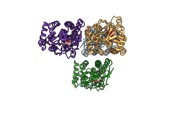 |
Crystal Structure Of A Trap Periplasmic Solute Binding Protein From Desulfovibrio Vulgaris (Deval_0042, Target Efi-510114) Bound To Copurified (R)-Pantoic Acid
Organism: Desulfovibrio vulgaris
Method: X-RAY DIFFRACTION Resolution:1.70 Å Release Date: 2015-01-28 Classification: TRANSPORT PROTEIN Ligands: PAF |
Organism: Desulfovibrio vulgaris
Method: X-RAY DIFFRACTION
Release Date: 2015-01-28
Ligands: PAF
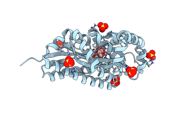 |
Crystal Structure Of A Trap Periplasmic Solute Binding Protein From Verminephrobacter Eiseniae Ef01-2 (Veis_3954, Target Efi-510324) A Nephridial Symbiont Of The Earthworm Eisenia Foetida, Bound To (R)-Pantoic Acid
Organism: Verminephrobacter eiseniae
Method: X-RAY DIFFRACTION Resolution:1.20 Å Release Date: 2014-05-07 Classification: SOLUTE-BINDING PROTEIN Ligands: SO4, PAF |
Organism: Verminephrobacter eiseniae
Method: X-RAY DIFFRACTION
Release Date: 2014-05-07
Ligands: SO4, PAF
 |
Crystal Structure Of A Trap Periplasmic Solute Binding Protein From Bordetella Bronchispeptica (Bb3421), Target Efi-510039, With Density Modeled As Pantoate
Organism: Bordetella bronchiseptica
Method: X-RAY DIFFRACTION Resolution:1.50 Å Release Date: 2014-01-01 Classification: PROTEIN TRANSPORT Ligands: PAF, SO4, CL |
Organism: Bordetella bronchiseptica
Method: X-RAY DIFFRACTION
Release Date: 2014-01-01
Ligands: PAF, SO4, CL
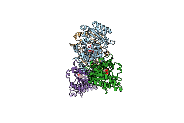 |
Pantoate-Beta-Alanine Ligase From Yersinia Pestis In Complex With Pantoate.
Organism: Yersinia pestis
Method: X-RAY DIFFRACTION Resolution:1.58 Å Release Date: 2011-02-02 Classification: LIGASE Ligands: PAF, CL |
Organism: Yersinia pestis
Method: X-RAY DIFFRACTION
Release Date: 2011-02-02
Ligands: PAF, CL
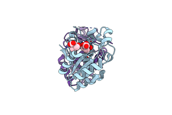 |
Structural And Substrate-Binding Studies Of Pantothenate Synthenate (Ps)Provide Insights Into Homotropic Inhibition By Pantoate In Ps'S
Organism: Escherichia coli
Method: X-RAY DIFFRACTION Resolution:1.67 Å Release Date: 2010-02-09 Classification: LIGASE Ligands: PAF |
Organism: Escherichia coli
Method: X-RAY DIFFRACTION
Release Date: 2010-02-09
Ligands: PAF
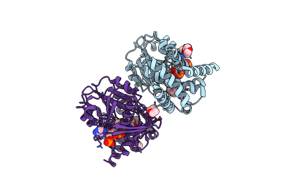 |
Crystal Structure Of Escherichia Coli Ketopantoate Reductase In A Ternary Complex With Nadp+ And Pantoate
Organism: Escherichia coli k12
Method: X-RAY DIFFRACTION Resolution:2.30 Å Release Date: 2007-01-16 Classification: OXIDOREDUCTASE Ligands: NAP, PAF, DIO, ACT |
Organism: Escherichia coli k12
Method: X-RAY DIFFRACTION
Release Date: 2007-01-16
Ligands: NAP, PAF, DIO, ACT
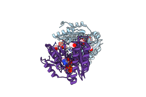 |
Crystal Structure Of A Pantothenate Synthetase From M. Tuberculosis In Complex With Ampcpp And Pantoate, Higher Occupancy Of Pantoate And Lower Occupancy Of Ampcpp In Subunit A
Organism: Mycobacterium tuberculosis
Method: X-RAY DIFFRACTION Resolution:1.70 Å Release Date: 2003-04-22 Classification: LIGASE Ligands: MG, SO4, APC, PAF, GOL, EOH |
Organism: Mycobacterium tuberculosis
Method: X-RAY DIFFRACTION
Release Date: 2003-04-22
Ligands: MG, SO4, APC, PAF, GOL, EOH
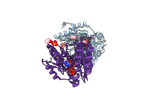 |
Crystal Structure Of A Pantothenate Synthetase From M. Tuberculosis In Complex With Ampcpp And Pantoate
Organism: Mycobacterium tuberculosis
Method: X-RAY DIFFRACTION Resolution:1.60 Å Release Date: 2003-04-22 Classification: LIGASE Ligands: SO4, APC, PAF, GOL, EOH, MG |
Organism: Mycobacterium tuberculosis
Method: X-RAY DIFFRACTION
Release Date: 2003-04-22
Ligands: SO4, APC, PAF, GOL, EOH, MG
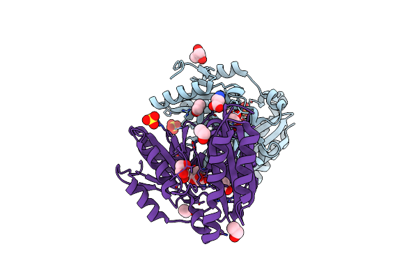 |
Crystal Structure Of A Pantothenate Synthetase From M. Tuberculosis In Complex With Pantoate
Organism: Mycobacterium tuberculosis
Method: X-RAY DIFFRACTION Resolution:1.80 Å Release Date: 2003-04-22 Classification: LIGASE Ligands: SO4, PAF, BAL, GOL, EOH |
Organism: Mycobacterium tuberculosis
Method: X-RAY DIFFRACTION
Release Date: 2003-04-22
Ligands: SO4, PAF, BAL, GOL, EOH