

Search Count: 52
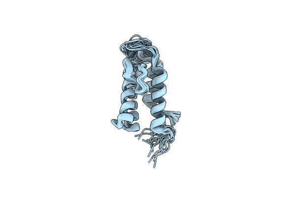 |
Solution Structure Of The Phosphatidylinositol 3-Phosphate Binding Domain From The Legionella Effector Seta
Organism: Legionella pneumophila
Method: SOLUTION NMR Release Date: 2023-02-08 Classification: LIPID BINDING PROTEIN |
Organism: Legionella pneumophila
Method: SOLUTION NMR
Release Date: 2023-02-08
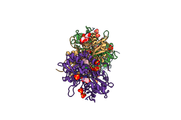 |
Crystal Structure Of The Glua2 Ligand-Binding Domain (S1S2J-L504Y-N775S) In Complex With Glutamate And Bpam121 At 1.78 A Resolution
Organism: Rattus norvegicus
Method: X-RAY DIFFRACTION Resolution:1.78 Å Release Date: 2018-01-03 Classification: MEMBRANE PROTEIN Ligands: SO4, 7M6, CL, GOL, GLU, EDO, CIT, PEG |
Organism: Rattus norvegicus
Method: X-RAY DIFFRACTION
Release Date: 2018-01-03
Ligands: SO4, 7M6, CL, GOL, GLU, EDO, CIT, PEG
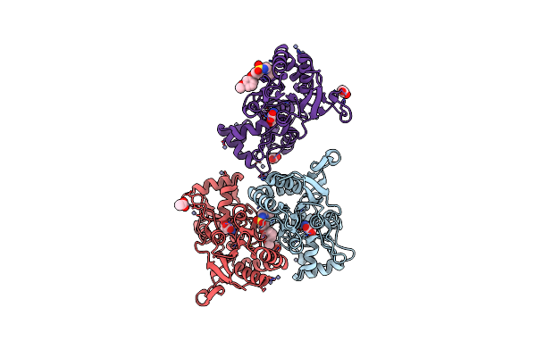 |
Crystal Structure Of The Glua2 Ligand-Binding Domain (S1S2J) In Complex With Glutamate And Positive Allosteric Modulator Bpam538
Organism: Rattus norvegicus
Method: X-RAY DIFFRACTION Resolution:2.00 Å Release Date: 2018-01-03 Classification: MEMBRANE PROTEIN Ligands: GLU, ZN, ACT, 9TE |
Organism: Rattus norvegicus
Method: X-RAY DIFFRACTION
Release Date: 2018-01-03
Ligands: GLU, ZN, ACT, 9TE
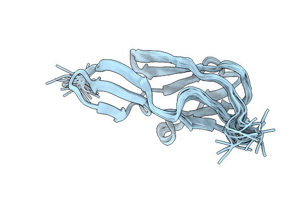 |
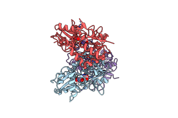
Solution Structure Of The Terminal Ig-Like Domain From Leptospira Interrogans Ligb
Organism: Leptospira interrogans
Method: SOLUTION NMR Release Date: 2014-08-13 Classification: CELL ADHESION |
Organism: Leptospira interrogans
Method: SOLUTION NMR
Release Date: 2014-08-13
 |
Nitrowillardiine Bound To The Ligand Binding Domain Of Glua2 At Ph 3.5
Organism: Rattus norvegicus
Method: X-RAY DIFFRACTION Resolution:2.03 Å Release Date: 2014-06-04 Classification: TRANSPORT PROTEIN Ligands: NWD, ZN |
Organism: Rattus norvegicus
Method: X-RAY DIFFRACTION
Release Date: 2014-06-04
Ligands: NWD, ZN
 |
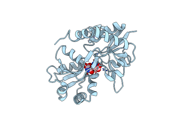
Ligand Binding Domain Of Glua2 (Ampa/Glutamate Receptor) Bound To (-)-Kaitocephalin
Organism: Rattus norvegicus
Method: X-RAY DIFFRACTION Resolution:1.96 Å Release Date: 2012-10-17 Classification: MEMBRANE PROTEIN Ligands: 0YS, ZN |
Organism: Rattus norvegicus
Method: X-RAY DIFFRACTION
Release Date: 2012-10-17
Ligands: 0YS, ZN
 |
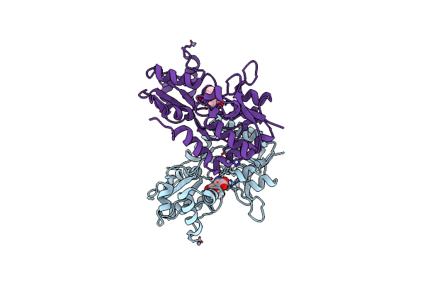
Quisqualate Bound To The D655A Mutant Of The Ligand Binding Domain Of Glua3
Organism: Rattus norvegicus
Method: X-RAY DIFFRACTION Resolution:2.20 Å Release Date: 2012-05-30 Classification: TRANSPORT PROTEIN/AGONIST Ligands: QUS, ZN |
Organism: Rattus norvegicus
Method: X-RAY DIFFRACTION
Release Date: 2012-05-30
Ligands: QUS, ZN
 |
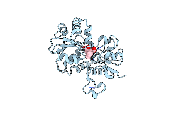
Kainate Bound To The D655A Mutant Of The Ligand Binding Domain Of Glua3
Organism: Rattus norvegicus
Method: X-RAY DIFFRACTION Resolution:2.29 Å Release Date: 2012-05-30 Classification: TRANSPORT PROTEIN/AGONIST Ligands: KAI, ZN |
Organism: Rattus norvegicus
Method: X-RAY DIFFRACTION
Release Date: 2012-05-30
Ligands: KAI, ZN
 |
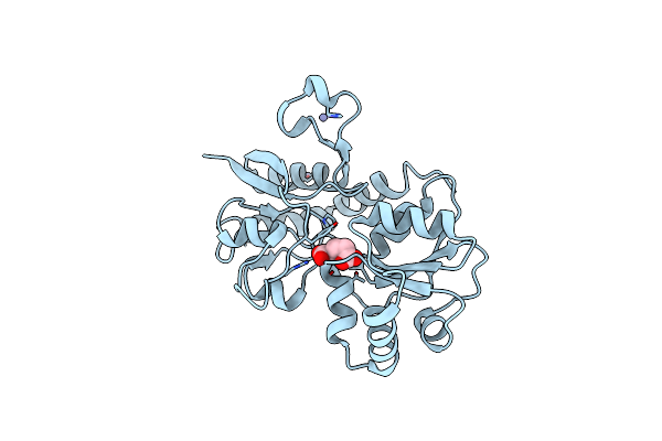
Kainate Bound To The Ligand Binding Domain Of Glua3
Organism: Rattus norvegicus
Method: X-RAY DIFFRACTION Resolution:1.83 Å Release Date: 2012-05-30 Classification: TRANSPORT PROTEIN/AGONIST Ligands: KAI, ZN |
Organism: Rattus norvegicus
Method: X-RAY DIFFRACTION
Release Date: 2012-05-30
Ligands: KAI, ZN
 |
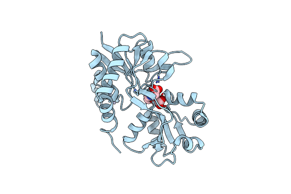
Glutamate Bound To The D655A Mutant Of The Ligand Binding Domain Of Glua3
Organism: Rattus norvegicus
Method: X-RAY DIFFRACTION Resolution:1.82 Å Release Date: 2012-05-30 Classification: TRANSPORT PROTEIN/SUBSTRATE Ligands: GLU, ZN |
Organism: Rattus norvegicus
Method: X-RAY DIFFRACTION
Release Date: 2012-05-30
Ligands: GLU, ZN
 |
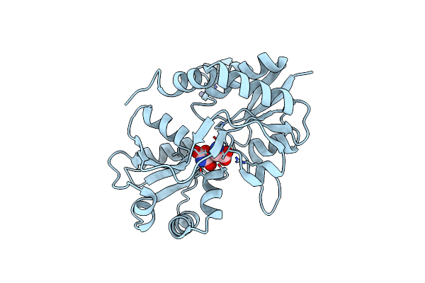
Kainate Bound To The Ligand Binding Domain Of Glua3I
Organism: Rattus norvegicus
Method: X-RAY DIFFRACTION Resolution:2.06 Å Release Date: 2012-05-30 Classification: TRANSPORT PROTEIN/AGONIST Ligands: KAI, ZN |
Organism: Rattus norvegicus
Method: X-RAY DIFFRACTION
Release Date: 2012-05-30
Ligands: KAI, ZN
 |
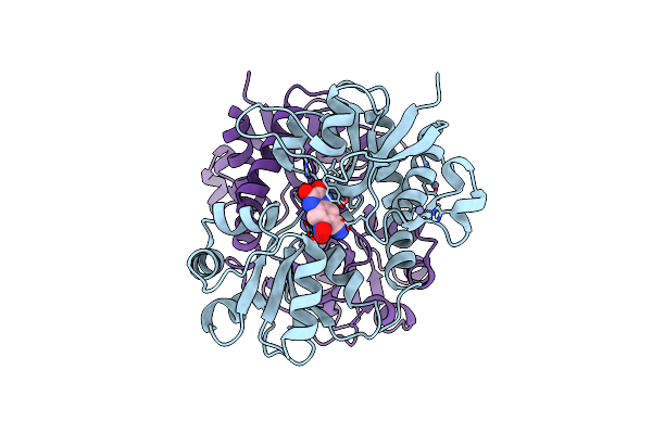
Quisqualate Bound To The D655A Mutant Of The Ligand Binding Domain Of Glua3
Organism: Rattus norvegicus
Method: X-RAY DIFFRACTION Resolution:1.91 Å Release Date: 2012-05-23 Classification: TRANSPORT PROTEIN/AGONIST Ligands: QUS, ZN |
Organism: Rattus norvegicus
Method: X-RAY DIFFRACTION
Release Date: 2012-05-23
Ligands: QUS, ZN
 |
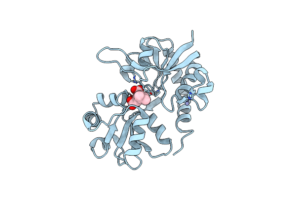
Cnqx Bound To The Ligand Binding Domain Of Glua3
Organism: Rattus norvegicus
Method: X-RAY DIFFRACTION Resolution:1.79 Å Release Date: 2012-05-16 Classification: TRANSPORT PROTEIN/AGONIST Ligands: CNI, ZN |
Organism: Rattus norvegicus
Method: X-RAY DIFFRACTION
Release Date: 2012-05-16
Ligands: CNI, ZN
 |
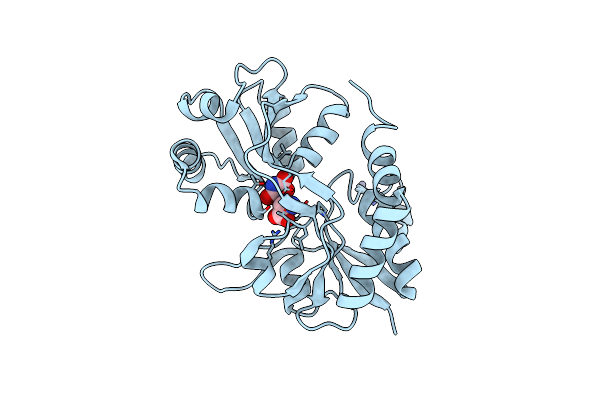
Kainate Bound To The K660A Mutant Of The Ligand Binding Domain Of Glua3
Organism: Rattus norvegicus
Method: X-RAY DIFFRACTION Resolution:2.06 Å Release Date: 2012-05-16 Classification: TRANSPORT PROTEIN/AGONIST Ligands: KAI, ZN |
Organism: Rattus norvegicus
Method: X-RAY DIFFRACTION
Release Date: 2012-05-16
Ligands: KAI, ZN
 |
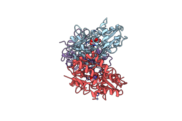
Quisqualate Bound To The Ligand Binding Domain Of Glua3I
Organism: Rattus norvegicus
Method: X-RAY DIFFRACTION Resolution:1.75 Å Release Date: 2012-05-16 Classification: TRANSPORT PROTEIN/AGONIST Ligands: QUS, ZN |
Organism: Rattus norvegicus
Method: X-RAY DIFFRACTION
Release Date: 2012-05-16
Ligands: QUS, ZN
 |
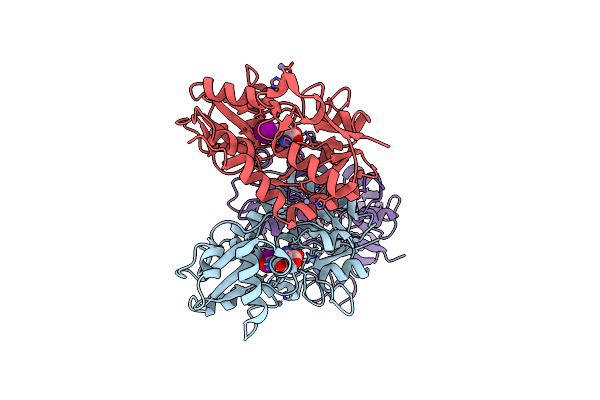
Glutamate Bound To A Double Cysteine Mutant (A452C/S652C) Of The Ligand Binding Domain Of Glua2
Organism: Rattus norvegicus
Method: X-RAY DIFFRACTION Resolution:1.91 Å Release Date: 2011-08-17 Classification: TRANSPORT PROTEIN Ligands: GLU, ZN |
Organism: Rattus norvegicus
Method: X-RAY DIFFRACTION
Release Date: 2011-08-17
Ligands: GLU, ZN
 |
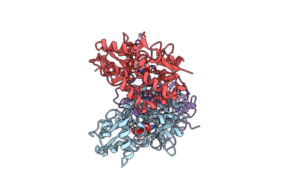
Iodowillardiine Bound To A Double Cysteine Mutant (A452C/S652C) Of The Ligand Binding Domain Of Glua2
Organism: Rattus norvegicus
Method: X-RAY DIFFRACTION Resolution:1.87 Å Release Date: 2011-08-17 Classification: TRANSPORT PROTEIN Ligands: IWD, ZN |
Organism: Rattus norvegicus
Method: X-RAY DIFFRACTION
Release Date: 2011-08-17
Ligands: IWD, ZN
 |
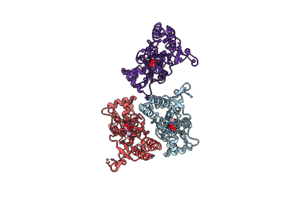
Kainate Bound To A Double Cysteine Mutant (A452C/S652C) Of The Ligand Binding Domain Of Glua2
Organism: Rattus norvegicus
Method: X-RAY DIFFRACTION Resolution:2.02 Å Release Date: 2011-08-17 Classification: TRANSPORT PROTEIN Ligands: KAI, ZN |
Organism: Rattus norvegicus
Method: X-RAY DIFFRACTION
Release Date: 2011-08-17
Ligands: KAI, ZN
 |
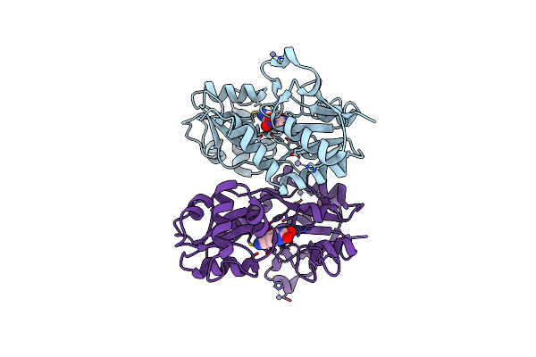
Cnqx Bound To An Oxidized Double Cysteine Mutant (A452C/S652C) Of The Ligand Binding Domain Of Glua2
Organism: Rattus norvegicus
Method: X-RAY DIFFRACTION Resolution:1.97 Å Release Date: 2011-08-17 Classification: TRANSPORT PROTEIN Ligands: CNI, ZN |
Organism: Rattus norvegicus
Method: X-RAY DIFFRACTION
Release Date: 2011-08-17
Ligands: CNI, ZN
 |
Cnqx Bound To A Reduced Double Cysteine Mutant (A452C/S652C) Of The Ligand Binding Domain Of Glua2
Organism: Rattus norvegicus
Method: X-RAY DIFFRACTION Resolution:1.98 Å Release Date: 2011-08-17 Classification: TRANSPORT PROTEIN Ligands: CNI, ZN |
Organism: Rattus norvegicus
Method: X-RAY DIFFRACTION
Release Date: 2011-08-17
Ligands: CNI, ZN