

Search Count: 74
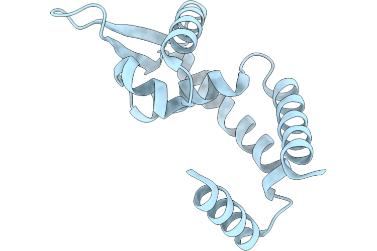 |
Hosa Transcriptional Regulator From Enteropathogenic Escherichia Coli O127:H6 (Strain E2348/69) Apo-Protein Structure At 2.03 Angstrom Resolution
Organism: Escherichia coli o127:h6 str. e2348/69
Method: X-RAY DIFFRACTION Resolution:2.03 Å Release Date: 2026-09-23 Classification: DNA BINDING PROTEIN |
Organism: Escherichia coli o127:h6 str. e2348/69
Method: X-RAY DIFFRACTION
Release Date: 2026-09-23
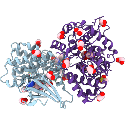 |
Crystal Structure Of Selective Inhibitor 16 Bound At The Active Site Of Cdk2
Organism: Homo sapiens
Method: X-RAY DIFFRACTION Resolution:1.59 Å Release Date: 2026-09-02 Classification: SIGNALING PROTEIN Ligands: MG, EDO, CL, DMS, A1DEQ, IPA |
Organism: Homo sapiens
Method: X-RAY DIFFRACTION
Release Date: 2026-09-02
Ligands: MG, EDO, CL, DMS, A1DEQ, IPA
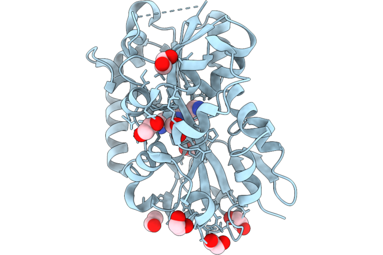 |
Crystal Structure Of A Spermidine Binding Protein Isolated From Synechococcus Cc9311
Organism: Synechococcus sp. cc9311
Method: X-RAY DIFFRACTION Resolution:1.80 Å Release Date: 2026-07-22 Classification: TRANSPORT PROTEIN Ligands: EDO, SPD, CL |
Organism: Synechococcus sp. cc9311
Method: X-RAY DIFFRACTION
Release Date: 2026-07-22
Ligands: EDO, SPD, CL
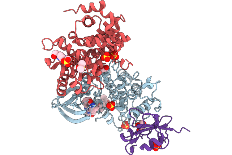 |
Crystal Structure Of Selective Inhibitor 16 Bound At The Active Site Of Cdk1
Organism: Homo sapiens
Method: X-RAY DIFFRACTION Resolution:2.40 Å Release Date: 2026-07-08 Classification: SIGNALING PROTEIN Ligands: A1DEQ, A1DJL, GOL, SO4, DMS, EPE |
Organism: Homo sapiens
Method: X-RAY DIFFRACTION
Release Date: 2026-07-08
Ligands: A1DEQ, A1DJL, GOL, SO4, DMS, EPE
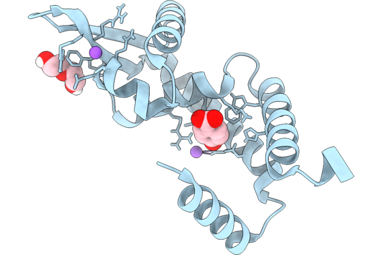 |
Hosa Transcriptional Regulator From Enteropathogenic Escherichia Coli O127:H6 (Strain E2348/69) Bound With 4-Hydroxy Benzoic Acid - L2' Conformation At 1.41 Angstrom Resolution
Organism: Escherichia coli o127:h6 str. e2348/69
Method: X-RAY DIFFRACTION Resolution:1.41 Å Release Date: 2026-06-17 Classification: DNA BINDING PROTEIN Ligands: PHB, PEG, CL, NA |
Organism: Escherichia coli o127:h6 str. e2348/69
Method: X-RAY DIFFRACTION
Release Date: 2026-06-17
Ligands: PHB, PEG, CL, NA
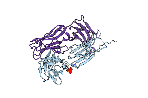 |
Chilob 7/4 H2 Hc-T219C/C224S Kappa Lc-E123C/C214S F(Ab')2
Organism: Homo sapiens
Method: X-RAY DIFFRACTION Resolution:2.67 Å Release Date: 2025-03-05 Classification: IMMUNE SYSTEM Ligands: SO4 |
Organism: Homo sapiens
Method: X-RAY DIFFRACTION
Release Date: 2025-03-05
Ligands: SO4
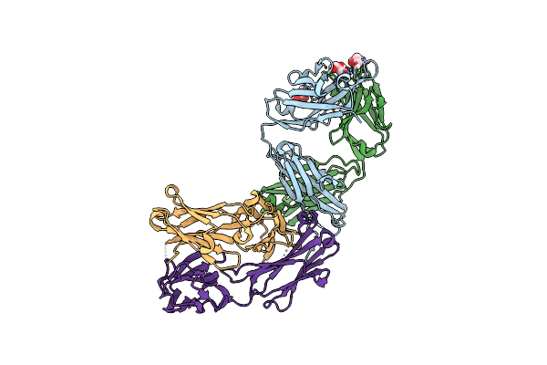 |
Chilob 7/4 H2 Hc-K223C/C224S Kappa Lc-C214S F(Ab')2
Organism: Homo sapiens
Method: X-RAY DIFFRACTION Resolution:1.93 Å Release Date: 2025-03-05 Classification: IMMUNE SYSTEM Ligands: GOL |
Organism: Homo sapiens
Method: X-RAY DIFFRACTION
Release Date: 2025-03-05
Ligands: GOL
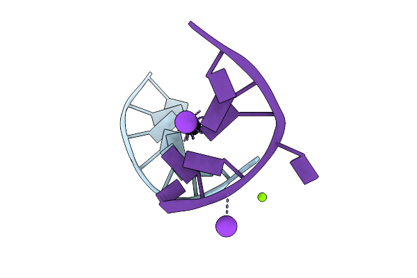 |
Crystal Structure Of Parallel G-Quadruplex Containing T-Tetrads And Tg-Octaplet
Organism: Synthetic construct
Method: X-RAY DIFFRACTION Resolution:1.15 Å Release Date: 2025-01-15 Classification: DNA Ligands: K, MG, NA |
Organism: Synthetic construct
Method: X-RAY DIFFRACTION
Release Date: 2025-01-15
Ligands: K, MG, NA
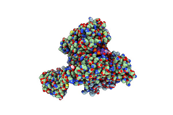 |
Experimental Localization Of Metal-Binding Sites Reveals The Role Of Metal Ions In The Delafloxacin-Stabilized Streptococcus Pneumoniae Topoisomerase Iv Dna Cleavage Complex
Organism: Streptococcus pneumoniae, Escherichia coli
Method: X-RAY DIFFRACTION Resolution:2.62 Å Release Date: 2024-10-16 Classification: DNA BINDING PROTEIN Ligands: MPD, MG, K, CL, ACT, TE9 |
Organism: Streptococcus pneumoniae, Escherichia coli
Method: X-RAY DIFFRACTION
Release Date: 2024-10-16
Ligands: MPD, MG, K, CL, ACT, TE9
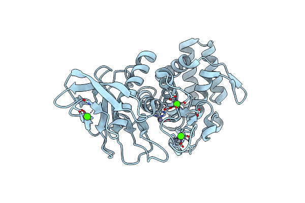 |
Identification Of Calcium Ions In Thermolysin.
Organism: Bacillus thermoproteolyticus
Method: X-RAY DIFFRACTION Resolution:2.10 Å Release Date: 2024-10-02 Classification: HYDROLASE Ligands: CA, ZN |
Organism: Bacillus thermoproteolyticus
Method: X-RAY DIFFRACTION
Release Date: 2024-10-02
Ligands: CA, ZN
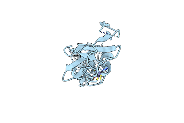 |
Identification Of Zinc Ions In Lmo4.
Organism: Homo sapiens
Method: X-RAY DIFFRACTION Resolution:1.80 Å Release Date: 2024-10-02 Classification: HYDROLASE Ligands: ZN |
Organism: Homo sapiens
Method: X-RAY DIFFRACTION
Release Date: 2024-10-02
Ligands: ZN
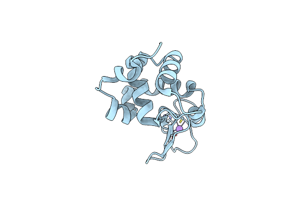 |
Identification Of Chloride Ions In Lysozyme At Long Wavelengths
Organism: Gallus gallus
Method: X-RAY DIFFRACTION Resolution:2.70 Å Release Date: 2024-10-02 Classification: HYDROLASE |
Organism: Gallus gallus
Method: X-RAY DIFFRACTION
Release Date: 2024-10-02
 |
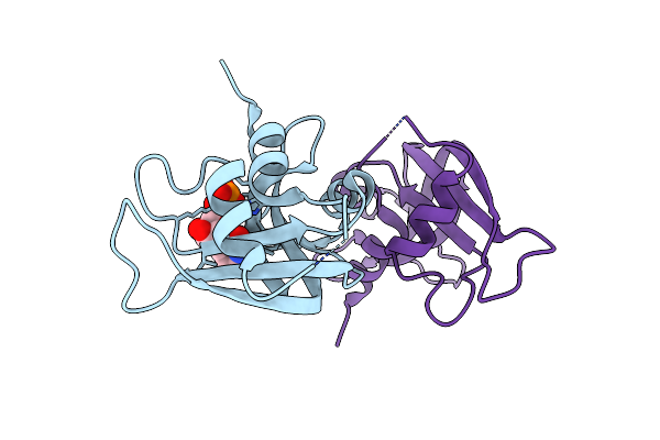
Structure Of A2A Adenosine Receptor A2Ar-Star2-Bril, Solved At Wavelength 2.75 A
Organism: Homo sapiens, Escherichia coli
Method: X-RAY DIFFRACTION Resolution:2.40 Å Release Date: 2023-10-25 Classification: MEMBRANE PROTEIN Ligands: TEP, OLA, CLR |
Organism: Homo sapiens, Escherichia coli
Method: X-RAY DIFFRACTION
Release Date: 2023-10-25
Ligands: TEP, OLA, CLR
 |
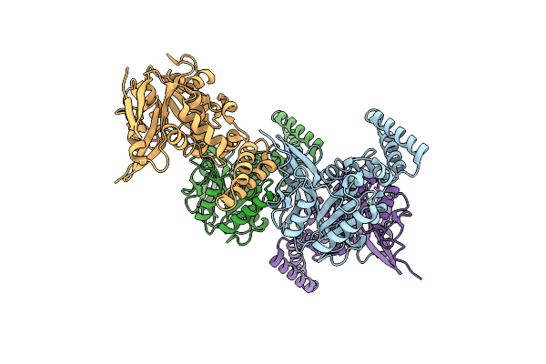
Structure Of Ribonuclease A, Solved At Wavelength 2.75 A
Organism: Bos taurus
Method: X-RAY DIFFRACTION Resolution:1.80 Å Release Date: 2023-10-25 Classification: MEMBRANE PROTEIN Ligands: 0U |
Organism: Bos taurus
Method: X-RAY DIFFRACTION
Release Date: 2023-10-25
Ligands: 0U
 |
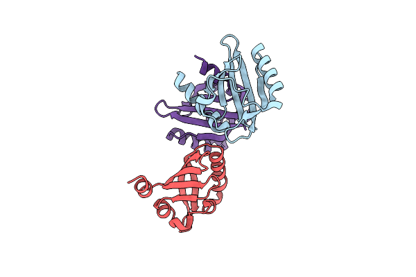
Structure Of Salmonella Effector Ssek3, Solved At Wavelength 2.75 A
Organism: Salmonella enterica subsp. enterica serovar typhimurium
Method: X-RAY DIFFRACTION Resolution:2.10 Å Release Date: 2023-10-25 Classification: TOXIN |
Organism: Salmonella enterica subsp. enterica serovar typhimurium
Method: X-RAY DIFFRACTION
Release Date: 2023-10-25
 |
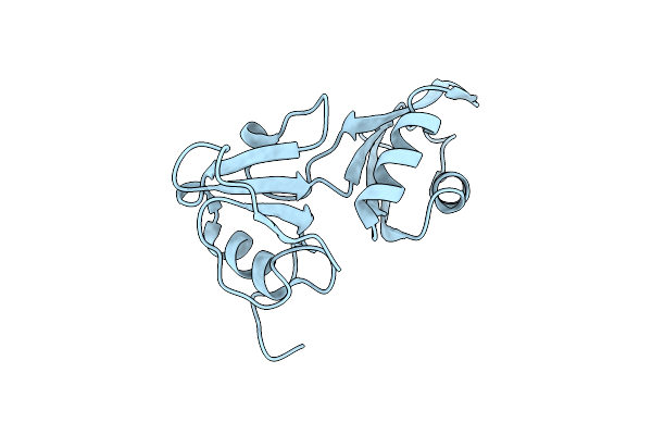
Structure Of The Pas Domain Code By The Lic_11128 Gene From Leptospira Interrogans Serovar Copenhageni Fiocruz, Solved At Wavelength 3.09 A
Organism: Leptospira interrogans serovar copenhageni
Method: X-RAY DIFFRACTION Resolution:2.50 Å Release Date: 2023-10-25 Classification: UNKNOWN FUNCTION |
Organism: Leptospira interrogans serovar copenhageni
Method: X-RAY DIFFRACTION
Release Date: 2023-10-25
 |
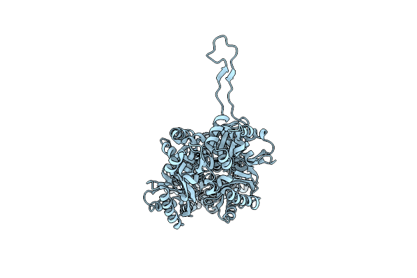
Structure Of The Rna Recognition Motif (Rrm) Of Seb1 From S. Pombe., Solved At Wavelength 2.75 A
Organism: Schizosaccharomyces pombe 972h-
Method: X-RAY DIFFRACTION Resolution:1.77 Å Release Date: 2023-10-25 Classification: TRANSCRIPTION |
Organism: Schizosaccharomyces pombe 972h-
Method: X-RAY DIFFRACTION
Release Date: 2023-10-25
 |
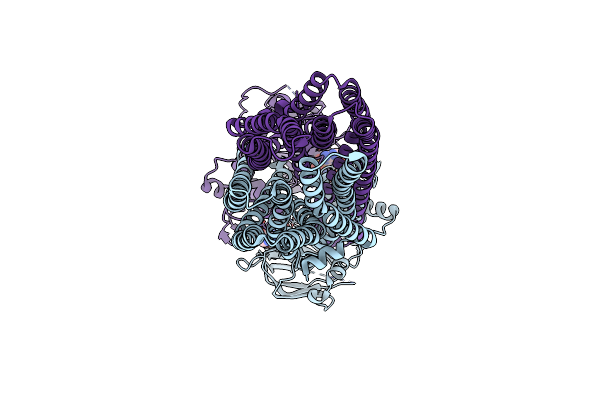
Structure Of Bacterial Multidrug Efflux Transporter Acrb, Solved At Wavelength 3.02 A
Organism: Escherichia coli k-12
Method: X-RAY DIFFRACTION Resolution:3.40 Å Release Date: 2023-10-25 Classification: MEMBRANE PROTEIN |
Organism: Escherichia coli k-12
Method: X-RAY DIFFRACTION
Release Date: 2023-10-25
 |
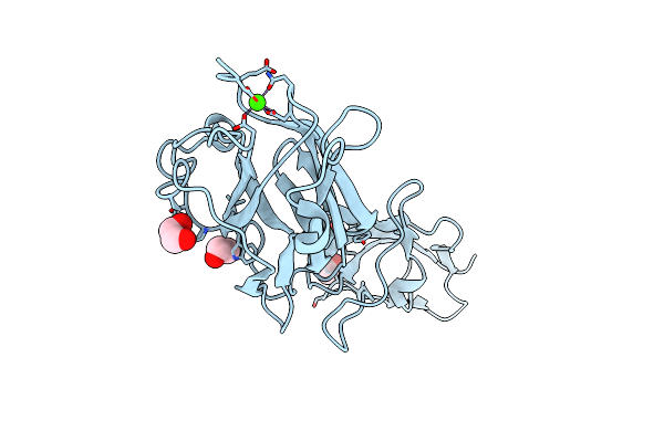
Structure Of The Antibacterial Peptide Abc Transporter Mcjd, Solved At Wavelength 2.75 A
Organism: Escherichia coli
Method: X-RAY DIFFRACTION Resolution:2.80 Å Release Date: 2023-10-25 Classification: MEMBRANE PROTEIN Ligands: MG, ANP |
Organism: Escherichia coli
Method: X-RAY DIFFRACTION
Release Date: 2023-10-25
Ligands: MG, ANP
 |
Structure Of Fap1, A Domain Of The Accessory Sec-Dependent Serine-Rich Glycoprotein Adhesin From Streptococcus Oralis, Solved At Wavelength 3.06 A
Organism: Streptococcus oralis
Method: X-RAY DIFFRACTION Resolution:1.97 Å Release Date: 2023-10-25 Classification: CELL ADHESION Ligands: CA, EDO |
Organism: Streptococcus oralis
Method: X-RAY DIFFRACTION
Release Date: 2023-10-25
Ligands: CA, EDO