
Search Count: 85,432
 |
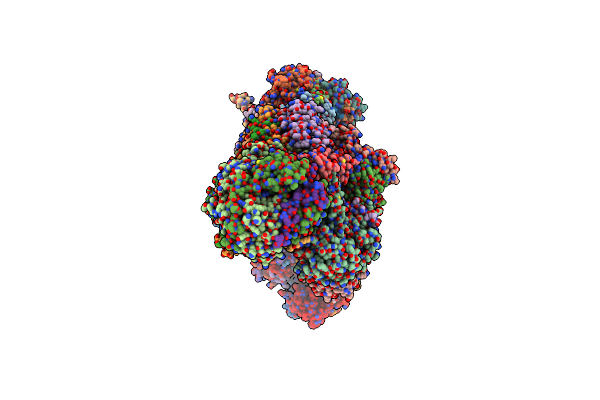
Organism: Bos taurus
Method: ELECTRON MICROSCOPY Release Date: 2022-05-18 Classification: OXIDOREDUCTASE Ligands: PC1, 3PE, FES, CDL, FMN, SF4, CU, HEA, MG, ZN, NAP, HEM, HEC, UQ2 |
 |
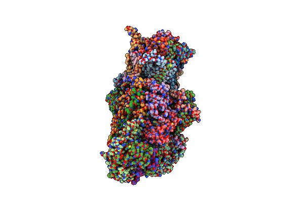
Organism: Bos taurus
Method: ELECTRON MICROSCOPY Release Date: 2022-05-18 Classification: OXIDOREDUCTASE Ligands: FES, CDL, 3PE, PC1, FMN, SF4, ZN, NAP, HEM, HEC, HEA, CU, MG |
 |
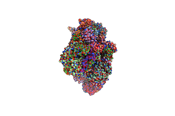
Organism: Bos taurus
Method: ELECTRON MICROSCOPY Release Date: 2022-05-18 Classification: OXIDOREDUCTASE Ligands: HEM, HEC, FES, 3PE, CDL, FMN, SF4, ZN, NAP, PC1, HEA, CU, MG |
 |
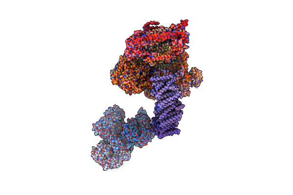
Organism: Bos taurus
Method: ELECTRON MICROSCOPY Release Date: 2022-05-18 Classification: OXIDOREDUCTASE Ligands: FES, 3PE, CDL, PC1, FMN, SF4, ZN, NAP, HEM, HEC, HEA, CU, MG |
 |
Organism: Homo sapiens, Bos taurus
Method: ELECTRON MICROSCOPY Release Date: 2017-09-13 Classification: OXIDOREDUCTASE/ELECTRON TRANSPORT Ligands: SF4, FMN, PLX, 8Q1, NDP, FES, CDL, PEE, CU, MG, HEA, ZN, HEC, HEM |
 |
Organism: Homo sapiens, Bos taurus
Method: ELECTRON MICROSCOPY Release Date: 2017-09-13 Classification: OXIDOREDUCTASE/ELECTRON TRANSPORT Ligands: SF4, FMN, PLX, 8Q1, NDP, FES, CDL, PEE, CU, MG, HEA, ZN, HEC, HEM |
 |
 |
 |
 |
Cryoem Structure Of Mitochondrial Complex I From Chaetomium Thermophilum (State 1)
Organism: Chaetomium thermophilum var. thermophilum dsm 1495
Method: ELECTRON MICROSCOPY Release Date: 2022-11-30 Classification: OXIDOREDUCTASE Ligands: 3PE, PC1, CDL, LMN, FES, SF4, FMN, NDP, ZN, ZMP |
 |
Cryoem Structure Of Mitochondrial Complex I From Chaetomium Thermophilum (Inhibited By Ddm)
Organism: Chaetomium thermophilum var. thermophilum dsm 1495
Method: ELECTRON MICROSCOPY Release Date: 2022-11-30 Classification: OXIDOREDUCTASE Ligands: PC1, LMT, CDL, 3PE, FES, SF4, FMN, NDP, ZN, ZMP |
 |
Cryoem Structure Of Mitochondrial Complex I From Chaetomium Thermophilum (State 2)
Organism: Chaetomium thermophilum var. thermophilum dsm 1495
Method: ELECTRON MICROSCOPY Release Date: 2022-11-30 Classification: OXIDOREDUCTASE Ligands: 3PE, PC1, CDL, LMN, FES, SF4, FMN, NDP, ZN, ZMP |
 |
Fitted Model For Bovine Mitochondrial Supercomplex I1Iii2Iv1 By Single Particle Cryo-Em (Emd-1876)
Organism: Thermus thermophilus, Bos taurus, Escherichia coli
Method: ELECTRON MICROSCOPY Resolution:19.00 Å Release Date: 2011-10-19 Classification: OXIDOREDUCTASE Ligands: SF4, FMN, NAI, MG, FES, CA, HEM, SMA, UQ1, HEC, CDL, HEA, CU, ZN |
 |
 |
Respiratory Complex Ci:Ciii2, Type Ib, Wild Type Mouse Under Cold Temperature
Organism: Mus musculus
Method: ELECTRON MICROSCOPY Release Date: 2024-09-18 Classification: ELECTRON TRANSPORT Ligands: SF4, UQ1, PC1, 3PE, FES, FMN, UQ9, CDL, ADP, NDP, ZN, EHZ, HEM, U10, UQ6, HEC |
 |
Respiratory Complex Ci:Ciii2, Type I, Perk -/- Mouse Under Cold Temperature
Organism: Mus musculus
Method: ELECTRON MICROSCOPY Release Date: 2024-09-18 Classification: ELECTRON TRANSPORT Ligands: SF4, UQ1, PC1, 3PE, FES, FMN, UQ9, CDL, ZN, ADP, NDP, EHZ, HEM, UQ6, HEC, U10 |
 |
Respiratory Complex Ci:Ciii2, Type Ii, Wild Type Mouse Under Cold Temperature
|
 |
 |
 |
Respiratory Complex Ci:Ciii2, Type Ia, Wild Type Mouse Under Cold Temperature
|