

Search Count: 99
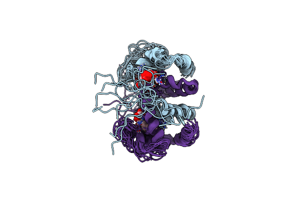 |
Nmr Study Of Holo Trp Repressor
Organism: Escherichia coli
Method: SOLUTION NMR Release Date: 1996-06-20 Classification: COMPLEX (OPERON REPRESSOR/PEPTIDE) Ligands: TRP |
Organism: Escherichia coli
Method: SOLUTION NMR
Release Date: 1996-06-20
Ligands: TRP
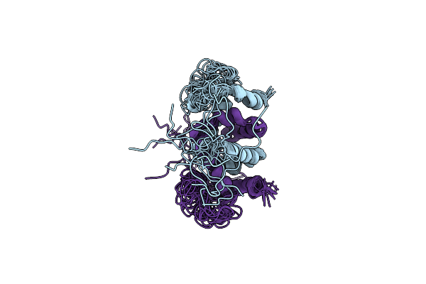 |
Nmr Study Of Apo Trp Repressor
Organism: Escherichia coli
Method: SOLUTION NMR Release Date: 1996-06-20 Classification: OPERON REPRESSOR |
Organism: Escherichia coli
Method: SOLUTION NMR
Release Date: 1996-06-20
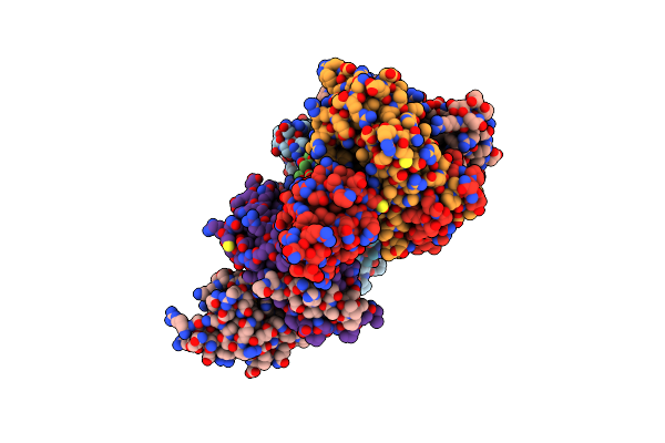 |
Crystal Structure Of The Mexr Repressor Of The Mexab-Oprm Multidrug Efflux Operon Of Pseudomonas Aeruginosa
Organism: Pseudomonas aeruginosa
Method: X-RAY DIFFRACTION Resolution:2.10 Å Release Date: 2002-09-11 Classification: TRANSCRIPTION REPRESSOR |
Organism: Pseudomonas aeruginosa
Method: X-RAY DIFFRACTION
Release Date: 2002-09-11
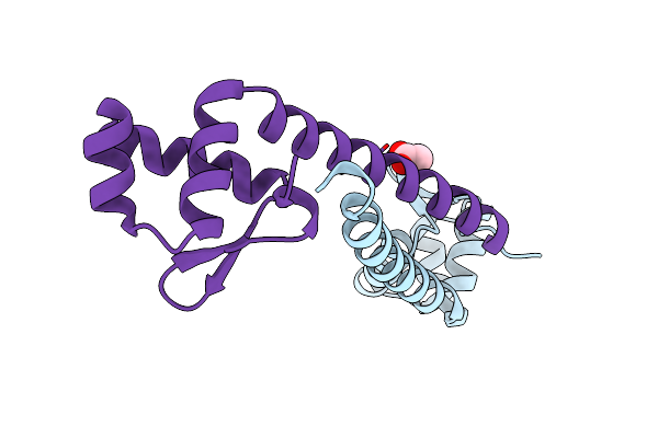 |
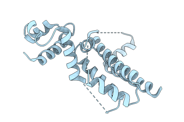
Crystal Structure Of The Putative Arsenical Resistance Operon Repressor From Archaeoglobus Fulgidus
Organism: Archaeoglobus fulgidus
Method: X-RAY DIFFRACTION Resolution:1.60 Å Release Date: 2004-12-28 Classification: TRANSCRIPTION REPRESSOR Ligands: ACT |
Organism: Archaeoglobus fulgidus
Method: X-RAY DIFFRACTION
Release Date: 2004-12-28
Ligands: ACT
 |
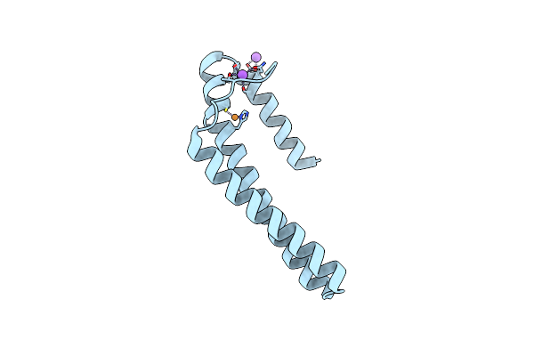
Crystal Structure Of The Engineered Sulfonylurea Repressor Esr L7-D01, Apo Form
Organism: Escherichia coli
Method: X-RAY DIFFRACTION Resolution:3.10 Å Release Date: 2025-10-08 Classification: DNA BINDING PROTEIN |
Organism: Escherichia coli
Method: X-RAY DIFFRACTION
Release Date: 2025-10-08
 |
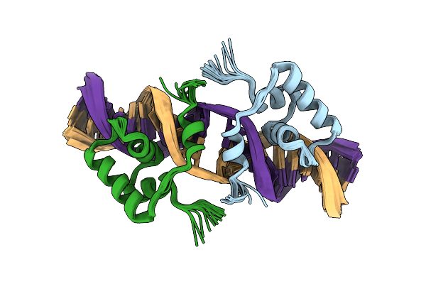
Crystal Structure Of The Copper-Sensing Repressor Csor With Cu(I) From Geobacillus Thermodenitrificans Ng80-2
Organism: Geobacillus thermodenitrificans
Method: X-RAY DIFFRACTION Resolution:2.56 Å Release Date: 2014-05-21 Classification: TRANSCRIPTION REPRESSOR Ligands: CU1, NA |
Organism: Geobacillus thermodenitrificans
Method: X-RAY DIFFRACTION
Release Date: 2014-05-21
Ligands: CU1, NA
 |
Nmr Structure Of A Protein-Dna Complex Of An Altered Specificity Mutant Of The Lac Repressor Headpiece That Mimics The Gal Repressor
Organism: Escherichia coli
Method: SOLUTION NMR Release Date: 2005-10-18 Classification: TRANSCRIPTION REGULATOR |
Organism: Escherichia coli
Method: SOLUTION NMR
Release Date: 2005-10-18
 |
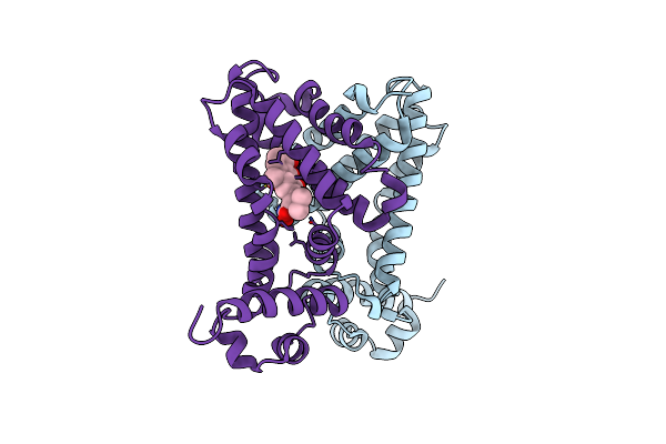
Trehalose Repressor From Escherichia Coli
Organism: Escherichia coli k-12
Method: X-RAY DIFFRACTION Resolution:2.40 Å Release Date: 2015-02-11 Classification: GENE REGULATION |
Organism: Escherichia coli k-12
Method: X-RAY DIFFRACTION
Release Date: 2015-02-11
 |
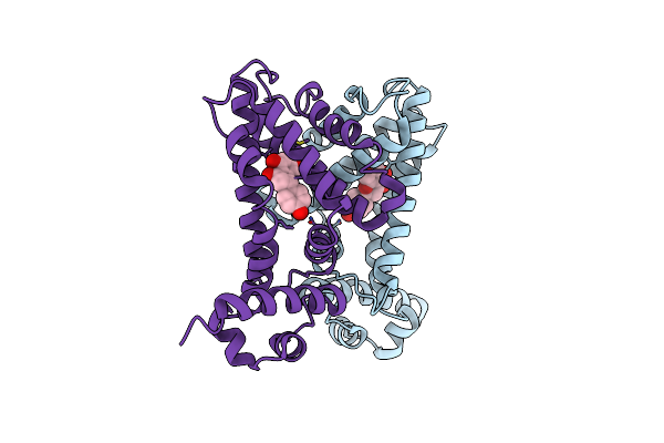
Ttgr In Complex With Tetracycline
Organism: Pseudomonas putida
Method: X-RAY DIFFRACTION Resolution:2.70 Å Release Date: 2007-05-08 Classification: TRANSCRIPTION Ligands: TAC |
Organism: Pseudomonas putida
Method: X-RAY DIFFRACTION
Release Date: 2007-05-08
Ligands: TAC
 |
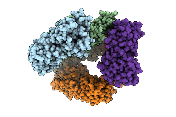
Ttgr In Complex With Naringenin
Organism: Pseudomonas putida
Method: X-RAY DIFFRACTION Resolution:2.30 Å Release Date: 2007-05-08 Classification: TRANSCRIPTION Ligands: NAR |
Organism: Pseudomonas putida
Method: X-RAY DIFFRACTION
Release Date: 2007-05-08
Ligands: NAR
 |
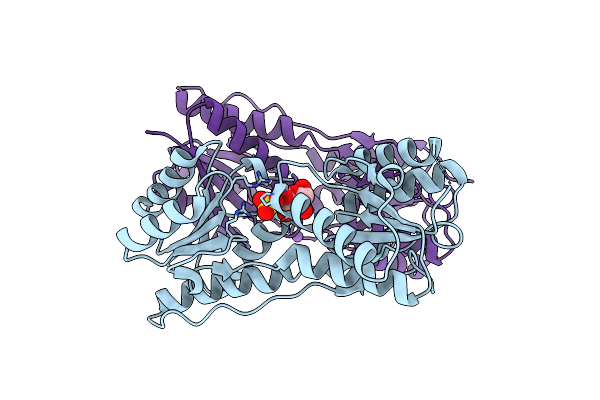
Crystal Structure Of The Ribose Operon Repressor, Rbsr, Bound To Ribose
Organism: Escherichia coli
Method: X-RAY DIFFRACTION Resolution:2.07 Å Release Date: 2025-07-16 Classification: DNA BINDING PROTEIN Ligands: RIP |
Organism: Escherichia coli
Method: X-RAY DIFFRACTION
Release Date: 2025-07-16
Ligands: RIP
 |
Crystal Structure Of The Escherichia Coli Transcriptional Repressor Ascg
Organism: Escherichia coli
Method: X-RAY DIFFRACTION Resolution:2.00 Å Release Date: 2008-01-22 Classification: TRANSCRIPTION REGULATOR Ligands: FRU, SO4, NA |
Organism: Escherichia coli
Method: X-RAY DIFFRACTION
Release Date: 2008-01-22
Ligands: FRU, SO4, NA
 |
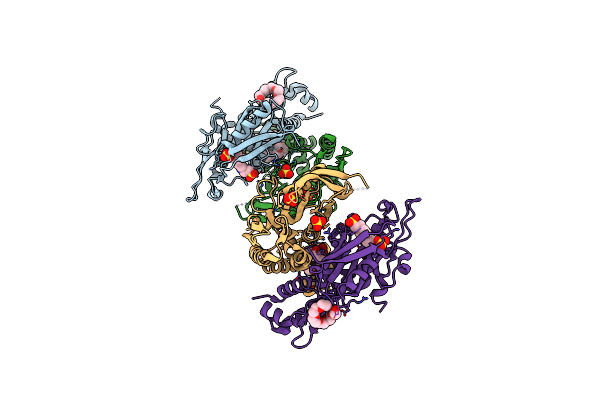
Crystal Structure Of The Ribose Operon Repressor, Rbsr, Bound To Ribose Operon
Organism: Escherichia coli
Method: X-RAY DIFFRACTION Resolution:2.10 Å Release Date: 2025-07-16 Classification: DNA BINDING PROTEIN/DNA Ligands: SO4 |
Organism: Escherichia coli
Method: X-RAY DIFFRACTION
Release Date: 2025-07-16
Ligands: SO4
 |
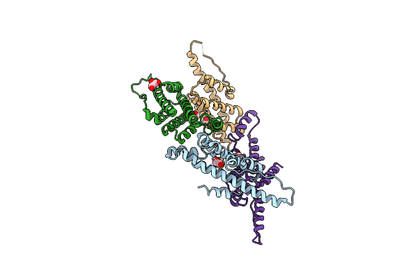
Crystal Structure Of The Purine Operon Repressor Of Bacillus Subtilis
Organism: Bacillus subtilis
Method: X-RAY DIFFRACTION Resolution:2.20 Å Release Date: 2003-08-26 Classification: DNA BINDING PROTEIN Ligands: SO4, EPE, P6G, 2PE, PG4, 1PE |
Organism: Bacillus subtilis
Method: X-RAY DIFFRACTION
Release Date: 2003-08-26
Ligands: SO4, EPE, P6G, 2PE, PG4, 1PE
 |
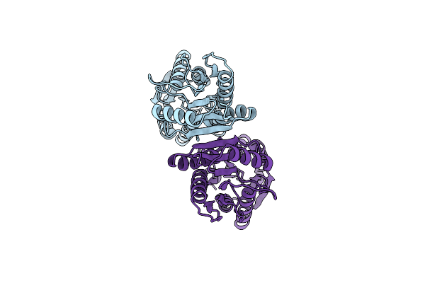
Crystal Structure Of Mannitol Operon Repressor (Mtlr) From Vibrio Parahaemolyticus Rimd 2210633
Organism: Vibrio parahaemolyticus rimd 2210633
Method: X-RAY DIFFRACTION Resolution:2.75 Å Release Date: 2008-01-15 Classification: TRANSCRIPTION Ligands: EDO, GOL |
Organism: Vibrio parahaemolyticus rimd 2210633
Method: X-RAY DIFFRACTION
Release Date: 2008-01-15
Ligands: EDO, GOL
 |
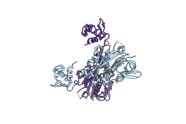
Crystal Structure Of Periplasmic Binding Ribose Operon Repressor Protein From Lactobacillus Acidophilus
Organism: Lactobacillus acidophilus
Method: X-RAY DIFFRACTION Resolution:1.60 Å Release Date: 2009-06-23 Classification: transcription regulator |
Organism: Lactobacillus acidophilus
Method: X-RAY DIFFRACTION
Release Date: 2009-06-23
 |
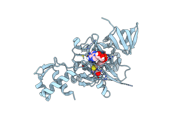
Bifunctional Ligase/Repressor Bira From Klebsiella Pneumoniae (Domain Swapped Dimer)
Organism: Klebsiella pneumoniae subsp. pneumoniae 1158
Method: X-RAY DIFFRACTION Resolution:2.85 Å Release Date: 2022-11-30 Classification: TRANSCRIPTION Ligands: IMD |
Organism: Klebsiella pneumoniae subsp. pneumoniae 1158
Method: X-RAY DIFFRACTION
Release Date: 2022-11-30
Ligands: IMD
 |
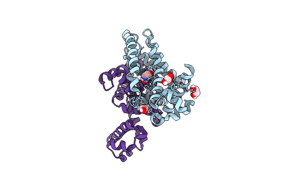
Structure Of E. Coli Bira G142A Bound To Biotinol-5'-Amp
Organism: Escherichia coli
Method: X-RAY DIFFRACTION Resolution:2.31 Å Release Date: 2014-10-22 Classification: LIGASE Ligands: BTX |
Organism: Escherichia coli
Method: X-RAY DIFFRACTION
Release Date: 2014-10-22
Ligands: BTX
 |
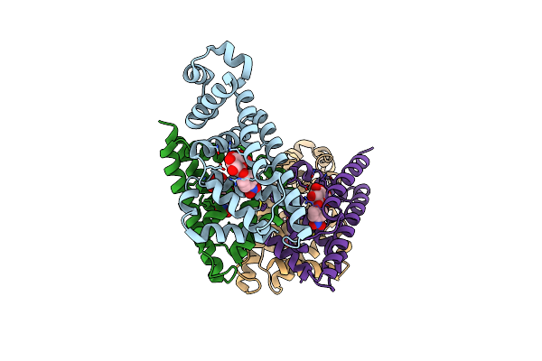
Structure Of K52-Acetylated Rutr In Complex With Uracil.
Organism: Escherichia coli k-12
Method: X-RAY DIFFRACTION Resolution:2.25 Å Release Date: 2022-06-01 Classification: TRANSCRIPTION Ligands: URA, EDO |
Organism: Escherichia coli k-12
Method: X-RAY DIFFRACTION
Release Date: 2022-06-01
Ligands: URA, EDO
 |
Escherichia Coli Gusr
Organism: Escherichia coli o157:h7
Method: X-RAY DIFFRACTION Resolution:2.09 Å Release Date: 2017-12-20 Classification: TRANSCRIPTION Ligands: C3G |
Organism: Escherichia coli o157:h7
Method: X-RAY DIFFRACTION
Release Date: 2017-12-20
Ligands: C3G