

Search Count: 10
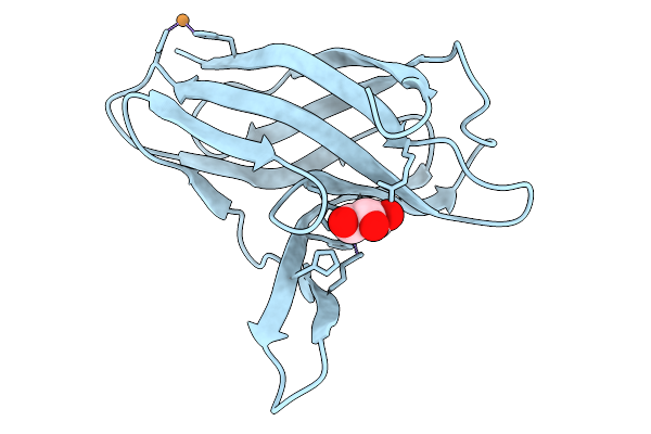 |
Crystal Structure Of Metallochaperone Acca From Neisseria Gonorrhoeae
Organism: Neisseria meningitidis serogroup a
Method: X-RAY DIFFRACTION Resolution:2.90 Å Release Date: 2026-03-25 Classification: METAL BINDING PROTEIN Ligands: CU, GOL |
Organism: Neisseria meningitidis serogroup a
Method: X-RAY DIFFRACTION
Release Date: 2026-03-25
Ligands: CU, GOL
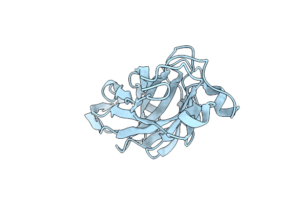 |
Crystal Structure Of The Ucad Lectin-Binding Domain
Organism: Proteus mirabilis
Method: X-RAY DIFFRACTION Resolution:1.62 Å Release Date: 2022-07-06 Classification: SUGAR BINDING PROTEIN Ligands: CL |
Organism: Proteus mirabilis
Method: X-RAY DIFFRACTION
Release Date: 2022-07-06
Ligands: CL
 |
Crystal Structure Of The Ucld Lectin-Binding Domain
Organism: Escherichia coli
Method: X-RAY DIFFRACTION Resolution:2.20 Å Release Date: 2022-07-06 Classification: SUGAR BINDING PROTEIN Ligands: IOD |
Organism: Escherichia coli
Method: X-RAY DIFFRACTION
Release Date: 2022-07-06
Ligands: IOD
 |
Crystal Structure Of The Ucad Lectin-Binding Domain In Complex With Fucose
Organism: Proteus mirabilis
Method: X-RAY DIFFRACTION Resolution:1.50 Å Release Date: 2022-07-06 Classification: SUGAR BINDING PROTEIN Ligands: FUL, CL |
Organism: Proteus mirabilis
Method: X-RAY DIFFRACTION
Release Date: 2022-07-06
Ligands: FUL, CL
 |
Crystal Structure Of The Ucad Lectin-Binding Domain In Complex With Glucose
Organism: Proteus mirabilis
Method: X-RAY DIFFRACTION Resolution:1.78 Å Release Date: 2022-07-06 Classification: SUGAR BINDING PROTEIN Ligands: BGC, CL |
Organism: Proteus mirabilis
Method: X-RAY DIFFRACTION
Release Date: 2022-07-06
Ligands: BGC, CL
 |
Crystal Structure Of The Ucad Lectin-Binding Domain In Complex With Galactose
Organism: Proteus mirabilis
Method: X-RAY DIFFRACTION Resolution:1.72 Å Release Date: 2022-07-06 Classification: SUGAR BINDING PROTEIN Ligands: GLA, CL |
Organism: Proteus mirabilis
Method: X-RAY DIFFRACTION
Release Date: 2022-07-06
Ligands: GLA, CL
 |
Crystal Structure Of The Molybdate-Binding Periplasmic Protein Moda From The Bacteria Pseudomonsa Aeruginosa In Ligand-Free Form
Organism: Pseudomonas aeruginosa pa1
Method: X-RAY DIFFRACTION Resolution:1.78 Å Release Date: 2022-05-04 Classification: METAL BINDING PROTEIN Ligands: GOL, NH4, SO4 |
Organism: Pseudomonas aeruginosa pa1
Method: X-RAY DIFFRACTION
Release Date: 2022-05-04
Ligands: GOL, NH4, SO4
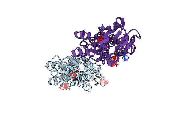 |
Crystal Structure Of The Molybdate-Binding Periplasmic Protein Moda From The Bacteria Pseudomonsa Aeruginosa In Chromate-Bound Form
Organism: Pseudomonas aeruginosa pa1
Method: X-RAY DIFFRACTION Resolution:1.90 Å Release Date: 2022-05-04 Classification: METAL BINDING PROTEIN Ligands: CQ4, NH4, GOL |
Organism: Pseudomonas aeruginosa pa1
Method: X-RAY DIFFRACTION
Release Date: 2022-05-04
Ligands: CQ4, NH4, GOL
 |
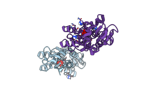
Crystal Structure Of The Molybdate-Binding Periplasmic Protein Moda From The Bacteria Pseudomonsa Aeruginosa In Molybdate-Bound Form
Organism: Pseudomonas aeruginosa pa1
Method: X-RAY DIFFRACTION Resolution:2.50 Å Release Date: 2022-05-04 Classification: METAL BINDING PROTEIN Ligands: NH4, MOO, GOL |
Organism: Pseudomonas aeruginosa pa1
Method: X-RAY DIFFRACTION
Release Date: 2022-05-04
Ligands: NH4, MOO, GOL
 |
Crystal Structure Of The Molybdate-Binding Periplasmic Protein Moda From The Bacteria Pseudomonsa Aeruginosa In Tungstate-Bound Form
Organism: Pseudomonas aeruginosa pa1
Method: X-RAY DIFFRACTION Resolution:2.16 Å Release Date: 2022-05-04 Classification: METAL BINDING PROTEIN Ligands: WO4, NH4 |
Organism: Pseudomonas aeruginosa pa1
Method: X-RAY DIFFRACTION
Release Date: 2022-05-04
Ligands: WO4, NH4