

Search Count: 25
 |
Crystal Structure Of Human Rac1-Gdp
Organism: Homo sapiens
Method: X-RAY DIFFRACTION Resolution:1.80 Å Release Date: 2025-09-03 Classification: SIGNALING PROTEIN Ligands: MG, GDP |
Organism: Homo sapiens
Method: X-RAY DIFFRACTION
Release Date: 2025-09-03
Ligands: MG, GDP
 |
Crystal Structure Of Human Rac1-Gdp-Pi
Organism: Homo sapiens
Method: X-RAY DIFFRACTION Resolution:2.27 Å Release Date: 2025-09-03 Classification: SIGNALING PROTEIN Ligands: GDP, MG, PO4 |
Organism: Homo sapiens
Method: X-RAY DIFFRACTION
Release Date: 2025-09-03
Ligands: GDP, MG, PO4
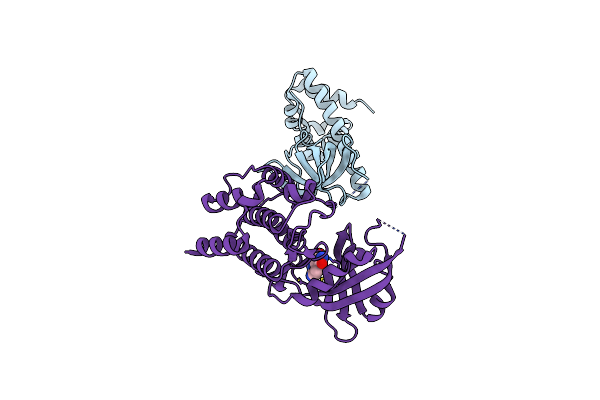 |
Pkr Kinase Domain- Eif2Alpha In Complex With Compound
Organism: Saccharomyces cerevisiae, Homo sapiens
Method: X-RAY DIFFRACTION Resolution:2.45 Å Release Date: 2023-09-13 Classification: RNA BINDING PROTEIN Ligands: UH3 |
Organism: Saccharomyces cerevisiae, Homo sapiens
Method: X-RAY DIFFRACTION
Release Date: 2023-09-13
Ligands: UH3
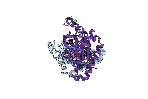 |
Crystal Structure Of Human Sting In Complex With Adu-S100
Organism: Homo sapiens
Method: X-RAY DIFFRACTION Resolution:2.17 Å Release Date: 2022-11-30 Classification: IMMUNE SYSTEM Ligands: GJF, CA, NA |
Organism: Homo sapiens
Method: X-RAY DIFFRACTION
Release Date: 2022-11-30
Ligands: GJF, CA, NA
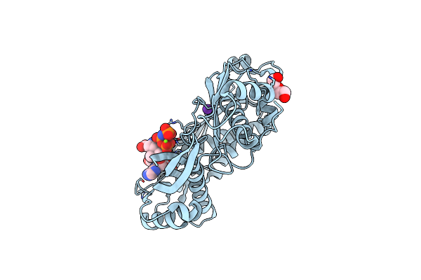 |
Crystal Structure Of Human Methionine Adenosyltransferase 2A (Mat2A) In Complex With S-Adenosylmethionine, Adenosin And Diphosphono-Aminophosphonic Acid.
Organism: Homo sapiens
Method: X-RAY DIFFRACTION Resolution:1.15 Å Release Date: 2022-10-05 Classification: TRANSFERASE Ligands: PPK, MG, K, SAM, ADN, PEG, GOL |
Organism: Homo sapiens
Method: X-RAY DIFFRACTION
Release Date: 2022-10-05
Ligands: PPK, MG, K, SAM, ADN, PEG, GOL
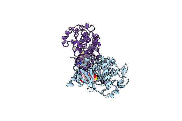 |
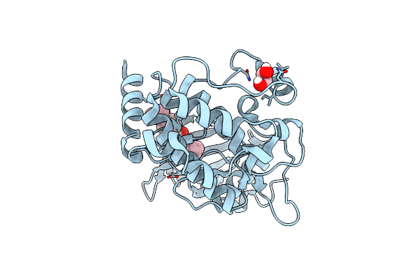
Crystal Structure Of Mst2 In Complex With Xmu-Mp-1
Organism: Homo sapiens
Method: X-RAY DIFFRACTION Resolution:1.90 Å Release Date: 2022-07-20 Classification: TRANSCRIPTION Ligands: 5BS, NA |
Organism: Homo sapiens
Method: X-RAY DIFFRACTION
Release Date: 2022-07-20
Ligands: 5BS, NA
 |
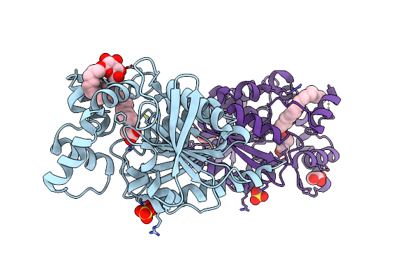
Structure Of Serine-Threonine Kinase Stk25 In Complex With Compound
Organism: Homo sapiens
Method: X-RAY DIFFRACTION Resolution:1.64 Å Release Date: 2022-04-06 Classification: ONCOPROTEIN Ligands: IB5, EDO |
Organism: Homo sapiens
Method: X-RAY DIFFRACTION
Release Date: 2022-04-06
Ligands: IB5, EDO
 |
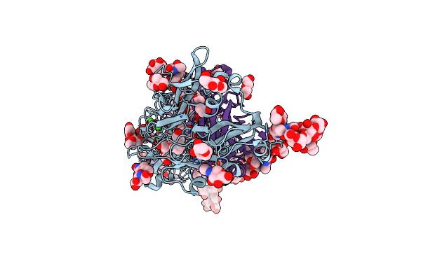
Crystal Structure Of Human Monoacylglycerol Lipase Abhd6 In Complex With Oleic Acid And Octyl Glucoside
Organism: Homo sapiens
Method: X-RAY DIFFRACTION Resolution:1.79 Å Release Date: 2021-06-23 Classification: HYDROLASE Ligands: BOG, OLA, SO4, GOL |
Organism: Homo sapiens
Method: X-RAY DIFFRACTION
Release Date: 2021-06-23
Ligands: BOG, OLA, SO4, GOL
 |
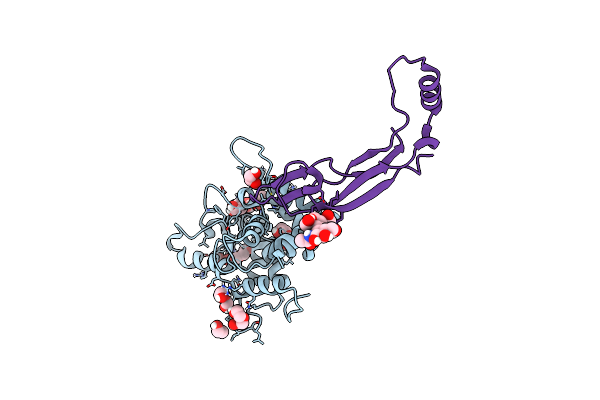
Zebrafish Ret Cadherin Like Domains 1 To 4.
Organism: Danio rerio
Method: X-RAY DIFFRACTION Resolution:2.20 Å Release Date: 2021-02-03 Classification: SIGNALING PROTEIN Ligands: CA, NAG, GOL, PGE, PEG, MES |
Organism: Danio rerio
Method: X-RAY DIFFRACTION
Release Date: 2021-02-03
Ligands: CA, NAG, GOL, PGE, PEG, MES
 |
Crystal Structure Of A Gdnf-Gfralpha1 Complex
Organism: Danio rerio
Method: X-RAY DIFFRACTION Resolution:2.20 Å Release Date: 2021-01-13 Classification: SIGNALING PROTEIN Ligands: EDO, PEG |
Organism: Danio rerio
Method: X-RAY DIFFRACTION
Release Date: 2021-01-13
Ligands: EDO, PEG
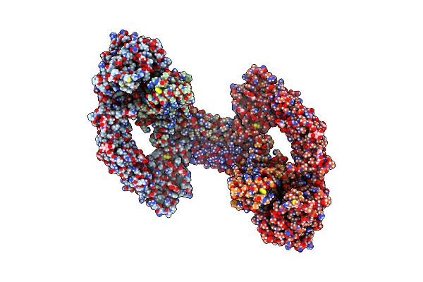 |
Ret/Gdnf/Gfra1 Extracellular Complex Cryo-Em Structure
Organism: Danio rerio
Method: ELECTRON MICROSCOPY Release Date: 2021-01-13 Classification: SIGNALING PROTEIN Ligands: CA, NAG |
Organism: Danio rerio
Method: ELECTRON MICROSCOPY
Release Date: 2021-01-13
Ligands: CA, NAG
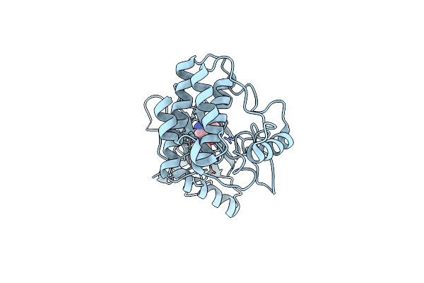 |
Structure Of Ddr2 Kinase Domain In Complex With Ibz3
Organism: Homo sapiens
Method: X-RAY DIFFRACTION Resolution:2.12 Å Release Date: 2020-12-16 Classification: MEMBRANE PROTEIN Ligands: 4VD |
Organism: Homo sapiens
Method: X-RAY DIFFRACTION
Release Date: 2020-12-16
Ligands: 4VD
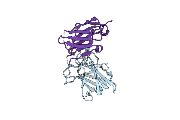 |
Structure Of Ddr2 Ds Domain In Complex With Vhh
Organism: Homo sapiens
Method: X-RAY DIFFRACTION Resolution:2.62 Å Release Date: 2020-11-25 Classification: MEMBRANE PROTEIN |
Organism: Homo sapiens
Method: X-RAY DIFFRACTION
Release Date: 2020-11-25
 |
Best Fitting Antiparallel Model For Volume 1 Of Truncated Dimeric Cytohesin-3 (Grp1; Amino Acids 14-399)
Organism: Homo sapiens
Method: ELECTRON MICROSCOPY Release Date: 2019-09-25 Classification: ENDOCYTOSIS Ligands: 4IP |
Organism: Homo sapiens
Method: ELECTRON MICROSCOPY
Release Date: 2019-09-25
Ligands: 4IP
 |
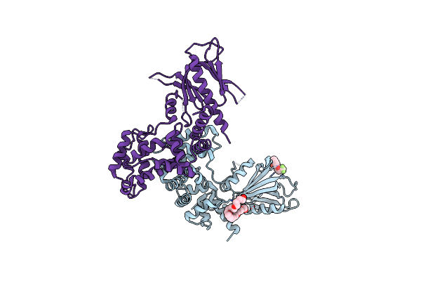
Best Fitting Antiparallel Model For Volume 2 Of Truncated Dimeric Cytohesin-3 (Grp1; Amino Acids 14-399)
Organism: Homo sapiens
Method: ELECTRON MICROSCOPY Release Date: 2019-09-25 Classification: ENDOCYTOSIS Ligands: 4IP |
Organism: Homo sapiens
Method: ELECTRON MICROSCOPY
Release Date: 2019-09-25
Ligands: 4IP
 |
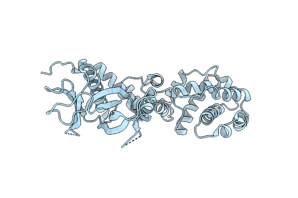
Structure Of Human Brag2 (Sec7-Ph Domains) With The Inhibitor Bragsin Bound To The Ph Domain
Organism: Homo sapiens
Method: X-RAY DIFFRACTION Resolution:2.50 Å Release Date: 2019-03-13 Classification: HYDROLASE Ligands: DY5, 2PE |
Organism: Homo sapiens
Method: X-RAY DIFFRACTION
Release Date: 2019-03-13
Ligands: DY5, 2PE
 |
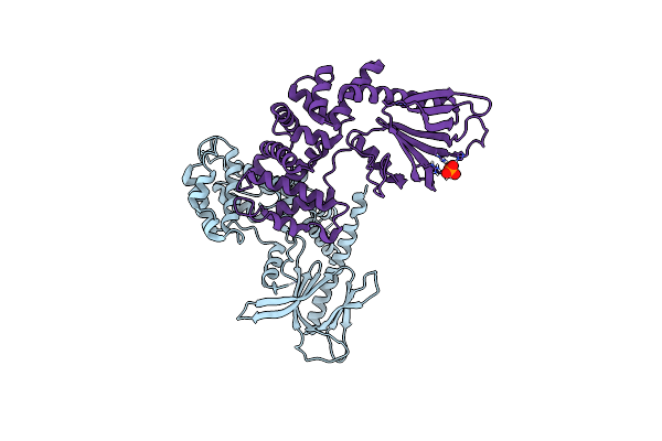
Brag2 Sec7-Ph (390-763)
Organism: Homo sapiens
Method: X-RAY DIFFRACTION Resolution:2.40 Å Release Date: 2017-09-27 Classification: HYDROLASE |
Organism: Homo sapiens
Method: X-RAY DIFFRACTION
Release Date: 2017-09-27
 |
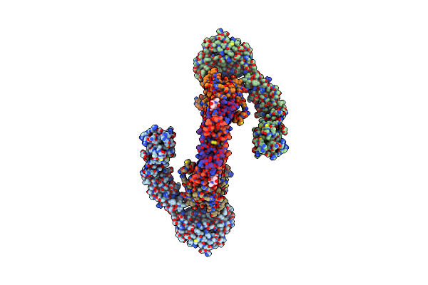
Brag2 Sec7-Ph (390-763), P212121
Organism: Homo sapiens
Method: X-RAY DIFFRACTION Resolution:2.00 Å Release Date: 2017-09-27 Classification: HYDROLASE Ligands: PO4 |
Organism: Homo sapiens
Method: X-RAY DIFFRACTION
Release Date: 2017-09-27
Ligands: PO4
 |
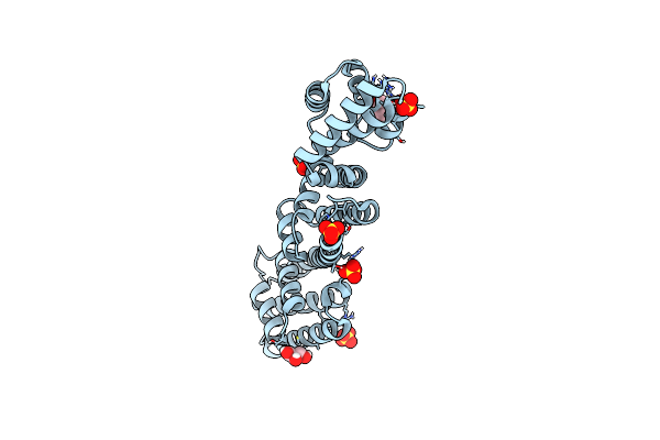
Ret Recognition Of Gdnf-Gfralpha1 Ligand By A Composite Binding Site Promotes Membrane-Proximal Self-Association
Organism: Homo sapiens, Rattus norvegicus
Method: ELECTRON MICROSCOPY Resolution:24.00 Å Release Date: 2014-10-01 Classification: SIGNALING PROTEIN Ligands: CA |
Organism: Homo sapiens, Rattus norvegicus
Method: ELECTRON MICROSCOPY
Release Date: 2014-10-01
Ligands: CA
 |
Chlamydia Pneumoniae Copn (D29 Construct)
Organism: Chlamydia pneumoniae
Method: X-RAY DIFFRACTION Resolution:1.77 Å Release Date: 2014-07-30 Classification: TRANSPORT PROTEIN Ligands: SO4, GOL |
Organism: Chlamydia pneumoniae
Method: X-RAY DIFFRACTION
Release Date: 2014-07-30
Ligands: SO4, GOL