

Search Count: 23
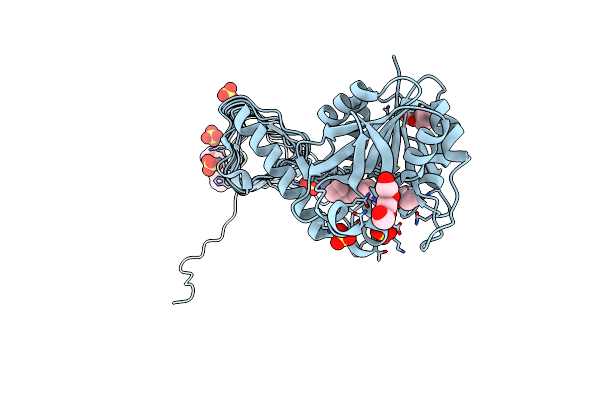 |
Crystal Structure Of Glmu From Haemophilus Influenzae In Complex With Quinazoline Inhibitor 1
Organism: Haemophilus influenzae
Method: X-RAY DIFFRACTION Resolution:1.99 Å Release Date: 2009-11-17 Classification: TRANSFERASE Ligands: LZR, PG4, PGE, SO4 |
Organism: Haemophilus influenzae
Method: X-RAY DIFFRACTION
Release Date: 2009-11-17
Ligands: LZR, PG4, PGE, SO4
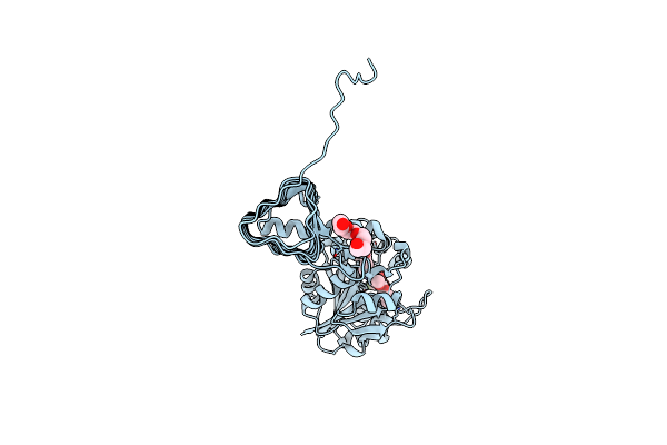 |
Crystal Structure Of Glmu From Haemophilus Influenzae In Complex With Quinazoline Inhibitor 2
Organism: Haemophilus influenzae
Method: X-RAY DIFFRACTION Resolution:2.59 Å Release Date: 2009-11-17 Classification: TRANSFERASE Ligands: LZS, PG4 |
Organism: Haemophilus influenzae
Method: X-RAY DIFFRACTION
Release Date: 2009-11-17
Ligands: LZS, PG4
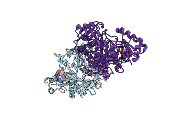 |
Crystal Structure Of Biotin Carboxylase From E. Coli In Complex With Amino-Oxazole Fragment Series
Organism: Escherichia coli
Method: X-RAY DIFFRACTION Resolution:2.00 Å Release Date: 2009-05-19 Classification: LIGASE Ligands: OA1, CL |
Organism: Escherichia coli
Method: X-RAY DIFFRACTION
Release Date: 2009-05-19
Ligands: OA1, CL
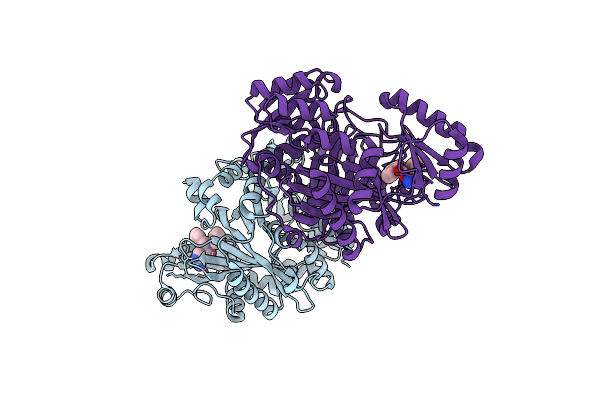 |
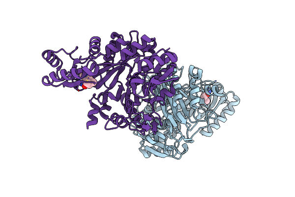
Crystal Structure Of Biotin Carboxylase From E. Coli In Complex With Amino-Oxazole Fragment Series
Organism: Escherichia coli
Method: X-RAY DIFFRACTION Resolution:1.87 Å Release Date: 2009-05-19 Classification: LIGASE Ligands: OA2, CL |
Organism: Escherichia coli
Method: X-RAY DIFFRACTION
Release Date: 2009-05-19
Ligands: OA2, CL
 |
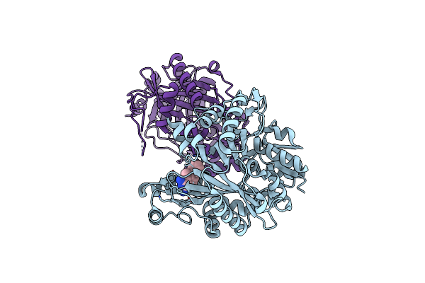
Crystal Structure Of Biotin Carboxylase From E. Coli In Complex With 4-Amino-7,7-Dimethyl-7,8-Dihydro-Quinazolinone Fragment
Organism: Escherichia coli
Method: X-RAY DIFFRACTION Resolution:2.50 Å Release Date: 2009-05-19 Classification: LIGASE Ligands: OA3, CL |
Organism: Escherichia coli
Method: X-RAY DIFFRACTION
Release Date: 2009-05-19
Ligands: OA3, CL
 |
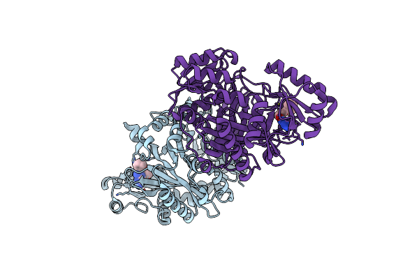
Crystal Structure Of Biotin Carboxylase From E. Coli In Complex With 5-Methyl-6-Phenyl-Quinazoline-2,4-Diamine
Organism: Escherichia coli
Method: X-RAY DIFFRACTION Resolution:1.85 Å Release Date: 2009-05-19 Classification: LIGASE Ligands: OA4 |
Organism: Escherichia coli
Method: X-RAY DIFFRACTION
Release Date: 2009-05-19
Ligands: OA4
 |
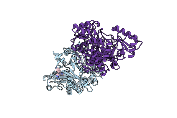
Crystal Structure Of Biotin Carboxylase From E. Coli In Complex With The Triazine-2,4-Diamine Fragment
Organism: Escherichia coli
Method: X-RAY DIFFRACTION Resolution:2.05 Å Release Date: 2009-05-19 Classification: LIGASE Ligands: OA5, CL |
Organism: Escherichia coli
Method: X-RAY DIFFRACTION
Release Date: 2009-05-19
Ligands: OA5, CL
 |
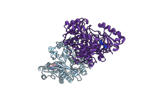
Crystal Structure Of Biotin Carboxylase From E. Coli In Complex With The 3-(3-Methyl-But-2-Enyl)-3H-Purin-6-Ylamine Fragment
Organism: Escherichia coli
Method: X-RAY DIFFRACTION Resolution:1.90 Å Release Date: 2009-05-19 Classification: LIGASE Ligands: L21, CL |
Organism: Escherichia coli
Method: X-RAY DIFFRACTION
Release Date: 2009-05-19
Ligands: L21, CL
 |
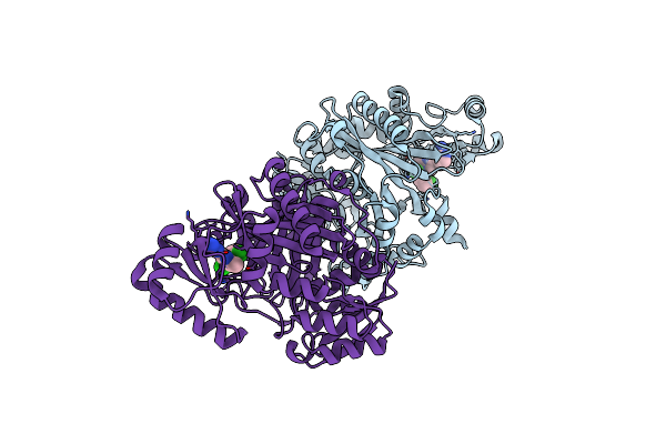
Crystal Structure Of Biotin Carboxylase From E. Coli In Complex With The Amino-Thiazole-Pyrimidine Fragment
Organism: Escherichia coli
Method: X-RAY DIFFRACTION Resolution:1.77 Å Release Date: 2009-05-19 Classification: LIGASE Ligands: L22, CL |
Organism: Escherichia coli
Method: X-RAY DIFFRACTION
Release Date: 2009-05-19
Ligands: L22, CL
 |
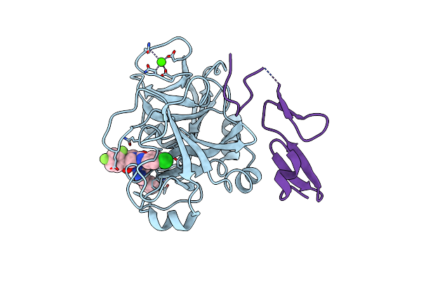
Crystal Structure Of Biotin Carboxylase From E. Coli In Complex With The Imidazole-Pyrimidine Inhibitor
Organism: Escherichia coli
Method: X-RAY DIFFRACTION Resolution:1.99 Å Release Date: 2009-05-19 Classification: LIGASE Ligands: CL, L23 |
Organism: Escherichia coli
Method: X-RAY DIFFRACTION
Release Date: 2009-05-19
Ligands: CL, L23
 |
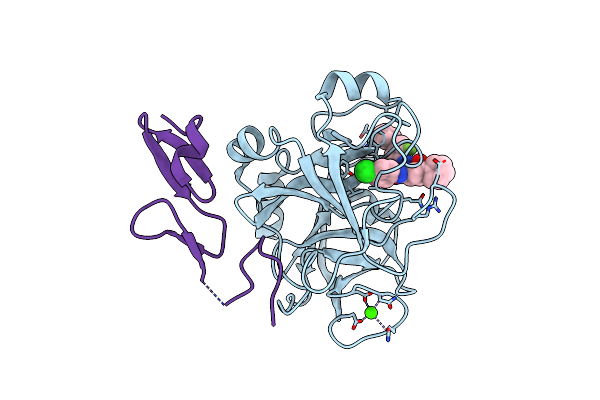
Crystal Structure Of Fxa In Complex With 4,4-Disubstituted Pyrrolidine-1,2-Dicarboxamide Inhibitor 2
Organism: Homo sapiens
Method: X-RAY DIFFRACTION Resolution:1.90 Å Release Date: 2009-04-07 Classification: HYDROLASE Ligands: CA, L1C |
Organism: Homo sapiens
Method: X-RAY DIFFRACTION
Release Date: 2009-04-07
Ligands: CA, L1C
 |
Crystal Structure Of Fxa In Complex With 4,4-Disubstituted Pyrrolidine-1,2-Dicarboxamide Inhibitor 1
Organism: Homo sapiens
Method: X-RAY DIFFRACTION Resolution:2.05 Å Release Date: 2009-04-07 Classification: HYDROLASE Ligands: L1D, CA |
Organism: Homo sapiens
Method: X-RAY DIFFRACTION
Release Date: 2009-04-07
Ligands: L1D, CA
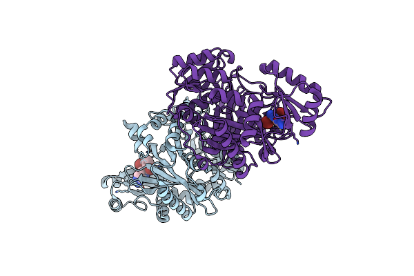 |
Crystal Structure Of Biotin Carboxylase From E.Coli In Complex With Potent Inhibitor 1
Organism: Escherichia coli
Method: X-RAY DIFFRACTION Resolution:2.10 Å Release Date: 2009-01-13 Classification: LIGASE Ligands: LZJ, CL |
Organism: Escherichia coli
Method: X-RAY DIFFRACTION
Release Date: 2009-01-13
Ligands: LZJ, CL
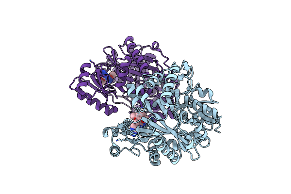 |
Crystal Structure Of Biotin Carboxylase From E.Coli In Complex With Potent Inhibitor 2
Organism: Escherichia coli
Method: X-RAY DIFFRACTION Resolution:2.40 Å Release Date: 2009-01-13 Classification: LIGASE Ligands: LZK |
Organism: Escherichia coli
Method: X-RAY DIFFRACTION
Release Date: 2009-01-13
Ligands: LZK
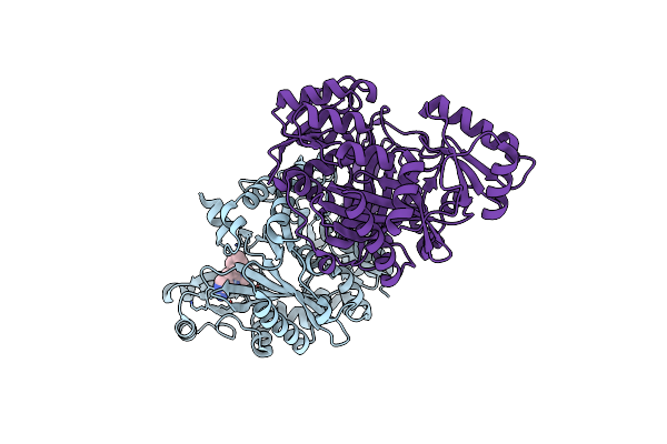 |
Crystal Structure Of Biotin Carboxylase From E.Coli In Complex With Potent Inhibitor 3
Organism: Escherichia coli
Method: X-RAY DIFFRACTION Resolution:2.31 Å Release Date: 2009-01-13 Classification: LIGASE Ligands: LZL, CL |
Organism: Escherichia coli
Method: X-RAY DIFFRACTION
Release Date: 2009-01-13
Ligands: LZL, CL
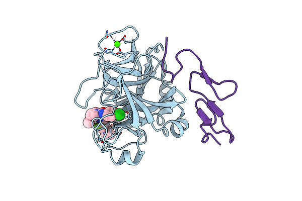 |
An Orally Efficacious Factor Xa Inhibitor
Organism: Homo sapiens
Method: X-RAY DIFFRACTION Resolution:2.30 Å Release Date: 2008-03-25 Classification: BLOOD CLOTTING Ligands: CA, 230 |
Organism: Homo sapiens
Method: X-RAY DIFFRACTION
Release Date: 2008-03-25
Ligands: CA, 230
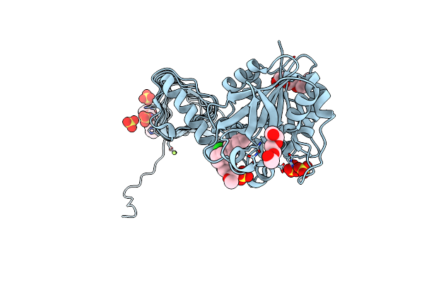 |
Structure Of Small-Molecule Inhibitor Of Glmu From Haemophilus Influenzae Reveals An Allosteric Binding Site
Organism: Haemophilus influenzae
Method: X-RAY DIFFRACTION Resolution:1.90 Å Release Date: 2008-01-15 Classification: TRANSFERASE Ligands: P21, SO4, PG4, PGE, MG |
Organism: Haemophilus influenzae
Method: X-RAY DIFFRACTION
Release Date: 2008-01-15
Ligands: P21, SO4, PG4, PGE, MG
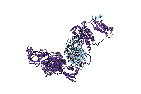 |
Phers From Staphylococcus Haemolyticus- Rational Protein Engineering And Inhibitor Studies
Organism: Staphylococcus haemolyticus
Method: X-RAY DIFFRACTION Resolution:2.20 Å Release Date: 2007-11-06 Classification: LIGASE Ligands: ZN, GAX |
Organism: Staphylococcus haemolyticus
Method: X-RAY DIFFRACTION
Release Date: 2007-11-06
Ligands: ZN, GAX
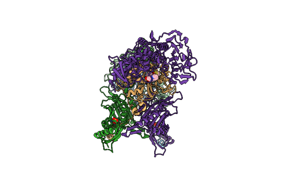 |
Phers From Staphylococcus Haemolyticus- Rational Protein Engineering And Inhibitor Studies
Organism: Staphylococcus haemolyticus
Method: X-RAY DIFFRACTION Resolution:2.20 Å Release Date: 2007-11-06 Classification: LIGASE Ligands: GAX, SO4, ZN |
Organism: Staphylococcus haemolyticus
Method: X-RAY DIFFRACTION
Release Date: 2007-11-06
Ligands: GAX, SO4, ZN
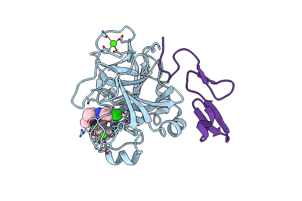 |
Factor Xa Inhibitor
Organism: Homo sapiens
Method: X-RAY DIFFRACTION Resolution:1.50 Å Release Date: 2007-08-14 Classification: BLOOD CLOTTING Ligands: CA, 237 |
Organism: Homo sapiens
Method: X-RAY DIFFRACTION
Release Date: 2007-08-14
Ligands: CA, 237