

Search Count: 27
 |
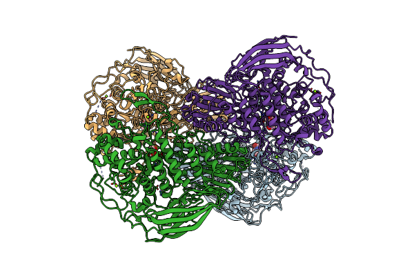
Beta-Glucosidase From Thermoascus Aurantiacus
Organism: Thermoascus aurantiacus
Method: X-RAY DIFFRACTION Resolution:2.15 Å Release Date: 2025-04-30 Classification: HYDROLASE Ligands: MG, NOJ |
Organism: Thermoascus aurantiacus
Method: X-RAY DIFFRACTION
Release Date: 2025-04-30
Ligands: MG, NOJ
 |
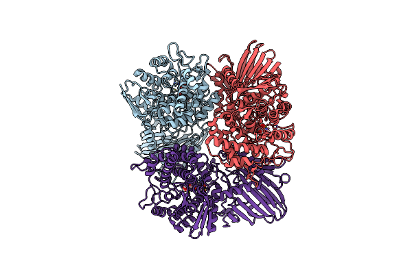
Beta-Glucosidase Mutant M279V_T308S_K361R_D433N_N514C
Organism: Thermoascus aurantiacus
Method: X-RAY DIFFRACTION Resolution:2.30 Å Release Date: 2025-04-30 Classification: HYDROLASE Ligands: MG, NOJ |
Organism: Thermoascus aurantiacus
Method: X-RAY DIFFRACTION
Release Date: 2025-04-30
Ligands: MG, NOJ
 |
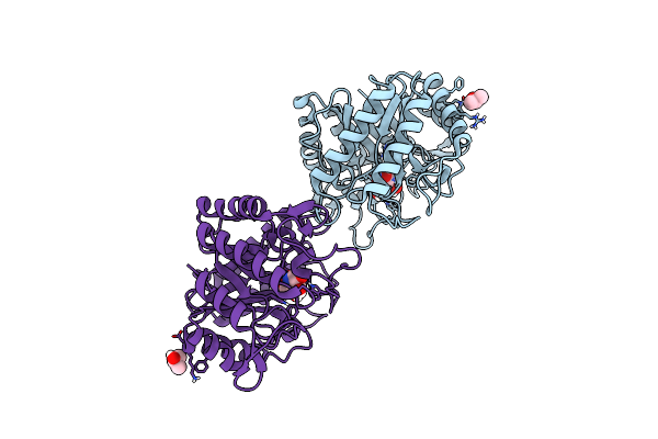
Crystal Structure Of Gh65 Alpha-1,2-Glucosidase From Flavobacterium Johnsoniae In Complex With 1-Deoxynojirimycin
Organism: Flavobacterium johnsoniae uw101
Method: X-RAY DIFFRACTION Resolution:1.90 Å Release Date: 2025-03-19 Classification: HYDROLASE Ligands: NOJ, EDO |
Organism: Flavobacterium johnsoniae uw101
Method: X-RAY DIFFRACTION
Release Date: 2025-03-19
Ligands: NOJ, EDO
 |
Structure Of The Thermotoga Maritima Family 5 Endo-Glucanase In Complex With 1-Deoxynojiromycin
Organism: Thermotoga maritima (strain atcc 43589 / msb8 / dsm 3109 / jcm 10099)
Method: X-RAY DIFFRACTION Resolution:1.80 Å Release Date: 2022-03-16 Classification: HYDROLASE Ligands: NOJ, IPA |
Organism: Thermotoga maritima (strain atcc 43589 / msb8 / dsm 3109 / jcm 10099)
Method: X-RAY DIFFRACTION
Release Date: 2022-03-16
Ligands: NOJ, IPA
 |
The Complex Structure Of Beta-1,2-Glucosyltransferase From Ignavibacterium Album With 1-Deoxynojirimycin
Organism: Ignavibacterium album (strain dsm 19864 / jcm 16511 / nbrc 101810 / mat9-16)
Method: X-RAY DIFFRACTION Resolution:2.00 Å Release Date: 2022-03-09 Classification: TRANSFERASE Ligands: NOJ, CA |
Organism: Ignavibacterium album (strain dsm 19864 / jcm 16511 / nbrc 101810 / mat9-16)
Method: X-RAY DIFFRACTION
Release Date: 2022-03-09
Ligands: NOJ, CA
 |
Bombyx Mori Gh13 Sucrose Hydrolase Complexed With 1-Deoxynojirimycin
Organism: Bombyx mori
Method: X-RAY DIFFRACTION Resolution:1.90 Å Release Date: 2020-05-20 Classification: HYDROLASE Ligands: MG, CA, NOJ, GOL |
Organism: Bombyx mori
Method: X-RAY DIFFRACTION
Release Date: 2020-05-20
Ligands: MG, CA, NOJ, GOL
 |
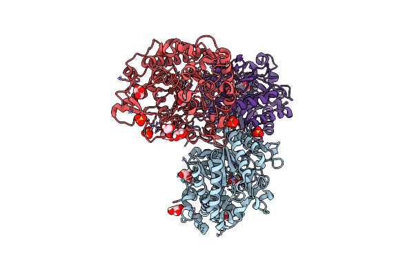
The Crystal Structure Of Glycoside Hydrolase Bglx From P. Aeruginosa In Complex With 1-Deoxynojirimycin
Organism: Pseudomonas aeruginosa (strain atcc 15692 / dsm 22644 / cip 104116 / jcm 14847 / lmg 12228 / 1c / prs 101 / pao1)
Method: X-RAY DIFFRACTION Resolution:2.00 Å Release Date: 2020-04-15 Classification: HYDROLASE Ligands: PGE, NOJ, MG |
 |
Crystal Structure Of Beta-Glucosidase Bglm-G1 Mutant H75R From Marine Metagenome In Complex With Inhibitor 1-Deoxynojirimycin
Organism: Marine metagenome
Method: X-RAY DIFFRACTION Resolution:1.55 Å Release Date: 2018-08-08 Classification: HYDROLASE Ligands: SO4, GOL, NOJ |
Organism: Marine metagenome
Method: X-RAY DIFFRACTION
Release Date: 2018-08-08
Ligands: SO4, GOL, NOJ
 |
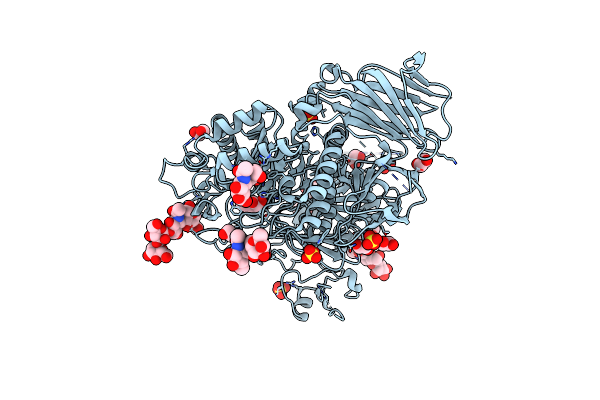
Crystal Structure Of Wild-Type Mall From Bacillus Subtilis With Ts Analogue 1-Deoxynojirimycin
Organism: Bacillus subtilis
Method: X-RAY DIFFRACTION Resolution:1.58 Å Release Date: 2018-03-28 Classification: HYDROLASE Ligands: NOJ, GOL |
Organism: Bacillus subtilis
Method: X-RAY DIFFRACTION
Release Date: 2018-03-28
Ligands: NOJ, GOL
 |
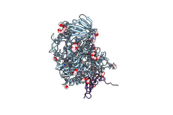
Crystal Structure Of Human Lysosomal Acid-Alpha-Glucosidase, Gaa, In Complex With 1-Deoxynojirimycin
Organism: Homo sapiens
Method: X-RAY DIFFRACTION Resolution:2.00 Å Release Date: 2017-10-25 Classification: HYDROLASE Ligands: NOJ, SO4, CL, EDO |
Organism: Homo sapiens
Method: X-RAY DIFFRACTION
Release Date: 2017-10-25
Ligands: NOJ, SO4, CL, EDO
 |
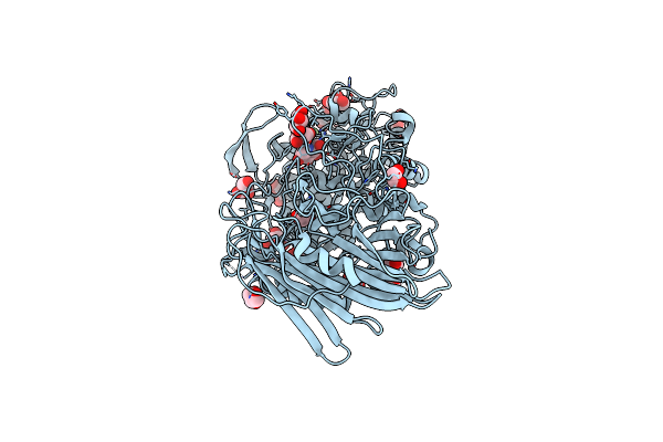
Murine Endoplasmic Reticulum Alpha-Glucosidase Ii With 1-Deoxynojirimycin
Organism: Mus musculus
Method: X-RAY DIFFRACTION Resolution:1.92 Å Release Date: 2016-07-27 Classification: HYDROLASE Ligands: PG4, P6G, EDO, FMT, NOJ, CA |
Organism: Mus musculus
Method: X-RAY DIFFRACTION
Release Date: 2016-07-27
Ligands: PG4, P6G, EDO, FMT, NOJ, CA
 |
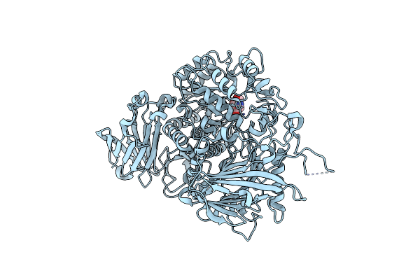
Crystal Structure Of Thermoanaerobacterium Xylanolyticum Gh116 Beta-Glucosidase With Deoxynojirimycin
Organism: Thermoanaerobacterium xylanolyticum lx-11
Method: X-RAY DIFFRACTION Resolution:1.96 Å Release Date: 2016-05-18 Classification: HYDROLASE Ligands: NOJ, GOL, CA, EDO |
Organism: Thermoanaerobacterium xylanolyticum lx-11
Method: X-RAY DIFFRACTION
Release Date: 2016-05-18
Ligands: NOJ, GOL, CA, EDO
 |
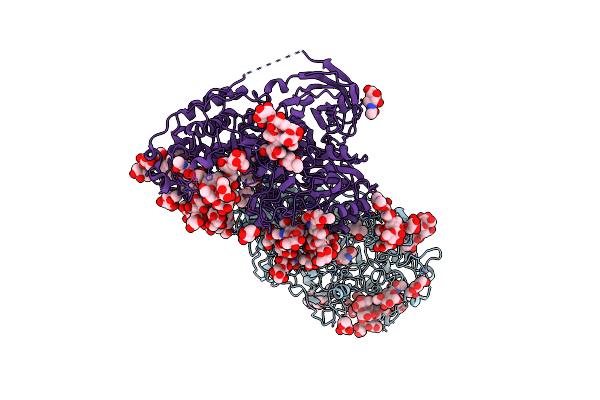
Crystal Structure Of Glucosidase Ii Alpha Subunit (Dnj-Bound From)
Organism: Chaetomium thermophilum (strain dsm 1495 / cbs 144.50 / imi 039719)
Method: X-RAY DIFFRACTION Resolution:1.60 Å Release Date: 2016-01-27 Classification: HYDROLASE Ligands: NOJ |
Organism: Chaetomium thermophilum (strain dsm 1495 / cbs 144.50 / imi 039719)
Method: X-RAY DIFFRACTION
Release Date: 2016-01-27
Ligands: NOJ
 |
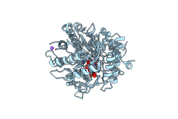
Crystal Structure Of Beta-Glucosidase 1 From Aspergillus Aculeatus In Complex With 1-Deoxynojirimycin
Organism: Aspergillus aculeatus
Method: X-RAY DIFFRACTION Resolution:2.30 Å Release Date: 2013-04-10 Classification: HYDROLASE Ligands: NAG, MRD, NOJ, MPD |
Organism: Aspergillus aculeatus
Method: X-RAY DIFFRACTION
Release Date: 2013-04-10
Ligands: NAG, MRD, NOJ, MPD
 |
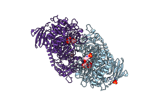
Crystal Structure Of Beta-Glucosidase From Termite Neotermes Koshunensis In Complex With 1-Deoxynojirimycin
Organism: Neotermes koshunensis
Method: X-RAY DIFFRACTION Resolution:0.99 Å Release Date: 2012-07-04 Classification: HYDROLASE/HYDROLASE INHIBITOR Ligands: NOJ, EPE, GOL, NA, CL |
Organism: Neotermes koshunensis
Method: X-RAY DIFFRACTION
Release Date: 2012-07-04
Ligands: NOJ, EPE, GOL, NA, CL
 |
Crystal Structure Of Cellvibrio Gilvus Cellobiose Phosphorylase Complexed With Sulfate And 1-Deoxynojirimycin
Organism: Cellvibrio gilvus
Method: X-RAY DIFFRACTION Resolution:2.39 Å Release Date: 2011-09-21 Classification: TRANSFERASE/TRANSFERASE INHIBITOR Ligands: BGC, SO4, NOJ, EPE |
Organism: Cellvibrio gilvus
Method: X-RAY DIFFRACTION
Release Date: 2011-09-21
Ligands: BGC, SO4, NOJ, EPE
 |
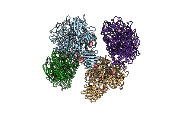
Crystal Structure Of Cellvibrio Gilvus Cellobiose Phosphorylase Complexed With Phosphate And 1-Deoxynojirimycin
Organism: Cellvibrio gilvus
Method: X-RAY DIFFRACTION Resolution:2.70 Å Release Date: 2011-09-21 Classification: TRANSFERASE/TRANSFERASE INHIBITOR Ligands: BGC, PO4, NOJ |
Organism: Cellvibrio gilvus
Method: X-RAY DIFFRACTION
Release Date: 2011-09-21
Ligands: BGC, PO4, NOJ
 |
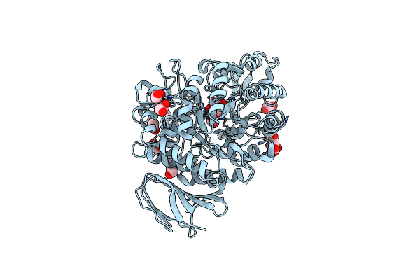
Crystal Structure Of The Gracilariopsis Lemaneiformis Alpha- 1,4-Glucan Lyase With Deoxynojirimycin
Organism: Gracilariopsis lemaneiformis
Method: X-RAY DIFFRACTION Resolution:2.35 Å Release Date: 2011-01-19 Classification: LYASE Ligands: NOJ, GOL, CL |
Organism: Gracilariopsis lemaneiformis
Method: X-RAY DIFFRACTION
Release Date: 2011-01-19
Ligands: NOJ, GOL, CL
 |
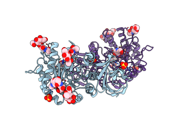
Crystal Structure Of The Isomaltulose Synthase Smua From Protaminobacter Rubrum In Complex With The Inhibitor Deoxynojirimycin
Organism: Protaminobacter rubrum
Method: X-RAY DIFFRACTION Resolution:1.70 Å Release Date: 2009-05-26 Classification: ISOMERASE Ligands: EDO, FLC, NOJ |
Organism: Protaminobacter rubrum
Method: X-RAY DIFFRACTION
Release Date: 2009-05-26
Ligands: EDO, FLC, NOJ
 |
Crystal Structure Of Alpha-Galactosidase A At Ph 4.5 Complexed With 1-Deoxygalactonijirimycin
Organism: Homo sapiens
Method: X-RAY DIFFRACTION Resolution:2.70 Å Release Date: 2009-05-26 Classification: HYDROLASE Ligands: SO4, NOJ, NAG |
Organism: Homo sapiens
Method: X-RAY DIFFRACTION
Release Date: 2009-05-26
Ligands: SO4, NOJ, NAG