

Search Count: 59
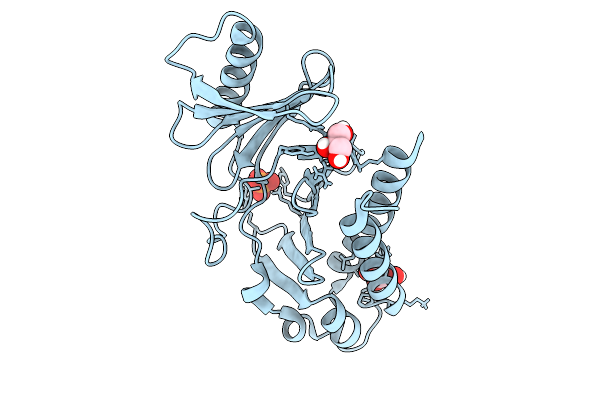 |
Plk1 Pbd Apo Protein At 1.6A
Organism: Homo sapiens
Method: X-RAY DIFFRACTION Resolution:1.60 Å Release Date: 2026-02-25 Classification: CELL CYCLE Ligands: PO4, GOL |
Organism: Homo sapiens
Method: X-RAY DIFFRACTION
Release Date: 2026-02-25
Ligands: PO4, GOL
 |
Crystal Structure Of The M18Bp1 Santa Domain As A Egfp-Santa-Nanobody Fusion
Organism: Homo sapiens
Method: X-RAY DIFFRACTION Resolution:2.42 Å Release Date: 2026-01-14 Classification: CELL CYCLE Ligands: PG4 |
Organism: Homo sapiens
Method: X-RAY DIFFRACTION
Release Date: 2026-01-14
Ligands: PG4
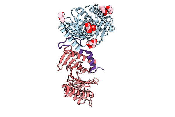 |
Two Plk1 Pbd Proteins Bound To Cenp-U(58-114) Phosphorylated At Thr98
Organism: Homo sapiens
Method: X-RAY DIFFRACTION Resolution:2.00 Å Release Date: 2025-09-24 Classification: CELL CYCLE Ligands: PG4, FLC, PEG |
Organism: Homo sapiens
Method: X-RAY DIFFRACTION
Release Date: 2025-09-24
Ligands: PG4, FLC, PEG
 |
Two Plk1 Pbd Proteins Bound To Cenp-U(58-114) Phosphorylated At Thr78 And Thr98
Organism: Homo sapiens
Method: X-RAY DIFFRACTION Resolution:2.15 Å Release Date: 2025-09-24 Classification: CELL CYCLE Ligands: PGE, PG4, PO4 |
Organism: Homo sapiens
Method: X-RAY DIFFRACTION
Release Date: 2025-09-24
Ligands: PGE, PG4, PO4
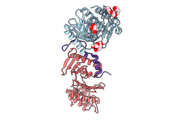 |
Two Plk1 Pbd Proteins Bound To Cenp-U(58-114) Phosphorylated At Thr98
Organism: Homo sapiens
Method: X-RAY DIFFRACTION Resolution:2.10 Å Release Date: 2025-09-24 Classification: CELL CYCLE Ligands: GOL, PG4, PEG |
Organism: Homo sapiens
Method: X-RAY DIFFRACTION
Release Date: 2025-09-24
Ligands: GOL, PG4, PEG
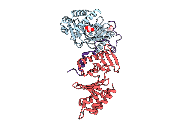 |
Two Plk1 Pbd Proteins Bound To Cenp-U(39-114) Phosphorylated At Thr78 And Thr98
Organism: Homo sapiens
Method: X-RAY DIFFRACTION Resolution:2.00 Å Release Date: 2025-09-24 Classification: CELL CYCLE Ligands: PEG, GOL |
Organism: Homo sapiens
Method: X-RAY DIFFRACTION
Release Date: 2025-09-24
Ligands: PEG, GOL
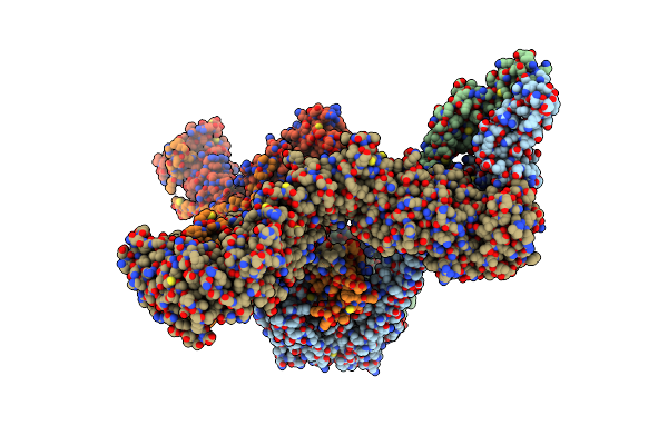 |
Human Condensin Ii - M18Bp1 Complex
Organism: Homo sapiens, Escherichia coli (strain k12)
Method: ELECTRON MICROSCOPY Release Date: 2025-07-02 Classification: DNA BINDING PROTEIN |
Organism: Homo sapiens, Escherichia coli (strain k12)
Method: ELECTRON MICROSCOPY
Release Date: 2025-07-02
 |
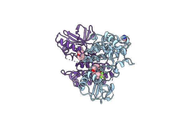
Crystal Structure Of Human Ripk1 With Compound 1
Organism: Homo sapiens
Method: X-RAY DIFFRACTION Resolution:2.53 Å Release Date: 2025-05-28 Classification: TRANSFERASE/INHIBITOR Ligands: A1BU4, IOD |
Organism: Homo sapiens
Method: X-RAY DIFFRACTION
Release Date: 2025-05-28
Ligands: A1BU4, IOD
 |
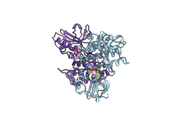
Crystal Structure Of Human Ripk1 With Compound 22
Organism: Homo sapiens
Method: X-RAY DIFFRACTION Resolution:2.32 Å Release Date: 2025-05-28 Classification: TRANSFERASE/INHIBITOR Ligands: A1BU6, IOD, GLY, CL |
Organism: Homo sapiens
Method: X-RAY DIFFRACTION
Release Date: 2025-05-28
Ligands: A1BU6, IOD, GLY, CL
 |
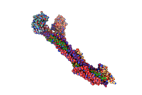
Crystal Structure Of Ripk1 With Compound 36
Organism: Homo sapiens
Method: X-RAY DIFFRACTION Resolution:2.68 Å Release Date: 2025-05-28 Classification: TRANSFERASE/INHIBITOR Ligands: A1BU5, IOD |
Organism: Homo sapiens
Method: X-RAY DIFFRACTION
Release Date: 2025-05-28
Ligands: A1BU5, IOD
 |
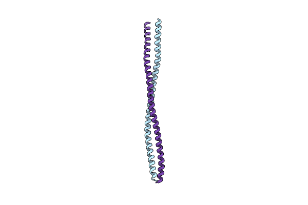
Human Kmn Network (Outer Kinetochore)
Organism: Homo sapiens
Method: ELECTRON MICROSCOPY Release Date: 2024-02-14 Classification: CELL CYCLE |
Organism: Homo sapiens
Method: ELECTRON MICROSCOPY
Release Date: 2024-02-14
 |
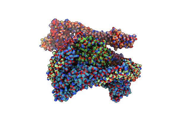
Crystal Structure Of The N-Terminal Parallel Dimeric Coiled-Coil Region Of The Human Kinetochore Associated Protein Spindly
Organism: Homo sapiens
Method: X-RAY DIFFRACTION Resolution:2.80 Å Release Date: 2022-09-07 Classification: CELL CYCLE |
Organism: Homo sapiens
Method: X-RAY DIFFRACTION
Release Date: 2022-09-07
 |
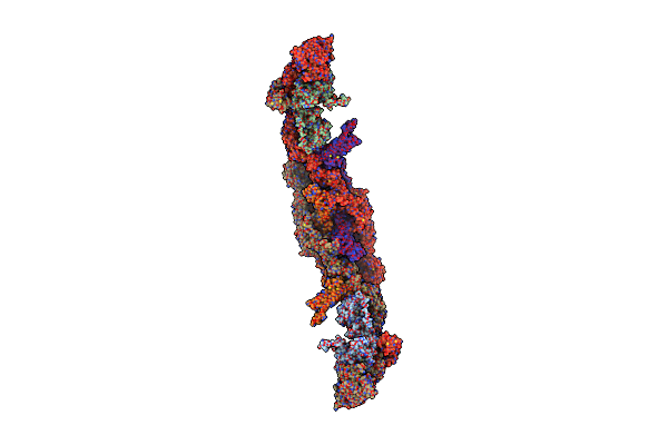
Structure Of The Human Inner Kinetochore Ccan Complex
Organism: Homo sapiens
Method: ELECTRON MICROSCOPY Release Date: 2022-06-08 Classification: CELL CYCLE |
Organism: Homo sapiens
Method: ELECTRON MICROSCOPY
Release Date: 2022-06-08
 |
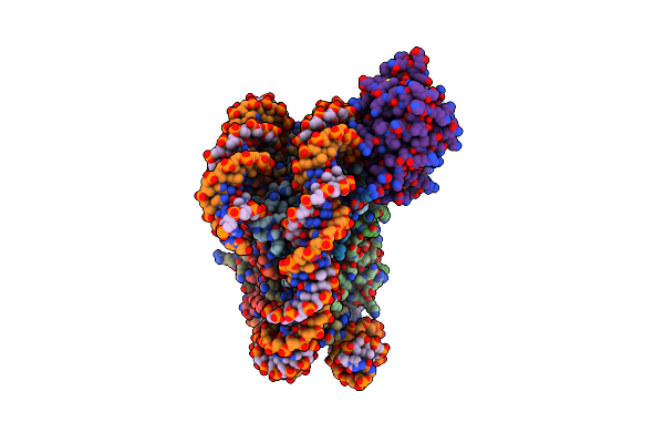
Human Rzz Kinetochore Corona Complex.
Organism: Homo sapiens
Method: ELECTRON MICROSCOPY Release Date: 2022-03-16 Classification: CELL CYCLE |
Organism: Homo sapiens
Method: ELECTRON MICROSCOPY
Release Date: 2022-03-16
 |
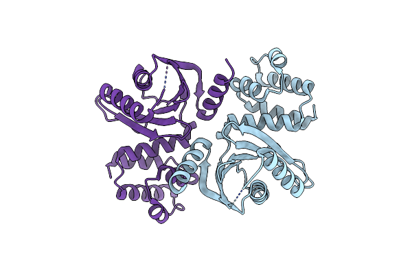
Cryo-Em Structure Of Human Kinetochore Protein Cenp-N With The Centromeric Nucleosome Containing Cenp-A
Organism: Homo sapiens, Escherichia coli
Method: ELECTRON MICROSCOPY Release Date: 2018-01-17 Classification: STRUCTURAL PROTEIN/DNA |
Organism: Homo sapiens, Escherichia coli
Method: ELECTRON MICROSCOPY
Release Date: 2018-01-17
 |
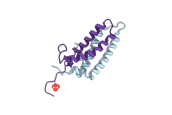
Crystal Structure Of The Human Kinetochore Protein Cenp-N
Organism: Homo sapiens
Method: X-RAY DIFFRACTION Resolution:2.74 Å Release Date: 2018-01-17 Classification: DNA BINDING PROTEIN |
Organism: Homo sapiens
Method: X-RAY DIFFRACTION
Release Date: 2018-01-17
 |
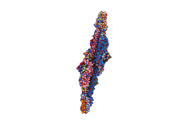
Crystal Structure Of The Kinetochore Mis12 Complex Head2 Subdomain Containing Dsn1 And Nsl1 Fragments
Organism: Homo sapiens
Method: X-RAY DIFFRACTION Resolution:2.00 Å Release Date: 2016-11-16 Classification: CELL CYCLE Ligands: SO4 |
Organism: Homo sapiens
Method: X-RAY DIFFRACTION
Release Date: 2016-11-16
Ligands: SO4
 |
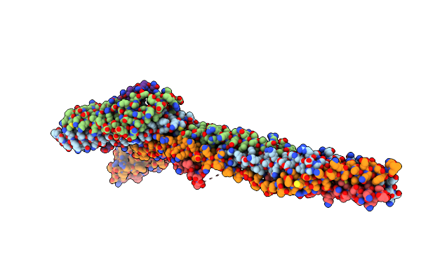
Crystal Structure Of The Human Kinetochore Mis12-Cenp-C Delta-Head2 Complex
Organism: Homo sapiens
Method: X-RAY DIFFRACTION Resolution:3.25 Å Release Date: 2016-11-16 Classification: CELL CYCLE |
Organism: Homo sapiens
Method: X-RAY DIFFRACTION
Release Date: 2016-11-16
 |
Crystal Structure Of The Human Kinetochore Mis12-Cenp-C Complex
Organism: Homo sapiens
Method: X-RAY DIFFRACTION Resolution:3.50 Å Release Date: 2016-11-16 Classification: CELL CYCLE |
Organism: Homo sapiens
Method: X-RAY DIFFRACTION
Release Date: 2016-11-16
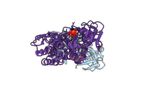 |
Structure Of Human Bub1 Kinase Domain Phosphorylated At Ser969
Organism: Homo sapiens
Method: X-RAY DIFFRACTION Resolution:2.40 Å Release Date: 2015-12-16 Classification: TRANSFERASE Ligands: ADP, MG |
Organism: Homo sapiens
Method: X-RAY DIFFRACTION
Release Date: 2015-12-16
Ligands: ADP, MG