
Search Count: 43
All
Selected
 |
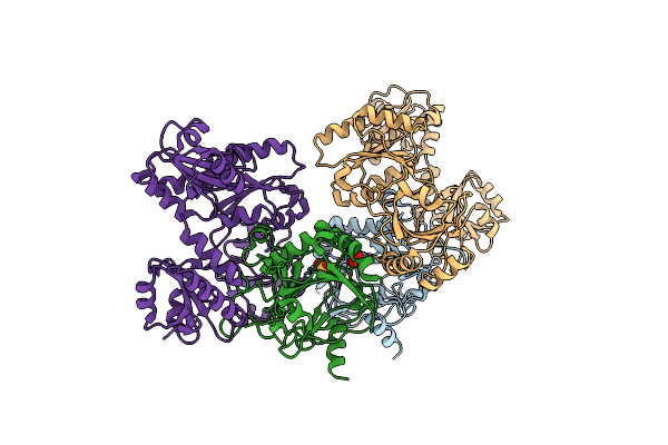
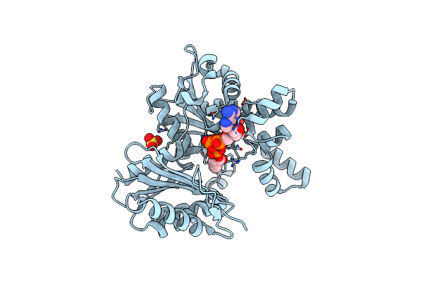
Organism: Homo sapiens
Method: ELECTRON MICROSCOPY Resolution:2.50 Å Release Date: 2026-01-28 Classification: LIGASE Ligands: ZN, A1CED |
 |
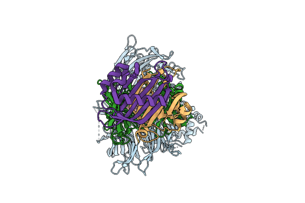
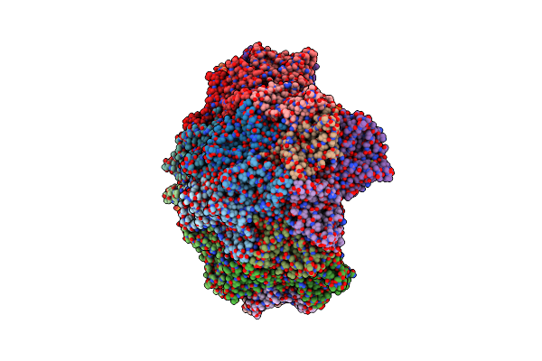
Structure Of Undecaprenyl-Phosphate 4-Deoxy-4-Formamido-L-Arabinose Transferase Embedded In Nanodisc
Organism: Escherichia coli
Method: ELECTRON MICROSCOPY Release Date: 2026-01-14 Classification: MEMBRANE PROTEIN |
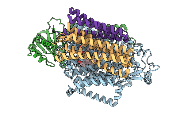 |
Organism: Escherichia coli
Method: ELECTRON MICROSCOPY Release Date: 2026-01-14 Classification: ELECTRON TRANSPORT Ligands: HEO, HEM, CU, ZN |
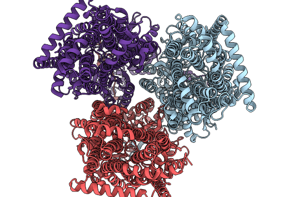 |
Organism: Escherichia coli
Method: ELECTRON MICROSCOPY Release Date: 2026-01-14 Classification: TRANSPORT PROTEIN |
 |
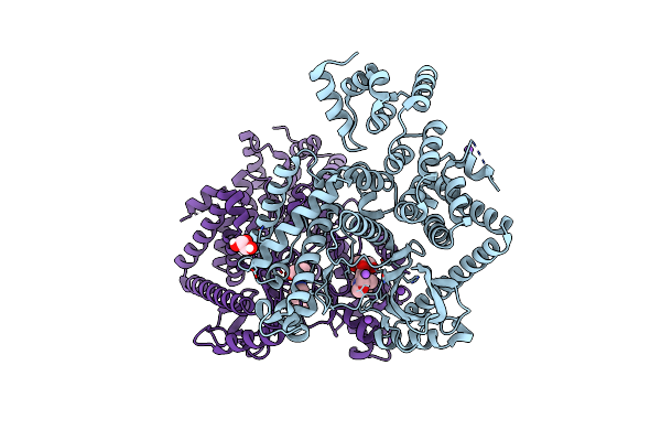
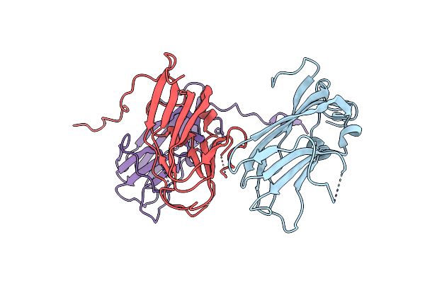
Crystal Structure Of The Heterodimeric Human C-P4H-Ii With Truncated Alpha Subunit (C-P4H-Ii Delta281)
Organism: Homo sapiens
Method: X-RAY DIFFRACTION Resolution:3.85 Å Release Date: 2022-11-09 Classification: HYDROLASE Ligands: SO4 |
 |
Organism: Homo sapiens
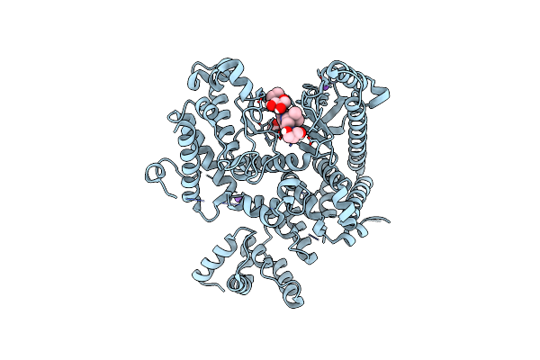
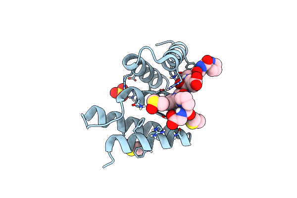
Method: X-RAY DIFFRACTION Resolution:1.88 Å Release Date: 2021-11-24 Classification: TRANSFERASE/TRANSFERASE inhibitor |
 |
Organism: Homo sapiens
Method: X-RAY DIFFRACTION Resolution:1.67 Å Release Date: 2021-11-24 Classification: TRANSFERASE/TRANSFERASE inhibitor Ligands: GOL, 7IK |
 |
Organism: Homo sapiens
Method: X-RAY DIFFRACTION Resolution:1.78 Å Release Date: 2021-11-24 Classification: TRANSFERASE/TRANSFERASE inhibitor Ligands: 7IQ, GOL, NA |
 |
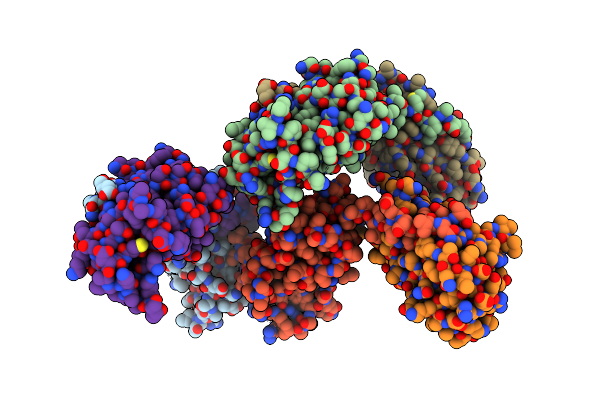
Crystal Structure Of Se-Met Labelled Mce Domain Of Mce4A From Mycobacterium Tuberculosis H37Rv
Organism: Mycobacterium tuberculosis h37rv
Method: X-RAY DIFFRACTION Resolution:3.61 Å Release Date: 2021-08-25 Classification: TRANSPORT PROTEIN |
 |
Crystal Structure Of Mce Domain Of Mce4A From Mycobacterium Tuberculosis H37Rv
Organism: Mycobacterium tuberculosis h37rv
Method: X-RAY DIFFRACTION Resolution:2.90 Å Release Date: 2021-08-25 Classification: TRANSPORT PROTEIN |
 |
Crystal Structure Of An Unlignaded Peptide-Substrate-Binding Domain Of Human Type Ii Collagen Prolyl 4-Hydroxylase
Organism: Homo sapiens
Method: X-RAY DIFFRACTION Resolution:1.87 Å Release Date: 2018-09-12 Classification: HYDROLASE Ligands: SO4, DMS, GLY |
 |
Crystal Structure Of A Pro-9 Complexed Peptide-Substrate-Binding Domain Of Human Type Ii Collagen Prolyl 4-Hydroxylase
Organism: Homo sapiens, Synthetic construct
Method: X-RAY DIFFRACTION Resolution:2.00 Å Release Date: 2018-09-12 Classification: HYDROLASE Ligands: SO4, DMS |
 |
Crystal Structure Of Peptide-Substrate-Binding Domain Of Human Type Ii Collagen Prolyl 4-Hydroxylase Complex With Pro-Pro-Gly-Pro-Ala-Gly-Pro-Pro-Gly.
Organism: Homo sapiens, Synthetic construct
Method: X-RAY DIFFRACTION Resolution:1.48 Å Release Date: 2018-09-12 Classification: HYDROLASE Ligands: SO4, DMS |
 |
Crystal Structure The Peptide-Substrate-Binding Domain Of Human Type Ii Collagen Prolyl 4-Hydroxylase Complexed With Pro-Pro-Gly-Pro-Arg-Gly-Pro-Pro-Gly.
Organism: Homo sapiens, Synthetic construct
Method: X-RAY DIFFRACTION Resolution:1.55 Å Release Date: 2018-09-12 Classification: HYDROLASE Ligands: SO4, DMS |
 |
Crystal Structure The Peptide-Substrate-Binding Domain Of Human Type Ii Collagen Prolyl 4-Hydroxylase Complexed With Pro-Pro-Gly-Pro-Glu-Gly-Pro-Pro-Gly.
Organism: Homo sapiens, Synthetic construct
Method: X-RAY DIFFRACTION Resolution:1.68 Å Release Date: 2018-09-12 Classification: HYDROLASE Ligands: SO4, DMS |
 |
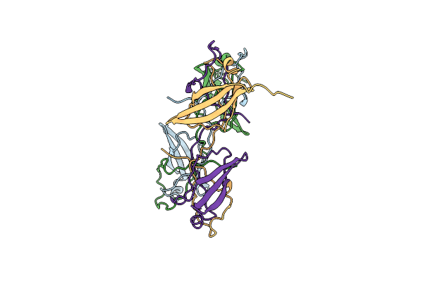
T=1 Capsid Structure Of Semv Ndel65Cp Fused With B-Domain Of S. Aureus Protein Spa At The N-Terminus (C2 Crystal Form)
Organism: Staphylococcus aureus, Sesbania mosaic virus
Method: X-RAY DIFFRACTION Resolution:3.00 Å Release Date: 2016-01-20 Classification: VIRUS Ligands: SO4 |
 |
T=1 Capsid Structure Of Semv Ndel65Cp Fused With B-Domain Of S. Aureus Protein Spa At The N-Terminus (P1 Crystal Form)
Organism: Staphylococcus aureus, Sesbania mosaic virus
Method: X-RAY DIFFRACTION Resolution:2.95 Å Release Date: 2016-01-20 Classification: VIRUS Ligands: SO4 |
 |
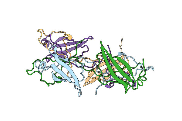
Structure Of Tobacco Streak Virus Coat Protein Dimer At 2.4 Angstroms Resolution
Organism: Tobacco streak virus
Method: X-RAY DIFFRACTION Resolution:2.40 Å Release Date: 2016-01-20 Classification: STRUCTURAL PROTEIN Ligands: EDO |
 |
Structure Of Tobacco Streak Virus Coat Protein At 2.1 Angstroms Resolution (C2 Crystal Form)
Organism: Tobacco streak virus
Method: X-RAY DIFFRACTION Resolution:2.10 Å Release Date: 2016-01-20 Classification: STRUCTURAL PROTEIN |
 |
Crystal Structure Of Salmonella Typhimurium Propionate Kinase A88G Mutant, In Complex With Amppnp And Propionate
Organism: Salmonella typhimurium (strain lt2 / sgsc1412 / atcc 700720)
Method: X-RAY DIFFRACTION Resolution:2.11 Å Release Date: 2015-09-23 Classification: TRANSFERASE Ligands: SO4, GOL, ANP, PPI |