

Search Count: 19
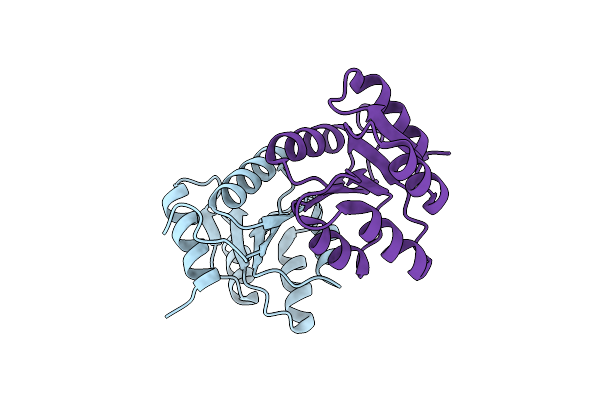 |
The Structure Of The N-Terminal Domain Of Rssb From E. Coli
Organism: Escherichia coli
Method: X-RAY DIFFRACTION Resolution:2.00 Å Release Date: 2020-07-29 Classification: STRUCTURAL PROTEIN |
Organism: Escherichia coli
Method: X-RAY DIFFRACTION
Release Date: 2020-07-29
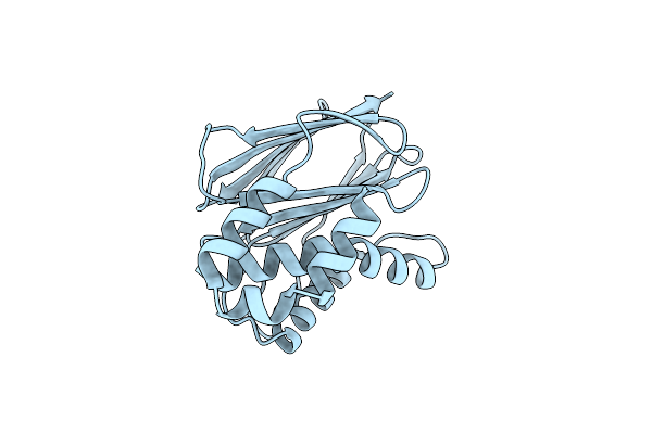 |
The Structure Of The C-Terminal Domain Of Rssb From E. Coli
Organism: Escherichia coli
Method: X-RAY DIFFRACTION Resolution:2.00 Å Release Date: 2020-07-29 Classification: STRUCTURAL PROTEIN |
Organism: Escherichia coli
Method: X-RAY DIFFRACTION
Release Date: 2020-07-29
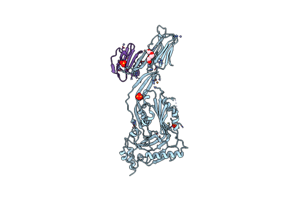 |
Toxin Receptor Complex
Organism: Streptococcus intermedius, Homo sapiens
Method: X-RAY DIFFRACTION Resolution:2.70 Å Release Date: 2016-08-24 Classification: TOXIN Ligands: PGE, SO4, ZN, CU |
Organism: Streptococcus intermedius, Homo sapiens
Method: X-RAY DIFFRACTION
Release Date: 2016-08-24
Ligands: PGE, SO4, ZN, CU
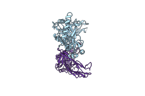 |
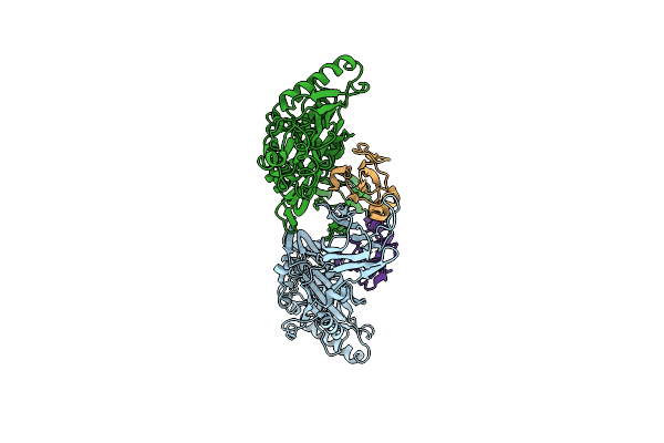
Trapped Toxin
Organism: Streptococcus intermedius
Method: X-RAY DIFFRACTION Resolution:2.89 Å Release Date: 2016-08-24 Classification: TOXIN |
Organism: Streptococcus intermedius
Method: X-RAY DIFFRACTION
Release Date: 2016-08-24
 |
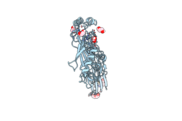
Trapped Toxin
Organism: Gardnerella vaginalis, Homo sapiens
Method: X-RAY DIFFRACTION Resolution:2.40 Å Release Date: 2016-08-24 Classification: toxin/toxin receptor |
Organism: Gardnerella vaginalis, Homo sapiens
Method: X-RAY DIFFRACTION
Release Date: 2016-08-24
 |
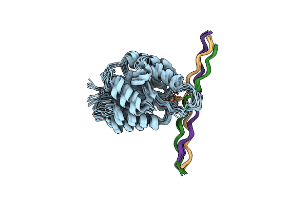
Structure Of Sugar Binding Protein Pneumolysin
Organism: Streptococcus pneumoniae
Method: X-RAY DIFFRACTION Resolution:2.90 Å Release Date: 2016-03-09 Classification: Sugar binding protein, toxin Ligands: EDO, PEG, AUC |
Organism: Streptococcus pneumoniae
Method: X-RAY DIFFRACTION
Release Date: 2016-03-09
Ligands: EDO, PEG, AUC
 |
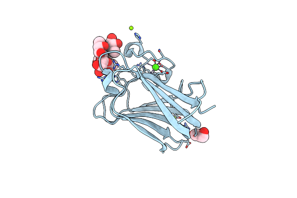
Alpha-1 Integrin I-Domain In Complex With Glogen Triple Helical Peptide
Organism: Homo sapiens
Method: SOLUTION NMR Release Date: 2013-11-06 Classification: CELL ADHESION Ligands: MG |
Organism: Homo sapiens
Method: SOLUTION NMR
Release Date: 2013-11-06
Ligands: MG
 |
His 62 Mutant Of The Lectin Binding Domain Of Lectinolysin Complexed With Lewis Y
Organism: Streptococcus mitis
Method: X-RAY DIFFRACTION Resolution:1.60 Å Release Date: 2012-11-21 Classification: SUGAR BINDING PROTEIN Ligands: MG, GOL, CA |
Organism: Streptococcus mitis
Method: X-RAY DIFFRACTION
Release Date: 2012-11-21
Ligands: MG, GOL, CA
 |
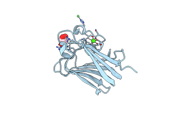
His 62 Mutant Of The Lectin Binding Domain Of Lectinolysin Complexed With Lewis B
Organism: Streptococcus mitis
Method: X-RAY DIFFRACTION Resolution:1.60 Å Release Date: 2012-11-21 Classification: SUGAR BINDING PROTEIN Ligands: MG, CA |
Organism: Streptococcus mitis
Method: X-RAY DIFFRACTION
Release Date: 2012-11-21
Ligands: MG, CA
 |
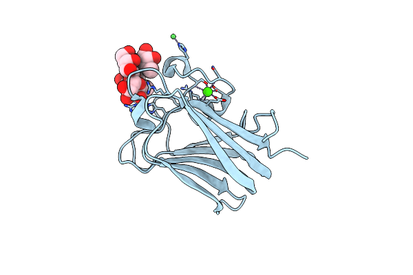
Lectin Domain Of Lectinolysin Complexed With Glycerol
Organism: Streptococcus mitis
Method: X-RAY DIFFRACTION Resolution:1.91 Å Release Date: 2010-12-29 Classification: BLOOD CLOTTING Ligands: CA, NI, GOL |
Organism: Streptococcus mitis
Method: X-RAY DIFFRACTION
Release Date: 2010-12-29
Ligands: CA, NI, GOL
 |
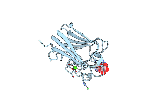
Lectin Domain Of Lectinolysin Complexed With Lewis Y Antigen
Organism: Streptococcus mitis
Method: X-RAY DIFFRACTION Resolution:2.01 Å Release Date: 2010-12-29 Classification: BLOOD CLOTTING Ligands: CA, NI |
Organism: Streptococcus mitis
Method: X-RAY DIFFRACTION
Release Date: 2010-12-29
Ligands: CA, NI
 |
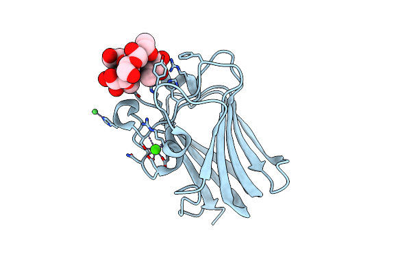
Lectin Domain Of Lectinolysin Complexed With Fucose
Organism: Streptococcus mitis
Method: X-RAY DIFFRACTION Resolution:1.90 Å Release Date: 2010-12-29 Classification: BLOOD CLOTTING Ligands: FUC, CA, NI |
Organism: Streptococcus mitis
Method: X-RAY DIFFRACTION
Release Date: 2010-12-29
Ligands: FUC, CA, NI
 |
Lectin Domain Of Lectinolysin Complexed With Lewis B Antigen
Organism: Streptococcus mitis
Method: X-RAY DIFFRACTION Resolution:2.00 Å Release Date: 2010-12-29 Classification: BLOOD CLOTTING Ligands: CA, NI |
Organism: Streptococcus mitis
Method: X-RAY DIFFRACTION
Release Date: 2010-12-29
Ligands: CA, NI
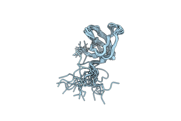 |
Nmr Structure Of The Sh3 Domain From The Tec Protein Tyrosine Kinase
|
Organism: Mus musculus
Method: SOLUTION NMR
Release Date: 2001-11-28
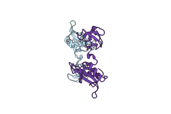 |
Crystal Structure Of Fyn Sh3-Sh2
Organism: Homo sapiens
Method: X-RAY DIFFRACTION Resolution:2.60 Å Release Date: 2001-05-30 Classification: TRANSFERASE |
Organism: Homo sapiens
Method: X-RAY DIFFRACTION
Release Date: 2001-05-30
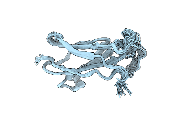 |
Nmr Structure Of The Ligand Binding Domain Of The Common Beta-Chain In The Gm-Csf, Il-3 And Il-5 Receptors
Organism: Homo sapiens
Method: SOLUTION NMR Release Date: 2000-06-15 Classification: MEMBRANE PROTEIN |
Organism: Homo sapiens
Method: SOLUTION NMR
Release Date: 2000-06-15
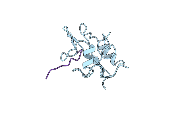 |
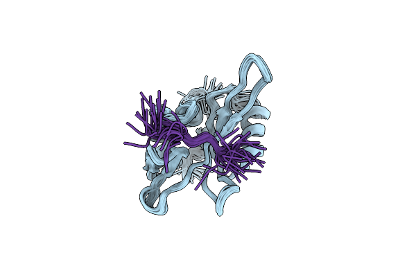
Nmr Structure Of The Fyn Sh2 Domain Complexed With A Phosphotyrosyl Peptide, Minimized Average Structure
Organism: Homo sapiens, Hamster polyomavirus
Method: SOLUTION NMR Release Date: 1998-01-14 Classification: COMPLEX (PROTO-ONCOGENE/EARLY PROTEIN) |
Organism: Homo sapiens, Hamster polyomavirus
Method: SOLUTION NMR
Release Date: 1998-01-14
 |
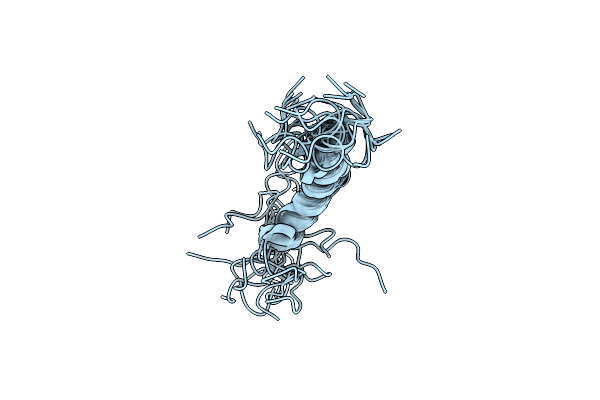
Nmr Structure Of The Fyn Sh2 Domain Complexed With A Phosphotyrosyl Peptide, 22 Structures
Organism: Homo sapiens, Hamster polyomavirus
Method: SOLUTION NMR Release Date: 1998-01-14 Classification: COMPLEX (PROTO-ONCOGENE/EARLY PROTEIN) |
Organism: Homo sapiens, Hamster polyomavirus
Method: SOLUTION NMR
Release Date: 1998-01-14
 |
Solution Structure Of A Polypeptide Containing Four Heptad Repeats From A Merozoite Surface Antigen Of Plasmodium Falciparum
Organism: Plasmodium falciparum
Method: SOLUTION NMR Release Date: 1995-02-07 Classification: POLYMORPHIC ANTIGEN |
Organism: Plasmodium falciparum
Method: SOLUTION NMR
Release Date: 1995-02-07