

Search Count: 25
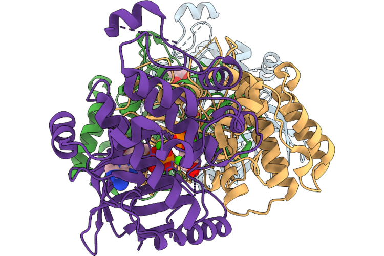 |
Crystal Structure Of Braf(Val600):Mek1(Ps222) Complex With Asymmetric Dimer Interface Bound To Adp
Organism: Homo sapiens
Method: X-RAY DIFFRACTION Resolution:2.45 Å Release Date: 2026-08-05 Classification: SIGNALING PROTEIN Ligands: ADP, CA, GOL, NA |
Organism: Homo sapiens
Method: X-RAY DIFFRACTION
Release Date: 2026-08-05
Ligands: ADP, CA, GOL, NA
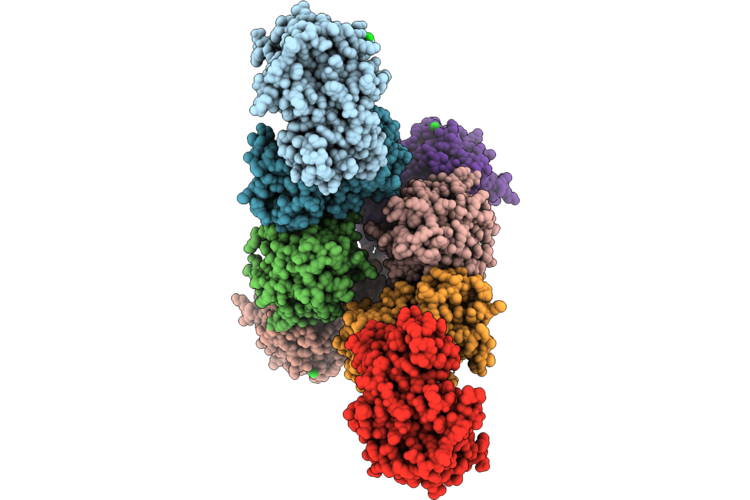 |
Crystal Structure Of Braf:Mek1 Complex With Symmetric Dimer Interface Bound To Amppnp
Organism: Homo sapiens
Method: X-RAY DIFFRACTION Resolution:2.91 Å Release Date: 2026-07-15 Classification: SIGNALING PROTEIN Ligands: ANP, CA, GOL, CL, PEG |
Organism: Homo sapiens
Method: X-RAY DIFFRACTION
Release Date: 2026-07-15
Ligands: ANP, CA, GOL, CL, PEG
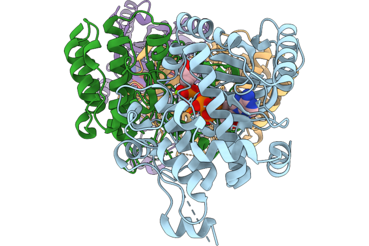 |
Crystal Structure Of Braf:Mek1 Complex With Asymmetric Dimer Interface Bound To Amppnp
Organism: Homo sapiens
Method: X-RAY DIFFRACTION Resolution:2.60 Å Release Date: 2026-07-15 Classification: SIGNALING PROTEIN Ligands: ANP, MG |
Organism: Homo sapiens
Method: X-RAY DIFFRACTION
Release Date: 2026-07-15
Ligands: ANP, MG
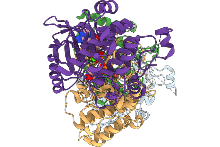 |
Crystal Structure Of Braf:Mek1(Ps222) Complex With Asymmetric Dimer Interface Bound To Adp
Organism: Homo sapiens
Method: X-RAY DIFFRACTION Resolution:2.30 Å Release Date: 2026-07-15 Classification: SIGNALING PROTEIN Ligands: ADP, CA, GOL, NA |
Organism: Homo sapiens
Method: X-RAY DIFFRACTION
Release Date: 2026-07-15
Ligands: ADP, CA, GOL, NA
 |
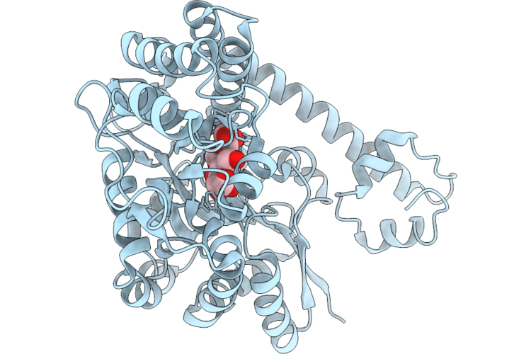
The Structure Of Egalitarian In Complex With The K10 Mrna Localization Signal Reveals A Modular Binding Surface Required For Function
Organism: Drosophila melanogaster
Method: X-RAY DIFFRACTION Resolution:3.08 Å Release Date: 2026-04-29 Classification: RNA BINDING PROTEIN Ligands: CA |
Organism: Drosophila melanogaster
Method: X-RAY DIFFRACTION
Release Date: 2026-04-29
Ligands: CA
 |
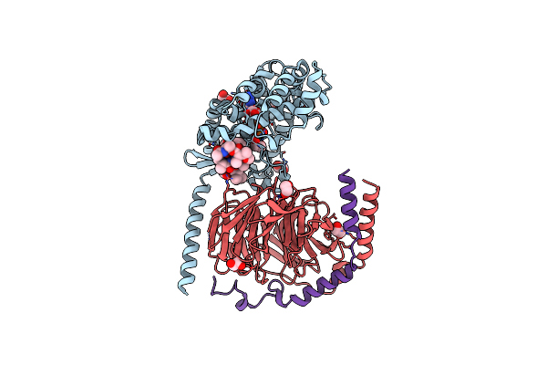
The Structure Of Egalitarian In Complex With The K10 Mrna Localization Signal Reveals A Modular Binding Surface Required For Function
Organism: Escherichia coli (strain k12), Drosophila melanogaster
Method: X-RAY DIFFRACTION Resolution:2.26 Å Release Date: 2026-04-29 Classification: RNA BINDING PROTEIN |
Organism: Escherichia coli (strain k12), Drosophila melanogaster
Method: X-RAY DIFFRACTION
Release Date: 2026-04-29
 |
Crystal Structure Of The G11 Protein Heterotrimer Bound To Ym-254890 Inhibitor
Organism: Homo sapiens
Method: X-RAY DIFFRACTION Resolution:1.70 Å Release Date: 2025-03-19 Classification: SIGNALING PROTEIN Ligands: GDP, EDO, DAM, HF2, THC, OTH, HL2, MAA, ALA, ACE |
Organism: Homo sapiens
Method: X-RAY DIFFRACTION
Release Date: 2025-03-19
Ligands: GDP, EDO, DAM, HF2, THC, OTH, HL2, MAA, ALA, ACE
 |
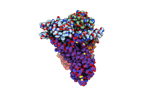
Crystal Structure Of The G11 Protein Heterotrimer Bound To Fr900359 Inhibitor
Organism: Homo sapiens
Method: X-RAY DIFFRACTION Resolution:1.43 Å Release Date: 2025-03-19 Classification: SIGNALING PROTEIN Ligands: EDO, GDP, ZN, CL, DAM, HF2, UDL, OTH, HL2, MAA, ALA, PPI, DMS |
 |
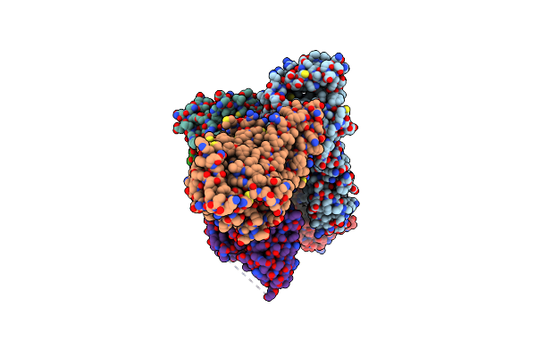
Cryo-Em Structure Of Rhodopsin-Gi Bound To Antibody Fragment Fab13
Organism: Bos taurus, Homo sapiens, Mus musculus
Method: ELECTRON MICROSCOPY Release Date: 2024-11-20 Classification: SIGNALING PROTEIN |
Organism: Bos taurus, Homo sapiens, Mus musculus
Method: ELECTRON MICROSCOPY
Release Date: 2024-11-20
 |
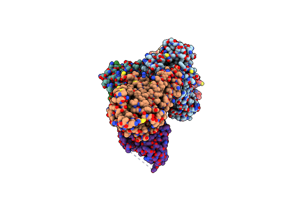
Cryo-Em Structure Of Rhodopsin-Gi Bound With Antibody Fragments Scfv16 And Fab79, Conformation 1
Organism: Bos taurus, Homo sapiens, Mus musculus
Method: ELECTRON MICROSCOPY Release Date: 2024-11-20 Classification: SIGNALING PROTEIN |
Organism: Bos taurus, Homo sapiens, Mus musculus
Method: ELECTRON MICROSCOPY
Release Date: 2024-11-20
 |
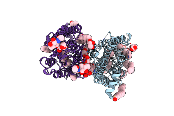
Cryo-Em Structure Of Rhodopsin-Gi Bound With Antibody Fragments Scfv16 And Fab79, Conformation 2
Organism: Bos taurus, Homo sapiens, Mus musculus
Method: ELECTRON MICROSCOPY Release Date: 2024-11-20 Classification: SIGNALING PROTEIN |
Organism: Bos taurus, Homo sapiens, Mus musculus
Method: ELECTRON MICROSCOPY
Release Date: 2024-11-20
 |
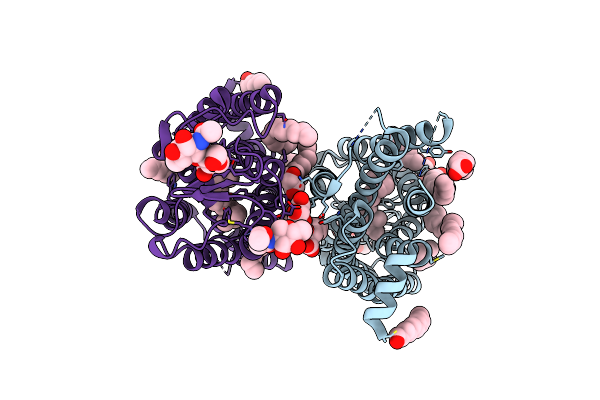
Dark State Crystal Structure Of Bovine Rhodopsin In Lipidic Cubic Phase (Sacla)
Organism: Bos taurus
Method: X-RAY DIFFRACTION Resolution:1.80 Å Release Date: 2023-03-29 Classification: MEMBRANE PROTEIN Ligands: ACE, RET, NAG, DAO, OLC, PLM |
Organism: Bos taurus
Method: X-RAY DIFFRACTION
Release Date: 2023-03-29
Ligands: ACE, RET, NAG, DAO, OLC, PLM
 |
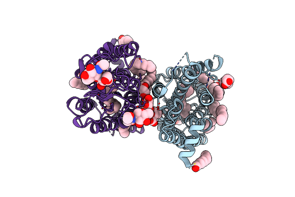
Dark State Crystal Structure Of Bovine Rhodopsin In Lipidic Cubic Phase (Swissfel)
Organism: Bos taurus
Method: X-RAY DIFFRACTION Resolution:1.80 Å Release Date: 2023-03-29 Classification: MEMBRANE PROTEIN Ligands: ACE, RET, NAG, DAO, OLC, PLM |
Organism: Bos taurus
Method: X-RAY DIFFRACTION
Release Date: 2023-03-29
Ligands: ACE, RET, NAG, DAO, OLC, PLM
 |
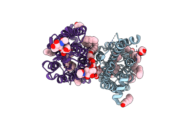
1 Picosecond Light Activated Crystal Structure Of Bovine Rhodopsin In Lipidic Cubic Phase
Organism: Bos taurus
Method: X-RAY DIFFRACTION Resolution:1.80 Å Release Date: 2023-03-29 Classification: MEMBRANE PROTEIN Ligands: ACE, RET, NAG, DAO, OLC, PLM |
Organism: Bos taurus
Method: X-RAY DIFFRACTION
Release Date: 2023-03-29
Ligands: ACE, RET, NAG, DAO, OLC, PLM
 |
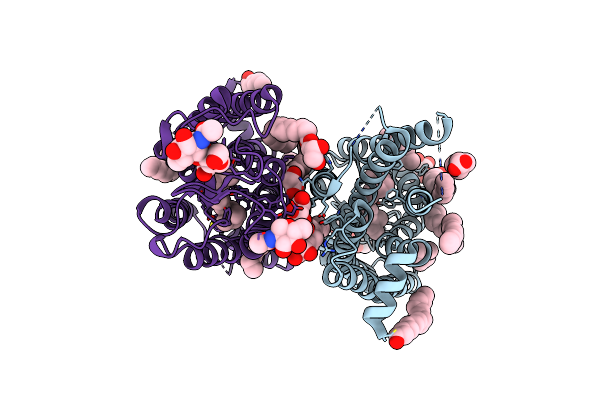
10 Picosecond Light Activated Crystal Structure Of Bovine Rhodopsin In Lipidic Cubic Phase
Organism: Bos taurus
Method: X-RAY DIFFRACTION Resolution:1.80 Å Release Date: 2023-03-29 Classification: MEMBRANE PROTEIN Ligands: ACE, RET, NAG, DAO, OLC, PLM |
Organism: Bos taurus
Method: X-RAY DIFFRACTION
Release Date: 2023-03-29
Ligands: ACE, RET, NAG, DAO, OLC, PLM
 |
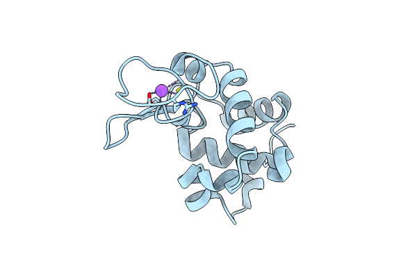
100 Picosecond Light Activated Crystal Structure Of Bovine Rhodopsin In Lipidic Cubic Phase (Sacla)
Organism: Bos taurus
Method: X-RAY DIFFRACTION Resolution:1.80 Å Release Date: 2023-03-29 Classification: MEMBRANE PROTEIN Ligands: RET, NAG, DAO, OLC, PLM |
Organism: Bos taurus
Method: X-RAY DIFFRACTION
Release Date: 2023-03-29
Ligands: RET, NAG, DAO, OLC, PLM
 |
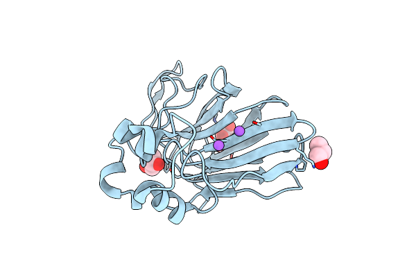
Structure Of Hen Egg White Lysozyme Collected By Rotation Serial Crystallography On A Coc Membrane At A Synchrotron Source
Organism: Gallus gallus
Method: X-RAY DIFFRACTION Resolution:1.51 Å Release Date: 2021-09-01 Classification: HYDROLASE Ligands: CL, NA |
Organism: Gallus gallus
Method: X-RAY DIFFRACTION
Release Date: 2021-09-01
Ligands: CL, NA
 |
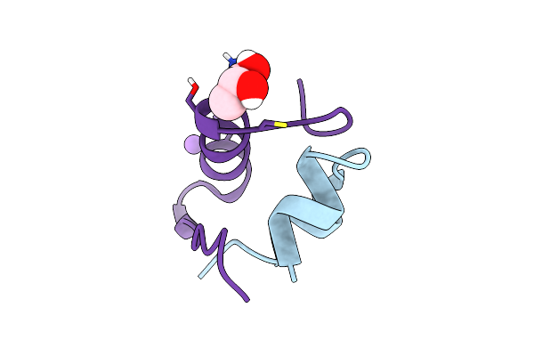
Structure Of Thaumatin Collected By Rotation Serial Crystallography On A Coc Membrane At A Synchrotron Source
Organism: Thaumatococcus daniellii
Method: X-RAY DIFFRACTION Resolution:1.65 Å Release Date: 2021-09-01 Classification: PLANT PROTEIN Ligands: TLA, PGO, NA |
Organism: Thaumatococcus daniellii
Method: X-RAY DIFFRACTION
Release Date: 2021-09-01
Ligands: TLA, PGO, NA
 |
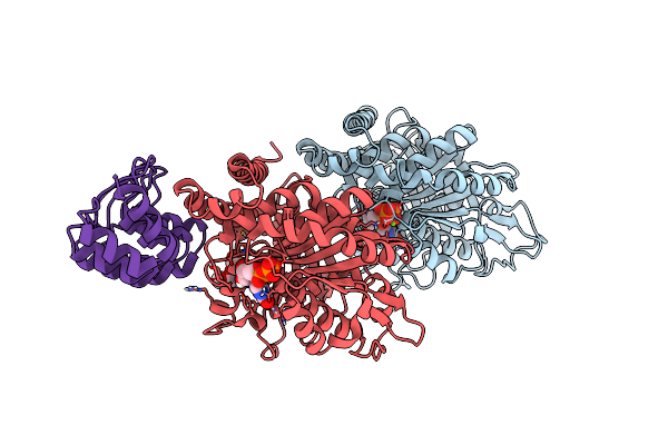
Structure Of Insulin Collected By Rotation Serial Crystallography On A Coc Membrane At A Synchrotron Source
Organism: Sus scrofa
Method: X-RAY DIFFRACTION Resolution:1.46 Å Release Date: 2021-09-01 Classification: HORMONE Ligands: PGR, NA |
Organism: Sus scrofa
Method: X-RAY DIFFRACTION
Release Date: 2021-09-01
Ligands: PGR, NA
 |
Structure Of Tubulin Darpin Complex 1 Collected By Rotation Serial Crystallography On A Coc Membrane At A Synchrotron Source
Organism: Bos taurus
Method: X-RAY DIFFRACTION Resolution:2.26 Å Release Date: 2021-09-01 Classification: CELL CYCLE Ligands: GTP, MG, GDP, PG0 |
Organism: Bos taurus
Method: X-RAY DIFFRACTION
Release Date: 2021-09-01
Ligands: GTP, MG, GDP, PG0