
Search Count: 45
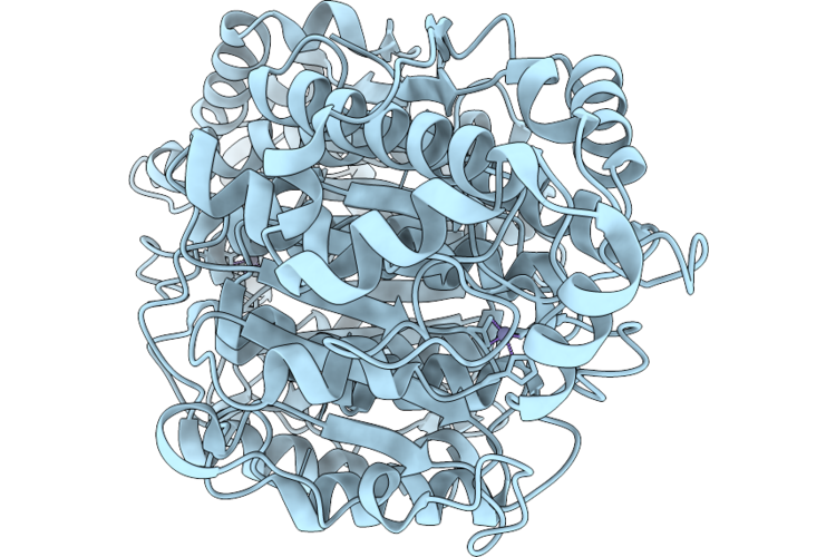 |
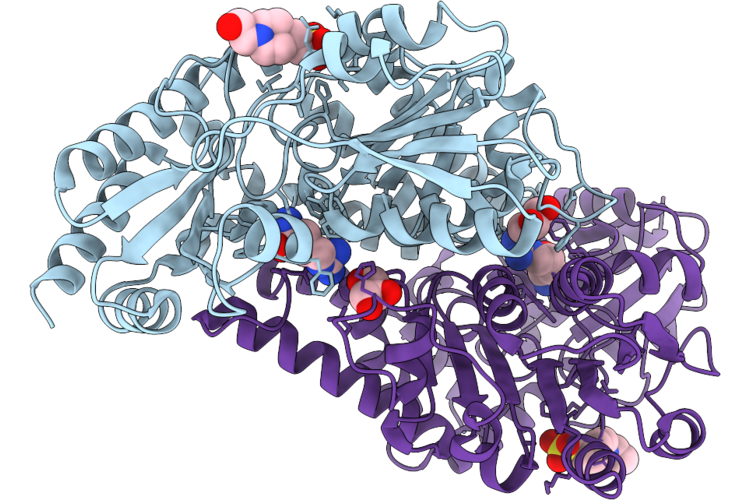
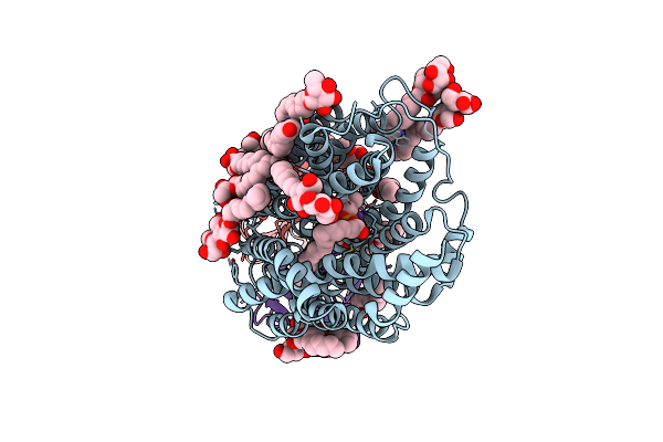
Cryo-Em Structure Of Human Histone Deacetylase 6 Tandem Catalytic Domain (Hdac6 Cd1-2)
Organism: Homo sapiens
Method: ELECTRON MICROSCOPY Release Date: 2026-07-15 Classification: HYDROLASE Ligands: ZN |
Organism: Homo sapiens
Method: ELECTRON MICROSCOPY
Release Date: 2026-07-15
Ligands: ZN
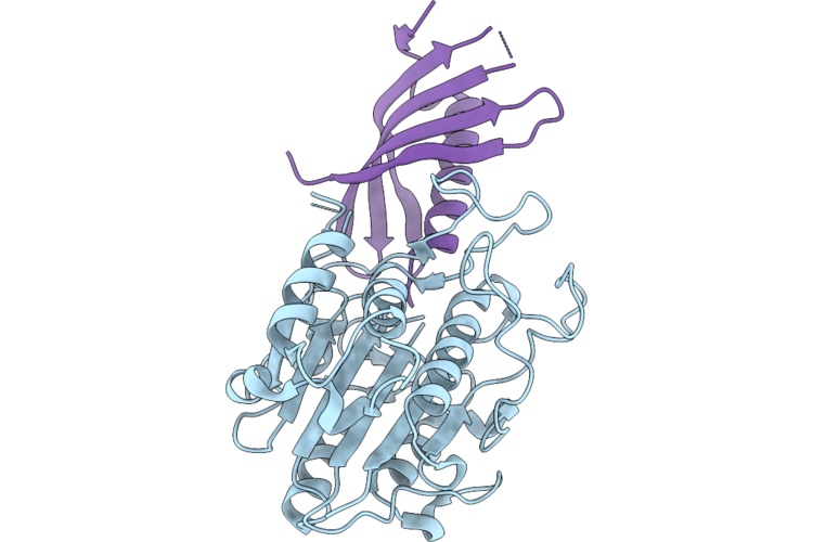 |
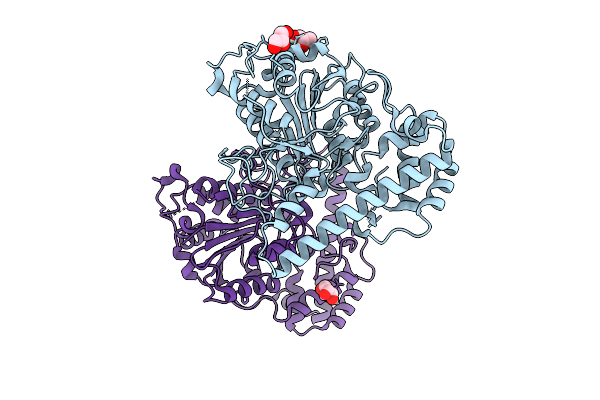
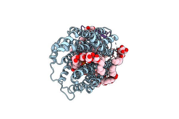
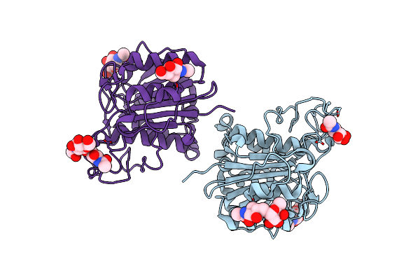
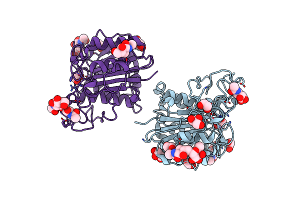
Crystal Structure Of Plant Legumain In Complex With Phytocystatin
Organism: Exallage chrysotricha, Clitoria ternatea
Method: X-RAY DIFFRACTION Resolution:2.30 Å Release Date: 2026-06-10 Classification: PLANT PROTEIN |
Organism: Exallage chrysotricha, Clitoria ternatea
Method: X-RAY DIFFRACTION
Release Date: 2026-06-10
 |
Crystal Structure Of Fgm3 In Complex With Plp
Organism: Gibberella zeae (strain atcc mya-4620 / cbs 123657 / fgsc 9075 / nrrl 31084 / ph-1)
Method: X-RAY DIFFRACTION Resolution:1.67 Å Release Date: 2026-05-20 Classification: BIOSYNTHETIC PROTEIN |
 |
Crystal Structure Of Fgm3 In Complex With Plp And L-Arg
Organism: Gibberella zeae (strain atcc mya-4620 / cbs 123657 / fgsc 9075 / nrrl 31084 / ph-1)
Method: X-RAY DIFFRACTION Resolution:1.90 Å Release Date: 2026-05-20 Classification: BIOSYNTHETIC PROTEIN Ligands: EQJ |
Organism: Gibberella zeae (strain atcc mya-4620 / cbs 123657 / fgsc 9075 / nrrl 31084 / ph-1)
Method: X-RAY DIFFRACTION
Release Date: 2026-05-20
Ligands: EQJ
 |
Crystal Structure Of Fgm3 In Complex With Plp And L-Arg
Organism: Fusarium graminearum ph-1
Method: X-RAY DIFFRACTION Resolution:1.86 Å Release Date: 2026-05-20 Classification: BIOSYNTHETIC PROTEIN Ligands: EQJ, GOL |
Organism: Fusarium graminearum ph-1
Method: X-RAY DIFFRACTION
Release Date: 2026-05-20
Ligands: EQJ, GOL
 |
Crystal Structure Of Fgm3 In Complex With Plp And L-Ala
Organism: Fusarium graminearum ph-1
Method: X-RAY DIFFRACTION Resolution:2.10 Å Release Date: 2026-05-20 Classification: BIOSYNTHETIC PROTEIN Ligands: GOL, 0JO |
Organism: Fusarium graminearum ph-1
Method: X-RAY DIFFRACTION
Release Date: 2026-05-20
Ligands: GOL, 0JO
 |
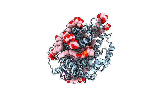
Crystal Structure Of Fgm3 In Complex With Plp And 4(S)-Oh-L-Arg
Organism: Fusarium graminearum ph-1
Method: X-RAY DIFFRACTION Resolution:1.48 Å Release Date: 2026-05-20 Classification: BIOSYNTHETIC PROTEIN Ligands: GOL, PLP, WYK |
Organism: Fusarium graminearum ph-1
Method: X-RAY DIFFRACTION
Release Date: 2026-05-20
Ligands: GOL, PLP, WYK
 |
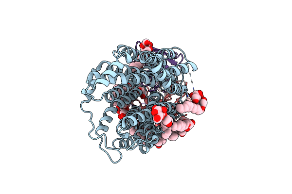
Crystal Structure Of Fgm3 In Complex With Plp And L-Arg
Organism: Fusarium graminearum ph-1
Method: X-RAY DIFFRACTION Resolution:2.18 Å Release Date: 2026-05-20 Classification: BIOSYNTHETIC PROTEIN Ligands: ARG, GOL, EPE |
Organism: Fusarium graminearum ph-1
Method: X-RAY DIFFRACTION
Release Date: 2026-05-20
Ligands: ARG, GOL, EPE
 |
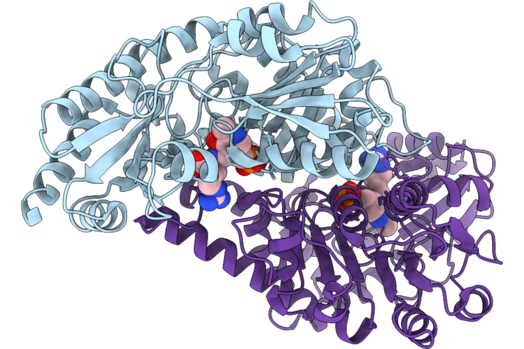
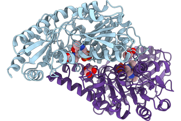
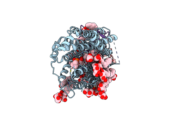
Peptide Asparaginyl Ligases From Viola Dissecta
Organism: Viola dissecta
Method: X-RAY DIFFRACTION Resolution:1.87 Å Release Date: 2026-03-11 Classification: LIGASE Ligands: GOL |
Organism: Viola dissecta
Method: X-RAY DIFFRACTION
Release Date: 2026-03-11
Ligands: GOL
 |
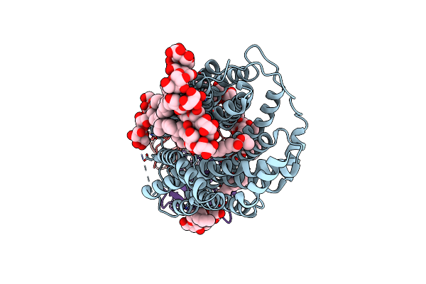
Crystal Structure Of Hydrogen Sulfide-Bound Superoxide Dismutase In Oxidized State
Organism: Bos taurus
Method: X-RAY DIFFRACTION Resolution:1.88 Å Release Date: 2023-09-06 Classification: OXIDOREDUCTASE Ligands: CU, ZN, EDO, GOL, SO4, CL, H2S |
Organism: Bos taurus
Method: X-RAY DIFFRACTION
Release Date: 2023-09-06
Ligands: CU, ZN, EDO, GOL, SO4, CL, H2S
 |
Crystal Structure Of Hydrogen Sulfide-Bound Superoxide Dismutase In Reduced State
Organism: Bos taurus
Method: X-RAY DIFFRACTION Resolution:1.80 Å Release Date: 2023-09-06 Classification: OXIDOREDUCTASE Ligands: CU, ZN, EDO, H2S, SO4, CL, GOL |
Organism: Bos taurus
Method: X-RAY DIFFRACTION
Release Date: 2023-09-06
Ligands: CU, ZN, EDO, H2S, SO4, CL, GOL
 |
Zebrafish Mfsd2A Isoform B In Inward Open Ligand Bound Conformation
Organism: Danio rerio, Mus musculus
Method: ELECTRON MICROSCOPY Release Date: 2023-05-10 Classification: LIPID TRANSPORT Ligands: ZGS, LMT, NA |
Organism: Danio rerio, Mus musculus
Method: ELECTRON MICROSCOPY
Release Date: 2023-05-10
Ligands: ZGS, LMT, NA
 |
Zebrafish Mfsd2A Isoform B In Inward Open Ligand-Free Conformation
Organism: Danio rerio, Mus musculus
Method: ELECTRON MICROSCOPY Release Date: 2023-05-10 Classification: LIPID TRANSPORT Ligands: LMT |
Organism: Danio rerio, Mus musculus
Method: ELECTRON MICROSCOPY
Release Date: 2023-05-10
Ligands: LMT
 |
Zebrafish Mfsd2A Isoform B In Inward Open Ligand 1A Conformation
Organism: Danio rerio, Mus musculus
Method: ELECTRON MICROSCOPY Release Date: 2023-05-10 Classification: LIPID TRANSPORT Ligands: ZGS, LMT, NA |
Organism: Danio rerio, Mus musculus
Method: ELECTRON MICROSCOPY
Release Date: 2023-05-10
Ligands: ZGS, LMT, NA
 |
Zebrafish Mfsd2A Isoform B In Inward Open Ligand 1B Conformation
Organism: Danio rerio, Mus musculus
Method: ELECTRON MICROSCOPY Release Date: 2023-05-10 Classification: LIPID TRANSPORT Ligands: ZGS, LMT, NA |
Organism: Danio rerio, Mus musculus
Method: ELECTRON MICROSCOPY
Release Date: 2023-05-10
Ligands: ZGS, LMT, NA
 |
Zebrafish Mfsd2A Isoform B In Inward Open Ligand 2B Conformation
Organism: Danio rerio, Mus musculus
Method: ELECTRON MICROSCOPY Release Date: 2023-05-10 Classification: LIPID TRANSPORT Ligands: ZGS, LMT |
Organism: Danio rerio, Mus musculus
Method: ELECTRON MICROSCOPY
Release Date: 2023-05-10
Ligands: ZGS, LMT
 |
Zebrafish Mfsd2A Isoform B In Inward Open Ligand 3C Conformation
Organism: Danio rerio, Mus musculus
Method: ELECTRON MICROSCOPY Release Date: 2023-05-10 Classification: LIPID TRANSPORT Ligands: ZGS, LMT |
Organism: Danio rerio, Mus musculus
Method: ELECTRON MICROSCOPY
Release Date: 2023-05-10
Ligands: ZGS, LMT
 |
L-Leucine Dehydrogenase From Exiguobacterium Sibiricum
Organism: Exiguobacterium sibiricum (strain dsm 17290 / cip 109462 / jcm 13490 / 255-15)
Method: X-RAY DIFFRACTION Resolution:3.02 Å Release Date: 2023-04-26 Classification: OXIDOREDUCTASE Ligands: GOL, CA |
Organism: Exiguobacterium sibiricum (strain dsm 17290 / cip 109462 / jcm 13490 / 255-15)
Method: X-RAY DIFFRACTION
Release Date: 2023-04-26
Ligands: GOL, CA
 |
The Crystal Structure Of Vypal2-C214A, A Dead Mutant Of Vypal2 Peptide Asparaginyl Ligase In Form Ii
Organism: Viola philippica
Method: X-RAY DIFFRACTION Resolution:1.80 Å Release Date: 2022-07-13 Classification: PLANT PROTEIN Ligands: NAG |
Organism: Viola philippica
Method: X-RAY DIFFRACTION
Release Date: 2022-07-13
Ligands: NAG
 |
The Crystal Structure Of Vypal2-I244V, A More Efficient Mutant Of Vypal2 Peptide Asparaginyl Ligase In Its Active Enzyme Form
Organism: Viola philippica
Method: X-RAY DIFFRACTION Resolution:1.59 Å Release Date: 2022-06-29 Classification: PLANT PROTEIN Ligands: NAG, EDO, GOL |
Organism: Viola philippica
Method: X-RAY DIFFRACTION
Release Date: 2022-06-29
Ligands: NAG, EDO, GOL