

Search Count: 12
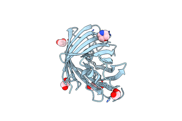 |
Structure Of Rscherry Crystallized In Anaerobic Conditions
Organism: Discosoma sp.
Method: X-RAY DIFFRACTION Resolution:1.55 Å Release Date: 2023-04-05 Classification: FLUORESCENT PROTEIN Ligands: PEG, MES, EDO, GOL |
Organism: Discosoma sp.
Method: X-RAY DIFFRACTION
Release Date: 2023-04-05
Ligands: PEG, MES, EDO, GOL
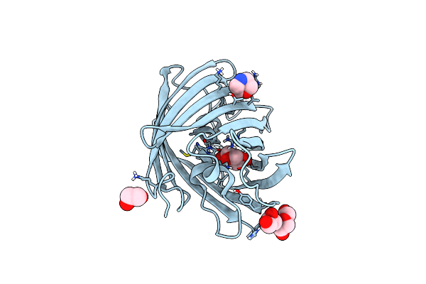 |
Structure Of Oxygen-Degraded Rscherry
Organism: Discosoma sp.
Method: X-RAY DIFFRACTION Resolution:1.30 Å Release Date: 2023-04-05 Classification: FLUORESCENT PROTEIN Ligands: GOL, MES, PEG, EDO |
Organism: Discosoma sp.
Method: X-RAY DIFFRACTION
Release Date: 2023-04-05
Ligands: GOL, MES, PEG, EDO
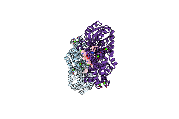 |
Crystal Structure Of A Halotag-Based Calcium Indicator, Halocamp V2, Bound To Jf635
Organism: Rhodococcus sp.
Method: X-RAY DIFFRACTION Resolution:2.00 Å Release Date: 2020-09-30 Classification: HYDROLASE Ligands: PUJ, CL, CA |
Organism: Rhodococcus sp.
Method: X-RAY DIFFRACTION
Release Date: 2020-09-30
Ligands: PUJ, CL, CA
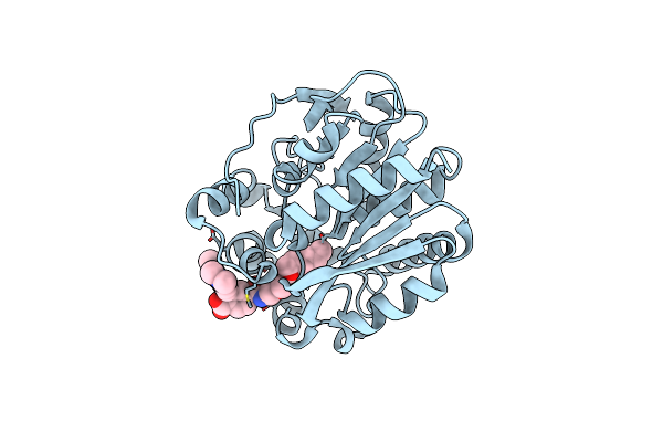 |
Crystal Structure Of Halotag Bound To Tetramethylrhodamine-Halotag Ligand
Organism: Rhodococcus sp.
Method: X-RAY DIFFRACTION Resolution:1.80 Å Release Date: 2020-09-30 Classification: HYDROLASE Ligands: PVY, CL |
Organism: Rhodococcus sp.
Method: X-RAY DIFFRACTION
Release Date: 2020-09-30
Ligands: PVY, CL
 |
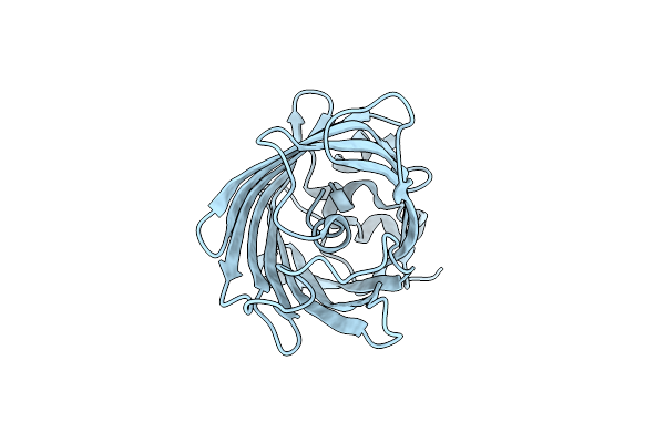
Structure Of Rsgreen0.7 In The Green-Off-State
Organism: Aequorea victoria
Method: X-RAY DIFFRACTION Resolution:1.20 Å Release Date: 2015-09-09 Classification: FLUORESCENT PROTEIN |
Organism: Aequorea victoria
Method: X-RAY DIFFRACTION
Release Date: 2015-09-09
 |
Structure Of Rsgreen0.7 In The Green-On-State
Organism: Aequorea victoria
Method: X-RAY DIFFRACTION Resolution:1.25 Å Release Date: 2015-09-09 Classification: FLUORESCENT PROTEIN |
Organism: Aequorea victoria
Method: X-RAY DIFFRACTION
Release Date: 2015-09-09
 |
Structure Of Pcdronpa-A69T Mutant
Organism: Echinophyllia
Method: X-RAY DIFFRACTION Resolution:2.15 Å Release Date: 2014-02-05 Classification: FLUORESCENT PROTEIN Ligands: K |
Organism: Echinophyllia
Method: X-RAY DIFFRACTION
Release Date: 2014-02-05
Ligands: K
 |
Crystal Structure Of A Green-To-Red Photoconvertible Dronpa, Pcdronpa In The Green-On-State
Organism: Echinophyllia
Method: X-RAY DIFFRACTION Resolution:1.95 Å Release Date: 2013-11-13 Classification: FLUORESCENT PROTEIN Ligands: EPE, PGE, PEG |
Organism: Echinophyllia
Method: X-RAY DIFFRACTION
Release Date: 2013-11-13
Ligands: EPE, PGE, PEG
 |
Crystal Structure Of A Green-To-Red Photoconvertible Dronpa, Pcdronpa In The Green-Off-State
Organism: Echinophyllia
Method: X-RAY DIFFRACTION Resolution:2.07 Å Release Date: 2013-11-13 Classification: FLUORESCENT PROTEIN Ligands: PEG, PGE, PG4 |
Organism: Echinophyllia
Method: X-RAY DIFFRACTION
Release Date: 2013-11-13
Ligands: PEG, PGE, PG4
 |
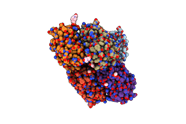
Crystal Structure Of A Green-To-Red Photoconvertible Dronpa, Pcdronpa In The Red-On-State
Organism: Echinophyllia
Method: X-RAY DIFFRACTION Resolution:2.05 Å Release Date: 2013-11-13 Classification: FLUORESCENT PROTEIN Ligands: PGE, PEG, PG4 |
Organism: Echinophyllia
Method: X-RAY DIFFRACTION
Release Date: 2013-11-13
Ligands: PGE, PEG, PG4
 |
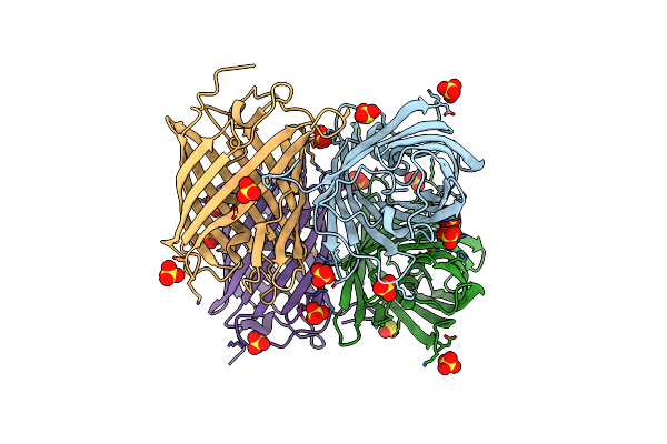
Crystal Structure Of A Single Mutant Of Dronpa, The Green-On-State Pdm1-4
Organism: Echinophyllia sp. sc22
Method: X-RAY DIFFRACTION Resolution:1.95 Å Release Date: 2012-11-21 Classification: FLUORESCENT PROTEIN Ligands: EPE, 1PE, PEG |
Organism: Echinophyllia sp. sc22
Method: X-RAY DIFFRACTION
Release Date: 2012-11-21
Ligands: EPE, 1PE, PEG
 |
Crystal Structure Of Meosfp In Its Green State
Organism: Lobophyllia hemprichii
Method: X-RAY DIFFRACTION Resolution:2.25 Å Release Date: 2011-10-19 Classification: FLUORESCENT PROTEIN Ligands: SO3, SO4 |
Organism: Lobophyllia hemprichii
Method: X-RAY DIFFRACTION
Release Date: 2011-10-19
Ligands: SO3, SO4